Received for publication 09/11/2015 - Accepted for publication 11/12/2015 The authors declare no conflict of interest.
Ophthalmic changes in cleft lip and palate
Alterações oftalmológicas na fissura lábio palatina
Luciano Sólia Násser1, Daniella Reis Barbosa Martelli1, Mário Sérgio Oliveira Swerts2, Daniela Araújo Veloso Popoff1, Letízia Monteiro de Barros2, Hercílio Martelli Júnior1,2
1 Health Science Programme, Universidade Estadual de Montes Claros, Montes Claros, MG, Brazil.
2 Centre for Rehabilitation of Craniofacial Anomalies, Universidade José do Rosário Vellano, Alfenas, MG, Brazil. Institution:Universidade Estadual de Montes Claros, Montes Claros, MG, Brazil.
R
ESUMOO presente estudo teve como objetivo analisar evidências de associação entre as alterações oculares e fissuras lábio palatinas não sindrômicas (FL/PNS), através de uma revisão da literatura. Foi realizada a revisão da literatura com pesquisa sistemática, observan-do o protocolo de colaboração com o Grupo Cochrane. PubMed, Scopus, Google Acadêmico e ISI-Web of Science. A partir de16 estudos acessados, 3 compuseram a amostra final. Todos os trabalhos da amostra final relataram alterações oculares em pacientes com FL/PNS. Os artigos relataram respectivamente alterações oculares em 6,21%, 17,54% e 1,03% dos pacientes. A presença de alterações oculares em pacientes com FL /PNS foi significativa nesta revisão sistemática, mas todos os três artigos sugerem que futuros estudos deverão explorar a possibilidade de que haja um aumento de alterações oculares em indivíduos com FL/PNS.
Descritores:Transtornos da visão; Manifestações oculares; Coloboma; Fissura palatina; Fissura labial
A
BSTRACTThe current study aimed to analyze through a literature review evidence of association between ocular changes and non-syndromic cleft lip and/or palate (NSCL/P). A literature review was carried out in accordance with the Cochrane Collaboration Group protocol. PubMed, Scopus, Academic Google and ISI Web of Science databases were systematically searched. A total of 16 studies were accessed, and three made up the final sample. All three studied ocular abnormalities in patients with NSCL/P. The articles found ocular abnormalities in 6.21%, 17.54% and 1.03% of patients respectively. The presence of ocular abnormalities in patients with NSCL/P was significant in this systematic review, but the articles all agreed that future studies should explore the possibility of a greater occurrence of ocular changes in individuals with NSCL/P.
I
NTRODUCTIONO
rofacial malformations are the most common form of congenital anomalies in the world(1). Among them, themost prevalent is the cleft lip with or without cleft palate (CL/P)(2-5), which may occur more commonly in an isolated and
non-syndromic form as a specific phenotype or, more rarely, composing several associations or syndromes(6,7). Embryologically,
clefts result from primary fusion defects of the craniofacial pro-cesses that form the primary and secondary palate in the first intrauterine trimester (8). The incidence of CL/P varies according
to geographical location, racial and ethnic groups, environmental exposures, and socioeconomic status, affecting approximately 1/ 700 live births with wide variability across geographic origin. Generally, Asian and Amerindian populations have the highest reported birth prevalence rates, often as high as 1/500, European-derived populations have intermediate prevalence rates at about 1/1,000, and African-derived populations have the lowest prevalence rates at about 1/2,500(5,9,10).
According to Vieira(7), the last decade was crucial in
clarifying issues concerning the etiology of CL/P when compared to other defects observed at birth. As this is a multifactorial trait, environmental risk factors such as smoking, alcohol, parental age, medications, birth order, inter-pregnancy interval, and folic acid deficiency are listed as modifiers. Risk factor identification is the first step to better understanding and preventing such craniofacial changes(7,11).
CL/P could be associated with many other structural abnormalities of the adjacent vital structures of the face like the ears, eyes, nose, teeth and brain. CL/P are inherently known to produce functional problems affecting the oropharynx (feeding and breathing), hearing, vision and speech; in addition, they have a negative cosmetic effect(12).Almost all skeletal and soft tissue
components of the craniofacial area are unbelievably derived from the neural crest cells (12). Because eyes originate as an
extension of the forebrain, malformations involving ocular structures invariably accompany those of the face and brain and vice versa (12).
The current study aimed to provide additional evidence by means of review of the literature.
M
ETHODSThe present review was carried out in accordance with the Cochrane Collaboration Group protocol for systematic reviews(13), including a literature search strategy, selection of
papers through the inclusion and exclusion criteria, data extraction, and quality assessment.
Literature search strategy
Our review was performed in July 2014 in order to obtain literature regarding NSCL/P and ocular changes. PubMed, Scopus, Google and ISI - Web of Science databases were systematically searched using the following approaches: the search strings “ocu-lar changes” AND “cleft lip and palate”, “ocu“ocu-lar findings” AND “cleft lip and palate”, “ocular features” AND “cleft lip and palate”, “ocular disorders” AND “cleft lip and palate”. The same happened with the terms “oral clefts” and “orofacial clefts” for studies published up to that time.
Selection of papers through the inclusion and exclusion criteria
The selection of papers is diagrammatically explained in Figure 1. Of the 16 studies originally found through the literature
search strategy and the references of the potential studies retrieved, three studies was excluded (for being in a language other than English with no full text available), and another was excluded because it did not describe the ocular abnormalities found. A further nine studies involving syndromic oral clefts were subsequently excluding. After wards, only articles presenting ocular changes with NSCL/P were included in the present study. Thus, three studies were selected (Figure 1).
Data extraction
Title and abstract screening was performed by two reviewers (LSN and HMJ) who worked independently to identify potentially relevant papers for which full text publications were retrieved. If, however, there was any discrepancy of opinion, the reviewers reexamined the paper together and arrived at a joint final decision. A standardized form was used to extract information, such as author and year of publication of the paper, origin of participants, study design, sample size, type of oral clefts and type of ocular changes associated with non-syndromic oral clefts.
R
ESULTSThe initial database search identified 16 citations published between 1977 and 2013. After screening, nine studies were excluded (because they described ocular changes in patients with syndromic CL/P), leaving seven papers. A second screening was performed on the remaining papers and four studies were excluded (three articles were in a language other than English with no full text available and one article did not describe the ocular changes found). Thus, three papers were selected for the systematic review.
The characteristics of the three selected papers are presented in Table 1. All three used a prospective study design and reported the frequency of ocular changes associated to
NSCL/P. The sample sizes and the population investigated varied considerably among the studies, not being homogeneous: one studied 322 patients, one studied 57 and the other studied 622 patients. Concerning the country of origin of articles: one was from the United States, one from Turkey and one from India (Table 1). Each study has its own classification of cleft type: one classified the clefts as unilateral or bilateral, one as CL, CP or CL/P and one did not separate the types of clefts. All three studied ocular abnormalities in patients with NSCL/P. The type of population studied was different among the three articles: one studied the ocular changes in patients with cleft lip and palate attending the three departments of K. S. Hegde Charitable Hospital, namely, Maxillofacial, Ophthalmology and Pediatrics. One studied patients with cleft lip and/or palate who sought orthodontic help at the Dental Hospital, Izmir, Turkey and one studied children admitted at the Jubilee Mission Medical College, Trichur for cleft lip or cleft palate repair. The age of the patients was similar in the three studies: one studied patients over the age of 20, one over 18 and the other over 19. Regarding ocular changes in each article, all classified them according to the modified anatomical region of the eye and its annexes (Table 2).
Two articles carried out a categorization relating to cleft type with associated ocular abnormalities(12,14). An article found
48.14% of ocular manifestations in patients with bilateral CL/P, 18.5% of changes in patients with unilateral CL/P, 7.4% in patients with facial clefts, 7.40% in patients with clefts with alveolus and 18.5% in patients with recognizable syndromes(12). The other
article found 64% of ocular manifestations in patients with bila-teral CL/P, 16% in patients with unilabila-teral CL/P, 10.5% in patients with isolated cleft palate and 8,7% in isolated cleft lip(14).
An article(12) found ocular abnormalities in 6.21% of patients.
Changes in the eyelid were the most frequent (30%), followed by orbital defects (20%), abnormalities of the lacrimal duct (10%), limb defects (10%), cataract (5%) and retinal coloboma (5%). Furthermore the article found refractive error in 15% of patients. The other article(14) found ocular abnormalities in 17.54%
of patients. The most frequent ocular disease was congenital nasolacrimal duct obstruction (50%), followed by bilateral iris coloboma (20%), eyelid (10%), dermoid tumor (10%), and esophoria (10%). The other article(15) found ocular changes in
1.03% of patients. The most frequent ocular disease was coloboma (57.14%), followed by eyelid (14.28%), congenital nasolacrimal duct obstruction (14.28%) and congenital esotropia (14.28%).
D
ISCUSSIONThis literature review assessed available studies describing ocular changes in patients with non-syndromic oral clefts. We demonstrated that there is a shortage of literature about ocular changes in patients with NSCL/P. Our results also demonstrated that the published studies disagree on the frequency of non-syndromic oral clefts. One article says: cleft lip and palate represents the second most frequently occurring congenital deformity after clubfoot deformity(12). Another article says: cleft
Table 2
Ocular changes in each of the three articles selected.
Ocular Total Eyelid Squint Orbital Nasolacrimal Limbal Cataract Colobomas changes defects abnormalities dermoids
% % % % % % % %
Anchlia et al., 6.21 30 20 20 10 10 5 5 2011
Yaman et al., 17.54 10 10 50 10 20 2011
Shobha et al., 1.03 14.28 14.28 14.28 57.14 2011
Author
Table 1
Main characteristics of the three selected papers
First author, Country Cleft Frequency of ocular Study Number of patients Age of year of publication of study classification abnormalities % design evaluated patients
Anchlia et al., EUA Bilateral CLP, 6.21 Prospective 322 Up to
2011 unilateral CLP 20 year Yaman et al., Turkey CL, CP, CLP 17.54 Prospective 57 Up to 2009 18 years
Shobha et al., India CLP, without 1.03 Prospective 674 6 months 2011 separating the to 19 years
lip and/or cleft palate are among the common congenital anomalies of the head and neck region(14). The other article says:
cleft lip and palate are common birth defects formed due to failure of fusion of the maxillary and mandibular processes between the fifth and ninth weeks of pregnancy. They are found in one in every 1,000 newborns(15).
Other studies about CL/P demonstrated that the studies published agree that non-syndromic oral clefts are one of the most common human malformations, with an average prevalence of 1 per 700 or 1,000 live births(2,4,5,7,16), as well as that its incidence
varies according to gender: 2:1 being the ratio of males to females for cleft lip and palate and 1:2 the approximate ratio of males to females for isolated cleft palate(1,10).
Other common information between the studies about CL/P is the fact that unilateral clefts are more common than bilateral clefts, and of the unilateral cases of non-syndromic cleft lip and palate, left-sided cleft lips occur more frequently than right-sided cleft lips(4,5,16,17). Many studies have also demonstrated
that genetic factors may play a role in the cause of non-syndromic oral clefts in addition to certain environmental and/or stochastic factors, meaning that this malformation is a multifactorial trait(2,16,18). Similarly, studies have shown that cleft lip, with or
without cleft palate, is entirely different from isolated cleft palate from both embryological and pathogenetic standpoints(18,19).
The pathogenesis of the ocular abnormalities associated with CL/P poses a problem. The most probable explanation is detective organogenesis in the early embryonic period when all of the structures involved are forming the facial structures, the anterior segment of the eye and the eyelids(20). The mechanisms
behind movement and fusion of mesodermal processes have been studied extensively in experimental oral clefting, but the findings may apply to other clefting anomalies(21,22). Genetic
regulation is critical to successful ocular embryogenesis. The two genes that have been described as most important in ocular development are the PAX6 gene (chromosome 11p13) and the Rx gene (chromosome 18). Both genes belong to a large family of factors that are related to the homeodomain region of the Drosophila paired protein. Early induction of both genes causes a series of gene activations and depressions which are tantamount to normal development of the mature eye. Rx and PAX6 are both expressed in proliferating cells. Mutations of PAX6 have been shown to lead to aniridia, congenital cataract, Peter’s anomaly, and midline fusion defects. Absence of the gene leads to anophthalmia. Rx is associated with retinal proliferation(23).
The defects in the eye may be viewed as showing similar defective mesenchymal movement and fusion. During the period between 17 and 22 weeks, three mesodermal tissue migrations normally occur from the rim of the optic cup(24). In turn, they
form the corneal endothelium, the cells of the corneal stroma and the mesodermal portion of the iris. These migrations have the additional effect of separating the developing lens from the cornea. Defective mesodermal migrations from the upper rim of the optic cup would account for lack of corneal development, failure of lens separation and incomplete aniridia superiorly. At the same time, the eyelids are forming above and below the eye as a fold of mesodermal covered by ectoderm and are advancing toward their partial fusion over the central cornea. The eyelid coloboma may represent an inappropriate fusion of the epithelium associated with cell death and disease characterized by CL / P, and eyelid coloboma is called oral clefts(20).
According to the Carnegier stages, the major congenital malformations of the eye and its attachments occur between the
fourth and eighth weeks of the embryonic period while the cleft lip and palate occur between the sixth and eighth weeks. That is, there is a common embryonic period from the sixth to the eighth week in which occur both ocular and oral alterations(24).
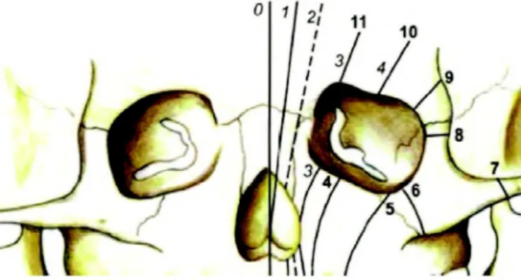
Tessier’s classification is used for patients with congenital clefts and lid coloboma. Tessier’s number 3, 4, 5 affect the eye (Figure 2).
Figure 2: Oro-ocular clefts and the Tessier’s classification.
Source: Shobha ME, Joseph A, Adenwalla HS, Narayanan PV, Kakkanat CV, Ocular findings in cleft lip and cleft palate patient. Kerala J Ophthalmol. 2011;23(4):358-60.
There was not a consensus in the reviewed articles regarding the type of ocular change most often related to non-syndromic CL/P, as well as any article deepened the study relating these changes to CL/ P, based on embryological and genetic concepts.
Thus the hypothesis that there is some correlation between ocular manifestations and non-syndromic CL/P cannot be confirmed by this systematic review.
To date, there is no known genetic or epigenetic explanation for the ocular changes described in the reviewed articles that can be correlated with the causative genes for non-syndromic CL/P. The articles are in agreement that future studies should explore the possibility that there is a preferential occurrence of ocular changes in individuals with non-syndromic CL/P and test the hypothesis that common genetic and epigenetic mechanisms are playing a role in both conditions. Through this information, future studies may be better able to identify the causes of non-syndromic orofacial clefts and ultimately to predict its occurrence and to facilitate genetic counseling of affected families(16).
Cleft lip and palate are usually repaired early in life. However, the ocular complications may be progressive and threaten sight. It is therefore important that these patients be under long-term ophthalmic supervision to try to prevent the sight threatening ocular complications(25). Thus, when ocular
disorders associated with CL/P are identified, the appropriate eye tracking is essential in the prevention of serious consequences, since the loss of vision can be more disabling in patients with CL/P.
A
CKNOWLEDGEMENTSR
EFERENCES1. Marazita L M. The evolution of human genetic studies of cleft lip and cleft palate. Annu Rev Genomics Hum Genet. 2012;13:263-83. 2. Taioli E, Ragin C, Robertson L, Linkov F, Thuman LE, Vieira AR. Cleft lip and palate in family members of cancer survivors. Can-cer Invest. 2010;28(9):958-62.
3. Klassen AF, Anthony SJ, Khan A, Sung L, Klaassen R. Identifying determinants of quality of life of children with cancer and child-hood cancer survivors: a systematic review. Support Care Cancer. 2011;19(9):1275-87.
4. Herkrath AP, Herkrath FJ, Rebelo MA, Vettore MV. Parental age as a risk for non syndromic oral clefts: a meta analysis. J Dent. 2012;40(1):3-14.
5. Dietz A, Pedersen DA, Jacobsen R, Wehby GL, Murray JC, Christensen K. Risk of breast cancer in females with cleft lip and palate. Ann Epidemiol. 2012;22(1):37-42.
6. Martelli Junior H, Porto LC, Barbosa DR, Bonan PR, Freitas AB, Coletta RD. Prevalence of nonsyndromic oral clefts in a refer-ence hospital in the state of Minas Gerais, Brazil, between 2000 2005. Braz Oral Res. 2007;21(4):314-7.
7. Vieira AR. Unraveling human cleft lip and palate research. J Dent Res. 2008;87(2):119-25.
8. Wantia N, Rettinger G. The current understanding of cleft lip malformations. Facial Plast Surg. 2002;18(3):147 53.
9. Cobourne MT. The complex genetics of cleft lip and palate. Eur J Orthod. 2004;26(1):7-16.
10. Dixon M, Marazita ML, Beaty TH, Murray JC. Cleft lip and pal-ate: synthesizing genetic and environmental influences. Nat Rev Genet. 2011;12(3):167–78.
11. Paranaiba LM, Miranda RT, Ribeiro LA, Barros LM, Martelli Júnior H. Frequency of congenital craniofacial malformations in a Brazil-ian Reference Center. Rev Bras Epidemiol. 2011;14:151 60. 12. Anchlia S, Rao KS, Bonanthaya K, Anupama B, Nayak LV.
Oph-thalmic considerations in cleft lip and palate patients. J Maxillofac Oral Surg. 2011;10(1):14-19.
13. Alderson P, Green S, Higgins JP. Cochrane Reviewers’ Handbook 4.2.2 [updated March 2004]. The Cochrane Library, Issue 1, 2004.
Corresponding author
Luciano Sólia Násser
Rua Walter Ferreira Barreto, 57- Zip code: 39401-347Montes Claros, Minas Gerais, Brazil
Tel: + 55 38 9132:5452
E-mail address: [email protected]
14. Yaman A, Saatçi P, Arýkan G, Soylu A, Saatçi AO, Kavukçu S.Ocular findings in children with nonsyndromic cleft lip and palate. TurkJ Pediatr. 2009;51(4):350-3.
15. Shobha ME, Joseph A, Adenwalla HS, Narayanan PV, Kakkanat CV. Ocular findings in cleft lip and cleft palate patient. Kerala J Ophthalmol. 2011;23(4):358-60.
16. Steinwachs EF, Amos C, Johnston D, Mulliken J, Stal S, Hecht JT. Nonsyndromic cleft lip and palate is not associated with cancer or other birth defects. Am J Med Genet. 2000;90(1):17-24. 17. Jugessur A, Farlie PG, Kilpatri ck N.The genetics of isolated
orofacial clefts: from genotypes to subphenotypes. Oral Dis. 2009;15(7):437-53.
18. Murray JC. Genetic environment causes of cleft lip and or palate. Clin Genet. 2002;61(4):248-56.
19. Kot M, Kruk Jeromini. Analysis of family incidence of cleft lip and or palate. J Med Sci Monit. 2007;13(5):231-4.
20. Kinsey JA, Streeten BA. Ocular abnormalities in the median cleft face syndrome. Am J Opthalmol. 1977;83(2):261-6. 21. Hassel JR, Orkin RW. Synthesis and distribution of collagen in
the rat palate during shelf elevation. Dev Biol. 1976;49(1):80-8. 22. Sedano HO, Cohen MM, Jirasek J, Gorlin RJ. Frontonasal
dyspla-sia. J Pediatr. 1970;76(6):906-13.
23. Edward DP, Kaufman LM. Anatomy, development, and physiol-ogy of the visual system. Pediatr Clin North Am. 2003;50(1):1-23. 24. Guercio JR, Martin LJ. Congenital malformations of the eye and
orbit. Otolaryngol Clin North Am. 2007;40(1):113-40.