Heglayne Pereira Vital da SILVA(a)
Thaynnan Thómaz Silva ARRUDA(a)
Karla Simone Costa de SOUZA(a)
João Felipe BEZERRA(a)
Gisele Correia Pacheco LEITE(b)
Maria Edinilma Felinto de BRITO(b)
Valéria Morgiana Gualberto Duarte Moreira LIMA(a)
André Ducati LUCHESSI(a)
Raul Hernandes BORTOLIN(a)
Marcela Abbott Galvão URURAHY(a)
Adriana Augusto de REZENDE(a)
(a) Universidade Federal do Rio Grande do Norte – UFRN, Department of Clinical and Toxicological Analyses, Natal, RN, Brazil.
(b) Universidade Federal do Rio Grande do Norte – UFRN, Department of Pediatrics, Natal, RN, Brazil.
Declaration of Interest: The authors certify that they have no commercial or associative interest that represents a conflict of interest in connection with the manuscript.
Corresponding Author:
Adriana Augusto de Rezende E-mail: adrirezende@yahoo.com
https://doi.org/10.1590/1807-3107bor-2018.vol32.0024
Submitted: September 13, 2017
Accepted for publication: January 31, 2018 Last revision: February 19, 2018
Risk factors and comorbidities in
Brazilian patients with orofacial clefts
Abstract: Considering that environmental risk factors substantially contribute to the etiology of orofacial clefts and that knowledge about the characteristics and comorbidities associated with oral clefts is fundamental to promoting better quality of life, this study aimed to describe the risk factors, main characteristics, and comorbidities of a group of patients with cleft lip and/or cleft palate (CL/P) from Rio Grande do Norte (RN), Brazil. Data were obtained from 173 patients with CL/P using a form from the Brazilian database on Orofacial Clefts. Most patients were male with cleft lip and palate and had a normal size and weight at birth; presented few neonatal intercurrent events; and had anemia and respiratory and cardiovascular diseases as main associated comorbidities. They also required timely surgical rehabilitation and multidisciplinary care to stimulate their neuropsychomotor development. In addition, a high frequency of familial recurrence and of parental consanguinity was evidenced in the studied population, especially for the cleft lip and cleft palate type. Other relevant findings were the considerable maternal exposure to alcohol, infections, smoking, and hypertension, as well as low supplementation with vitamins and minerals and deliberate consumption of analgesics, antibiotics, and antihypertensives during pregnancy. Characterization of the CL/P patient profile is essential for the planning of health services and integration among the health professionals involved in the diagnosis and treatment of these malformations. Our results reinforce the need for additional research to confirm the association between environmental factors and the development of orofacial clefts.
Keywords: Cleft Lip; Cleft Palate; Comorbidity; Risk Factors; Environmental Exposure.
Introduction
Cleft lip and/or cleft palate (CL/P) is a congenital malformation characterized by the lack of fusion of the upper lip and/or palate, which may be present in isolation or in association with a syndrome.1 The
highest prevalence at birth of CL/P is found in Asian and native American populations (1 in 500 live births), while the lowest prevalence is observed in populations of African descent, with approximately 1 in 2,500 live births.2
Northeast has the lowest one.3 In recent years, however,
there has been an upward trend in the reported CL/P prevalence in the northern and northeastern regions attributed mainly to improved notification to the National Health Information System or, alternatively, to changes in risk factors.4 In Rio Grande do Norte
(RN) state, in the northeastern region, a previous study reported a prevalence of 4.9 per 10,000 live births between 2000 and 2005, remaining within the incidence range of 4.82 to 5.50 per 10,000 live births between the years of 2009 and 2013.3,4,5
The etiology of CL/P is attributed to genetic susceptibility and to maternal exposure to environmental risk factors, including smoking, alcohol consumption, medications, and vitamin deficiencies during pregnancy.2
According to some studies, alcohol consumption can inhibit retinoic acid production, increasing the risk of CL/P.6,7 Smoking during pregnancy apparently doubles
the risk of orofacial cleft in newborns. Moreover, in vitro studies have shown that tobacco inhibits palatal fusion and affects cell proliferation, leading to medial edge epithelial cell death.1 In addition, some
drugs such as anticonvulsants with antifolate activity, antihypertensives, and corticosteroids administered during morphogenesis may lead to CL/P through different cellular mechanisms.8 By contrast, folic acid
supplementation, alone or in combination with vitamins and minerals, prevents the development of neural tube defects, and its use from before conception to 12 weeks’ gestation is recommended by the World Health Organization (WHO); however, there is no clear evidence of its preventive effect on CL/P.9
Family history is also an important factor associated with CL/P development; actually, it has been described as the most important factor in patients with clefts.10
Familial recurrence is very common among CL/P patients, and their relatives have a high risk compared to the general population, but the risk decreases with increasing genetic distance between relatives.11
The strong familial aggregation is ascribed to the multifactorial threshold model of inheritance that is characteristic of orofacial clefts, in which the probability of sharing alleles that are identical by descent is constant whether one, a few, or many genes control risk.12
In addition to facial deformity, CL/P patients usually present several associated comorbidities such
as feeding difficulties, speech problems, dentition defects, dental malocclusion, abnormal facial growth, middle ear infections, and psychological disorders, which can be minimized or prevented through timely surgical treatment and follow-up by a multidisciplinary team.13 Early identification of these abnormalities
and intervention are essential for the appropriate neuropsychomotor development of CL/P patients.
Considering the paucity of data on characteristics associated with the multifactorial nature of CL/P in RN, northeastern Brazil, and the need for early identification of the main CL/P complications for proper monitoring and intervention, the present study aimed to describe the characteristics, main risk factors, and associated comorbidities of a group of CL/P patients from RN, Brazil.
Methodology
Study participants
A total of 173 patients aged 1 month to 21 years presenting with CL/P either as a single entity or in combination with other diseases were recruited from the Pediatrics Unit of the Children’s Hospital of the Federal University of Rio Grande do Norte (UFRN), Natal, RN, Brazil, from April 2013 to May 2015. The patients were evaluated and diagnosed by the Orofacial Cleft Multidisciplinary Program, which included a group of pediatricians, radiologists, speech therapists, cardiologists, and geneticists. The CL/P patients were classified into three groups according to Fogh-Andersen: cleft lip and palate (CLP), cleft palate (CP), and cleft lip (CL).14
The study, which is an integral part of Brazil’s Craniofacial project,15 was conducted according to the
guidelines set by the Research Ethics Committee of the UFRN, in compliance with the Declaration of Helsinki (process number 328.230). An informed consent was obtained from all adult participants and from the parents or legal guardians of underage patients.
Data acquisition
were applied after routine pediatrician visits by trained pharmacists or undergraduate students in a private room in the Pediatrics Unit. All patients treated at the hospital during the study period were invited to participate, and those who agreed were included in the study. Those patients whose mothers or guardians did not sufficiently answer the questionnaire were excluded from the study (173 out of 180 participants remained). The form included retrospective patient information such as type and severity of cleft, gender, birth weight, birth length, head circumference at birth, neonatal and personal history, and neuropsychomotor development. Data on surgical lip and palate rehabilitation were also assessed. The questionnaire also covered retrospective parent information such as age at conception, educational level, mother’s occupation during pregnancy, family history of orofacial clefts, and parental consanguinity. Obstetrical data, alcohol intake, smoking, and illicit drug use at any time during pregnancy, diagnosis of gestational diabetes, and medications used during pregnancy were also retrieved.
Data analysis
The results were presented as absolute numbers (n) and as frequency (%).Weight, length, and head circumference at birth were grouped into lower, middle, or higher according to the WHO child growth standards. Differences between categorical variables were tested by χ2 analysis or Fisher’s exact
test. Significance was established at p < 0.05. Data were analyzed using SPSS version 15.0 (SPSS Inc., Chicago, IL, USA).
Results
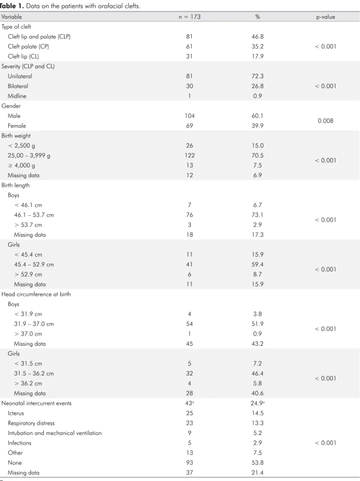
The characteristics of CL/P patients are shown in Table 1. There was a higher prevalence of CLP, followed by CP and CL (p < 0.001), regarding the type of cleft. Unilateral clefts prevailed over bilateral ones, and there was a higher frequency of CL/P in male than in female patients (p = 0.008). At birth, most patients (70.5%) weighed between 2,500 g and 3,999 g, and body lengths ranged from 46.1 cm to 53.7 cm among boys and 45.4 to 52.9 cm among girls. Most boys had a head circumference at birth
between 31.9 cm and 37.0 cm, compared to 31.5 cm to 36.2 cm in girls. Almost 25% of all patients had some neonatal intercurrent event, especially icterus and respiratory distress.
Associated comorbidities or complications were present in 45.7% of the patients (Table 1). The most frequent comorbidities included anemia (16.2%), followed by respiratory diseases such as asthma, rhinitis, cold, and influenza (12.1%); cardiovascular diseases such as patent foramen ovale and ventricular septal defect (9.8%); and neurological diseases such as autism and epilepsy (8.1%).
Eighty-two patients (47.4%) showed appropriate neuropsychomotor development for their age. However, 8.1% showed concomitant motor, speech, and behavioral delay or diagnosis of neuropsychomotor development delay, while 6.9% presented only speech delay. Half of the patients did not attend any supportive therapy. Almost 30% of those who underwent therapy attended speech therapy.
Lip and palate surgical repair outcomes are shown in Table 2. A total of 49 CLP or CP patients had already undergone their first palatoplasty and 67 had already undergone their first cheiloplasty. Most cheiloplasty patients were aged 6 to 12 years (46.9%), while palatoplasty patients were aged 1 to 2 months (53.7%). A high percentage of patients had not undergone any surgery and an even larger percentage exceeded the standard age for both cheiloplasty (72.7%) and palatoplasty (59%).
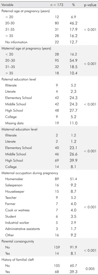
A higher frequency was found for conception at the ages of 20 to 30 years for both fathers and mothers (46.2% and 54.9%, respectively) (Table 3). Mean paternal age at conception was 29.4 ± 8.3 years, whereas mean maternal age at conception was 26.6 ± 6.5 years (data not shown). There were no differences between the mean maternal ages of syndromic (34.3 ± 6.9) and non-syndromic patients (33.9 ± 8.6) (data not shown).
Table 1. Data on the patients with orofacial clefts.
Variable n = 173 % p-value
Type of cleft
Cleft lip and palate (CLP) 81 46.8
< 0.001
Cleft palate (CP) 61 35.2
Cleft lip (CL) 31 17.9
Severity (CLP and CL)
Unilateral 81 72.3
< 0.001
Bilateral 30 26.8
Midline 1 0.9
Gender
Male 104 60.1
0.008
Female 69 39.9
Birth weight
< 2,500 g 26 15.0
< 0.001
25,00 – 3,999 g 122 70.5
≥ 4,000 g 13 7.5
Missing data 12 6.9
Birth length Boys
< 46.1 cm 7 6.7
< 0.001
46.1 – 53.7 cm 76 73.1
> 53.7 cm 3 2.9
Missing data 18 17.3
Girls
< 45.4 cm 11 15.9
< 0.001
45.4 – 52.9 cm 41 59.4
> 52.9 cm 6 8.7
Missing data 11 15.9
Head circumference at birth Boys
< 31.9 cm 4 3.8
< 0.001
31.9 – 37.0 cm 54 51.9
> 37.0 cm 1 0.9
Missing data 45 43.2
Girls
< 31.5 cm 5 7.2
< 0.001
31.5 – 36.2 cm 32 46.4
> 36.2 cm 4 5.8
Missing data 28 40.6
Neonatal intercurrent events 43a 24.9a
Icterus 25 14.5
< 0.001
Respiratory distress 23 13.3
Intubation and mechanical ventilation 9 5.2
Infections 5 2.9
Other 13 7.5
None 93 53.8
Missing data 37 21.4
in 8.1% of the cases, first cousins being the most common type of kinship. Familial history was found in 39.3% (Table 3).
The obstetric history and birth characteristics of studied patients (Table 4) evidences that most pregnancies were spontaneous (98.3%); Cesarean sections were the most frequent type of birth (41.6%); and most pregnancies reached full term (56.1%). Furthermore, the largest proportion of mothers had one or two pregnancies (31.8% and 32.4%, respectively) with the first child being the most frequently affected
by clefts (40.5%). Thirty mothers (17.3%) had at least one miscarriage, and only eight (4.6%) attempted to terminate pregnancy. Prenatal history revealed that 22.5% of the mothers had consumed alcohol during pregnancy, 13.9% had had urinary tract infection, 11.6% had smoked during pregnancy, and 11.0% had been diagnosed with gestational hypertension or preeclampsia. Bleeding and gestational diabetes were also frequently reported. As additional information, two mothers reported direct and daily contact with gases such as ammonia and those obtained from the
Comorbidities 79b 45.7b
Anemia 28 16.2
< 0.001
Respiratory diseases 21 12.1
Cardiovascular diseases 17 9.8
Neurological diseases 14 8.1
Otitis 13 7.5
Hearing loss 10 5.8
Pneumonia 9 5.2
Gastrointestinal diseases 6 3.5
Other 20 11.6
None 70 40.5
Missing data 24 13.9
Neuropsychomotor development
Normal for age 82 47.4
< 0.001
Motor, speech and behavioral delays and NPMDc 14 8.1
Speech delay only 12 6.9
Motor and speech delay 5 2.9
Motor delay only 4 2.3
Speech and behavior delay 3 1.7
Behavioral delay only 1 0.6
Not applicable 27 15.6
Missing data 25 14.5
Therapy
No 87 50.3
0.002
Yes 51 29.5
Missing data 35 20.2
Type of therapy
Speech Therapy 41 23.7
< 0.001
Physical therapy 8 4.6
Psychology 5 2.9
Occupational therapy 4 2.3
Other 4 2.3
Syndrome
Nonsyndromic 119 68.8
< 0.001
Syndromic 47 27.2
Not classified 7 4.0
aTotal number of neonatal complications and frequency relative to the whole study group; bTotal number of comorbidities and frequency relative to the whole study group; cNPMD, Neuropsychomotor developmental delay.
evaporation of paint and solvent while working in the industrial sector during the first trimester.
Folic acid and iron supplementation during pregnancy was reported by 24.3% and 21.4% of the mothers, respectively. In addition to these supplements, analgesics (17.3%) such as dipyrone and paracetamol, followed by antibiotics (16.8%) – most notably cephalexin and macrodantin, were taken. The use of antihypertensives (8.7%), especially methyldopa, to treat gestational hypertension was frequently reported. Vitamin supplements, corticosteroids, progesterone, and metoclopramide were also mentioned.
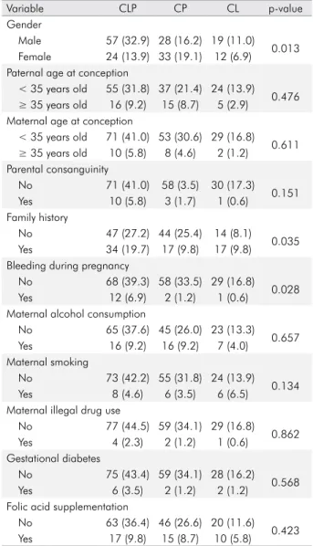
Table 5 shows the risk factors commonly associated with susceptibility to CL/P according to type of cleft. CLP was statistically more frequent in male than in female participants, among whom CP was more
Table 3. Data on parents of orofacial cleft patients.
Variable n = 173 % p-value
Paternal age at pregnancy (years)
< 20 12 6.9
< 0.001
20-30 80 46.2
31-35 31 17.9
> 35 28 16.2
No information 22 12.7
Maternal age at pregnancy (years)
< 20 28 16.2
< 0.001
20–30 95 54.9
31–35 32 18.5
> 35 18 10.4
Paternal education level
Illiterate 9 5.2
< 0.001
Literate 4 2.3
Elementary School 42 24.3
Middle School 42 24.3
High School 48 27.7
College 9 5.2
Missing data 19 11.0
Maternal education level
Illiterate 2 1.2
< 0.001
Literate 2 1.2
Elementary School 40 23.1
Middle School 46 26.6
High School 69 39.9
College 14 8.1
Maternal occupation during pregnancy
Homemaker 89 51.4
< 0.001
Salesperson 16 9.2
Housekeeper 15 8.7
Teacher 9 5.2
Farmer 7 4.0
Cook or waitress 7 4.0
Student 6 3.5
Industrial worker 5 2.9
Administrative assistants 3 1.7
Other 16 9.2
Parental consanguinity
No 159 91.9
< 0.001
Yes 14 8.1
History of familial cleft
No 105 60.7
0.005
Yes 68 39.3
Table 2. Data on lip and palate surgical repair.
Variable n = 173 % p-value
Age at first surgical repair Cheiloplasty
< 6 months old 18 26.9
0.001
6–12 months old 36 53.7
13 month–5 years old 13 19.4
> 5 years old 0 0.0
Total 67 100
Palatoplasty
< 1 year old 6 12.2
< 0.001
1–2 years old 23 46.9
2–5 years old 16 32.7
> 5 years old 4 8.2
Total 49 100
Delay in surgical repair Cheiloplasty
Waiting for surgery and
on time 9 27.3
0.009 Waiting for surgery and
delay 24 72.7
Total 33 100
Palatoplasty
Waiting for surgery and
on time 32 41.0
0.113 Waiting for surgery and
delay 46 59.0
prevalent (p = 0.013). Family history of clefts (19.7%; p = 0.035) and bleeding episodes during pregnancy (6.9%; p = 0.028) were most often found in the CLP group. There were no statistically significant differences among cleft types with regard to other risk factors.
Discussion
CL/P global prevalence, the need for an integrated long-term multidisciplinary treatment, and economic impact have prompted WHO to consider CL/P a public health problem.17
In the present study, aimed at describing the characteristics, comorbidities, and main risk factors
Table 4. Obstetric history and birth characteristics of studied patients.
Variable n = 173 % p-value
Conception method
Spontaneous 170 98.3
< 0.001
Assisted 3 1.7
Type of Birth
Vaginal 65 37.6
0.550
Cesarean 72 41.6
Missing data 36 20.8
Timing of birth
Preterm 24 13.9
< 0.001
Full-term 97 56.1
Post-term 17 9.8
Missing data 35 20.2
Number of pregnancies
1 55 31.8
0.003
2 56 32.4
3 31 17.9
> 03 31 17.9
Birth order
1st 70 40.5
< 0.001
2nd 51 29.5
3rd 26 15.0
> 3rd 26 15.0
Miscarriage
No 143 82.7
< 0.001
Yes 30 17.3
Termination of pregnancy attempts
No 163 94.2
< 0.001
Yes 8 4.6
Missing data 2 1.2
Prenatal history
Alcohol consumption 39 22.5
< 0.001 Urinary tract infection 24 13.9
Smoking 20 11.6
Hypertension 19 11.0
Bleeding 15 8.7
Diabetes Mellitus 10 5.8
Illegal drug use 7 4.0
Other 30 17.3
Medications used during pregnancy
Folic acid 42 24.3
< 0.001
Iron 37 21.4
Analgesic 30 17.3
Antibiotic 29 16.8
Antihypertensive 15 8.7
Vitamin supplementation 12 6.9
Anti-inflammatory 7 4.0
Anti-abortion drug 6 3.5
Antiemetic 4 2.3
Hypoglycemic agent 3 1.7
Other 22 12.7
None 70 40.5
Missing data 2 1.2
Table 5. Risk factors according to cleft types.
Variable CLP CP CL p-value
Gender
Male 57 (32.9) 28 (16.2) 19 (11.0) 0.013 Female 24 (13.9) 33 (19.1) 12 (6.9)
Paternal age at conception
< 35 years old 55 (31.8) 37 (21.4) 24 (13.9) 0.476
≥ 35 years old 16 (9.2) 15 (8.7) 5 (2.9)
Maternal age at conception
< 35 years old 71 (41.0) 53 (30.6) 29 (16.8) 0.611
≥ 35 years old 10 (5.8) 8 (4.6) 2 (1.2)
Parental consanguinity
No 71 (41.0) 58 (3.5) 30 (17.3) 0.151
Yes 10 (5.8) 3 (1.7) 1 (0.6)
Family history
No 47 (27.2) 44 (25.4) 14 (8.1)
0.035 Yes 34 (19.7) 17 (9.8) 17 (9.8)
Bleeding during pregnancy
No 68 (39.3) 58 (33.5) 29 (16.8) 0.028
Yes 12 (6.9) 2 (1.2) 1 (0.6)
Maternal alcohol consumption
No 65 (37.6) 45 (26.0) 23 (13.3) 0.657 Yes 16 (9.2) 16 (9.2) 7 (4.0)
Maternal smoking
No 73 (42.2) 55 (31.8) 24 (13.9) 0.134
Yes 8 (4.6) 6 (3.5) 6 (6.5)
Maternal illegal drug use
No 77 (44.5) 59 (34.1) 29 (16.8) 0.862
Yes 4 (2.3) 2 (1.2) 1 (0.6)
Gestational diabetes
No 75 (43.4) 59 (34.1) 28 (16.2) 0.568
Yes 6 (3.5) 2 (1.2) 2 (1.2)
Folic acid supplementation
No 63 (36.4) 46 (26.6) 20 (11.6) 0.423 Yes 17 (9.8) 15 (8.7) 10 (5.8)
of a group of patients with CL/P from RN, Brazil, we observed a higher prevalence of unilateral CLP followed by CP, and male participants were more affected than female ones. CLP was also more frequent in male participants whereas CP was more frequent in female participants. Most of the assessed patients had normal weight, height, and head circumference at birth and exhibited a low frequency of neonatal intercurrent events. This finding is in agreement with previous reports of higher prevalence of CL/P among male patients and of CLP as the most common type of cleft both in Brazil and worldwide.18,19 According
to the literature, gender differences in the risk for CL/P and CP are explained by the multifactorial threshold model in which the etiology of orofacial clefts is inserted.12 As in the present study, no evidence
of low birth weight and short stature was found in follow-up studies with CL/P children.20
A considerable number of associated comorbidities were found in the studied patients, especially anemia, followed by respiratory problems and cardiovascular diseases. These findings are in line with those observed by Dvivedi and Dvivedi,21 who identified anemia in
most of the 4,657 CL/P cases studied in India,21 and
by Nagalo et al.,22 who found anemia as the most
frequent comorbidity in 185 children with CL/P, followed by respiratory infections in Western Africa. The same results were obtained by Kulkarni et al. (2013).23 Anemia in CL/P patients is attributed mainly
to feeding difficulty, while respiratory problems are frequently associated with irritation of the nasal and respiratory cavities by food and saliva, which also predisposes to recurrent infections. The frequency of cardiovascular malformations found by Harry et al.24
in 10% of CL/Ps cases was similar to that of the present study. Cardiac anomalies are associated with the common development of both palate and heart between 5 and 9 weeks of gestation as part of cardiac and craniofacial development, which relies on complex signaling processes among interdependent embryonic tissues.24
Other complications commonly seen in CL/P patients are related to neuropsychomotor development. We observed concomitant motor, speech, and behavioral delay, followed by speech delay only. Moreover, 50% of the assessed children and adolescents
did not participate in any supportive therapy, and only 23.7% attended speech therapy. Similar results were found by Feragen et al.25 in a study with 754
children with CL/P in which 32% had alterations such as developmental delay, attention deficit/hyperactivity disorder, or a specific speech impairment or dyslexia. Despite early cleft repair, some children exhibit “cleft palate speech,” characterized by atypical consonant productions, abnormal nasal resonance, abnormal nasal airflow, altered laryngeal voice quality, and nasal or facial grimaces, demonstrating the importance of patient follow-up by a multidisciplinary team.26
Lip and palate surgical repair is an important aspect to be considered in the treatment of CL/P patients, but the ideal timing for the repair remains controversial. It has been recommended that cleft lip surgical repair be performed at least 3 months of age – preferably at 4 or 5 months, if possible, and that cleft palate not be corrected after the age of 18 months.27 At such ages, anesthesia is safer, the repair
is more accurate, and malformations are more easily accepted by parents.28 In the present study, most
patients underwent palatoplasty and cheiloplasty at the recommended repair time with a few months’ delay. However, there was a long delay among patients who had not undergone surgery yet. This indicates that poor access to surgical treatment and inappropriate management and planning of health services in Brazil are a hindrance, as pointed out previously.3 Delay in performing the surgery leads
to a series of consequences such as difficulty eating, speaking, and listening; psychological problems; stigmatization; social exclusion; and unemployment.3,29
By analyzing the characteristics of the parents of children with CL/P in the present study, there was no association between advanced age of mothers and fathers and the occurrence of orofacial clefts. Along the same line, Campos Neves et al.,6 in a study with
116 orofacial cleft patients from Mato Grosso, Brazil, found 60.34% of the mothers were aged 20 to 34 years and 82.76% of the fathers were aged 20 to 39 years at the onset of pregnancy. Studies performed in Canada, Iran, the Netherlands, and South America did not find an association between advanced maternal age and CL/P or CP.30 Advanced ages may be related
caused by environmental exposures or chromosomal alterations (lifelong medication use, prevalence of chronic diseases, and socioeconomic factors), as well as low selectivity of the uterus regarding defective embryos and higher placental permeability to teratogenic agents.31
Although the etiology of CL/P is still not fully understood, genetic susceptibility has been shown as one of the most important associated causes.2 Family
history of CL/P in the present study corroborates previous findings of high rates of familial recurrence.1,32
Brito et al.,32 in a study with 1,042 families from five
different locations in Brazil, observed a familial recurrence similar to that observed in the present study, in Barbalha (37%) and in Fortaleza (40%) in the Brazilian state of Ceará. Martelli-Junior et al.33
found that 35.1% of 185 non-syndromic CL/P patients from Minas Gerais, Brazil, had a positive family history of orofacial clefts. A 30% frequency was found by Figueiredo et al.34 in a study with 40 CL/P
patients from Cuiabá, Brazil. Consistent with Leite and Kofman ’s study of a Brazilian sample from Rio de Janeiro, the family history found in the present study was statistically more frequent in CLP cases.35
Cohort studies indicate that relatives of orofacial cleft patients were at a higher risk than the general population, showing a steep decrease in such risk as the genetic distance between relatives increases.11
The parental consanguinity rate found in the present study (8.1%) was close to that found by Brito et al.32 in Fortaleza, Brazil (11.5%), but it was
higher than the 4% reported by Leite and Koifman35
in Rio de Janeiro, Brazil and the 5% presented by Alvarez et al.36 in a study with 356 patients from São
Paulo, Brazil. The results found here reinforce the inheritable nature of this malformation, probably due to consanguineous marriages typical of the region where the study population is from, and also highlight the importance of genetic counseling for this population.
The prenatal history data presented here concur with those of previous publications: alcohol use and cigarette smoking may be associated with the development of craniofacial malformation.7
Frequencies of alcohol consumption during pregnancy slightly lower than in the present study (22.5%) were
reported by Campos Neves et al.6 (17.2%) and by
Bezerra et al.37 (15%), both for the Brazilian population.
A frequency of smoking higher than that observed in the present study (11.6%) was found by Nilsson et al.38 in a study of Swedish children with CL/P, in
which 23% of the mothers reported cigarette smoking during pregnancy. A 45% frequency of pregnant women who smoked at any time during pregnancy was found by Little et al.39 in a study with 190 CL/P
patients from Scotland and England. These findings make us hypothesize that maternal smoking has different effects on the risk of orofacial clefts.
Previous literature associated the use of drugs (especially phenytoin, phenobarbital, benzodiazepines, and corticosteroids) during pregnancy with CL/P occurrence; however, in the present study, these drugs were administered at low frequencies, and the most common drugs used were folic acid, iron, analgesics, antibiotics, and antihypertensives. Although folic acid was the most widely used drug, considering the whole sample, there was a low percentage of mothers on supplementation with this substance (24.3%). Higher folic acid supplementation during pregnancy was observed by McKinney et al.40 in a
study with 86 CL/P patients from Thailand (35.8%) and by Taghavi et al.41 in a study with 300 CL/P patients
from Iran with frequencies of 93.7% and 80.3% of folic acid and iron intake, respectively. Nevertheless, the effects of folic acid supplementation on orofacial clefts are paradoxical. While in the McKinney et al. (2013)40 study the use of this supplement did not
statistically decrease the risk of having an affected child, the Taghavi et al.41 study showed a lower risk
for orofacial clefts.
one of the disadvantages of ecological studies, as is the case of the present study. However, ecological studies are important as they allow an initial examination of the status and needs of communities, especially of health status.42
Conclusions
This study provided an overview of several aspects related to the development and monitoring of CL/P patients, highlighting the risk factors and comorbidities presented by this population in a developing country. Most patients were male with CLP type and born of normal size and weight and presented few neonatal intercurrent events and had or have anemia, respiratory, and cardiovascular diseases as the main associated comorbidities. They also needed timely surgical rehabilitation and multidisciplinary care to stimulate their neuropsychomotor development. Other relevant findings were the considerable maternal exposure to alcohol, infections, smoking, and hypertension,
as well as low supplementation of vitamins and minerals and use of analgesics, antibiotics, and antihypertensives during pregnancy. In addition, a high frequency of familial recurrence and mainly of parental consanguinity was evidenced in the studied population, especially in CLP patients. Knowledge of CL/P patient profiles is important to aid professionals with the better management and planning of local health services made available to CL/P patients. Furthermore, our findings reinforce the need for further confirmation of environmental risk factors associated with the development of orofacial clefts.
Acknowledgements
We are thankful for the technical support provided by students from the LABMULT/UFRN/RN. We thank all the physicians, nurses, and hospital staff at the Children’s Hospital of UFRN who were involved in this study. The authors also thank all children, adolescents, and young adults with CL/P and their parents who gave their consent and participated in the study.
1. Wehby GL, Uribe LM, Wilcox AJ, Christensen K, Romitti PA, Munger RG et al. Interaction between smoking and body mass index and risk of oral clefts. Ann Epidemiol. 2017 Feb;27(2):103-107.e2. https://doi.org/10.1016/j.annepidem.2016.11.009 2. Dixon MJ, Marazita ML, Beaty TH, Murray JC. Cleft lip and palate:
understanding genetic and environmental influences. Nat Rev Genet. 2011 Mar;12(3):167-78. https://doi.org/10.1038/nrg2933 3. Sousa GF, Roncalli AG. Orofacial clefts in Brazil and
surgical rehabilitation under the Brazilian National Health System. Braz Oral Res. 2017 Mar;31(0):e23. https://doi. org/10.1590/1807-3107bor-2017.vol31.0023
4. Abreu MH, Lee KH, Luquetti DV, Starr JR. Temporal trend in the reported birth prevalence of cleft lip and/or cleft palate in Brazil, 2000 to 2013. Birth Defects Res A Clin Mol Teratol. 2016 Sep;106(9):789-92. https://doi.org/10.1002/bdra.23528 5. Figueirêdo CJ, Vasconcelos WK, Maciel SS, Maciel
WV, Gondim LA, Tassitano RM. Prevalência de fissuras orais no Estado do Rio Grande do Norte, Brasil, entre 2000 e 2005. Rev Paul Pediatr. 2011 Mar;29(1):29-34. https://doi.org/10.1590/S0103-05822011000100005 6. Campos Neves AT, Volpato LE, Espinosa MM, Aranha AM,
Borges AH. Environmental factors related to the occurrence of oral clefts in a Brazilian subpopulation. Niger Med J. 2016 May-Jun;57(3):167-72. https://doi.org/10.4103/0300-1652.184064
7. DeRoo LA, Wilcox AJ, Lie RT, Romitti PA, Pedersen DA, Munger RG et al. Maternal alcohol binge-drinking in the first trimester and the risk of orofacial clefts in offspring: a large population-based pooling study. Eur J Epidemiol. 2016 Oct;31(10):1021-34. https://doi.org/10.1007/s10654-016-0171-5
8. Skuladottir H, Wilcox AJ, Ma C, Lammer EJ, Rasmussen SA, Werler MM et al. Corticosteroid use and risk of orofacial clefts. Birth Defects Res A Clin Mol Teratol. 2014 Jun;100(6):499-506. https://doi.org/10.1002/bdra.23248 9. De-Regil LM, Fernández-Gaxiola AC, Dowswell T,
Peña-Rosas JP. Effects and safety of periconceptional folate supplementation for preventing birth defects. Cochrane Database Syst Rev. 2010 Oct;(10):CD007950. https://doi. org/10.1002/14651858.CD007950.pub2.
10. Gil-da-Silva-Lopes VL, Monlleó IL. Risk factors and the prevention of oral clefts. Braz Oral Res. 2014 Jan 12;28(spe):1-5.
https://doi.org/10.1590/S1806-83242014.50000008 11. Grosen D, Chevrier C, Skytthe A, Bille C, Mølsted K,
Sivertsen A et al. A cohort study of recurrence patterns among more than 54,000 relatives of oral cleft cases in Denmark: support for the multifactorial threshold model of inheritance. J Med Genet. 2010 Mar;47(3):162-8. https://doi.org/10.1136/jmg.2009.069385
12. Beaty TH, Marazita ML, Leslie EJ. Genetic factors influencing risk to orofacial clefts: today’s challenges and tomorrow’s opportunities. F1000 Res. 2016 Nov;5(0):2800. https://doi.org/10.12688/f1000research.9503.1
13. Zhao YJ, Xiong YX, Wang Y. Three-dimensional accuracy of facial scan for facial deformities in clinics: a new evaluation method for facial scanner accuracy. PLoS One. 2017 Jan;12(1):e0169402. https://doi.org/10.1371/journal.pone.0169402
14. Fogh-Andersen P. Inheritance of harelip and cleft palate: contribution to the elucidation of the etiology of the congenital clefts of the face. København: Munksgaard; 1942.
15. Monlleó IL, Gil-da-Silva-Lopes VL. Brazil’s Craniofacial Project: genetic evaluation and counseling in
the reference network for craniofacial treatment. Cleft Palate Craniofac J. 2006 Sep;43(5):577-9. https://doi.org/10.1597/04-203 PMID:16986979 16. Monlleó IL, Fontes MÍ, Ribeiro EM, de Souza J, Leal
GF, Félix TM et al. Implementing the brazilian database on orofacial clefts. Plast Surg Int. 2013;2013:641570. https://doi.org/10.1155/2013/641570
17. Agbenorku P. Orofacial clefts: a worldwide review of the problem. ISRN Plast Surg. 2013;2013:ID348468. httpS://doi.org/10.5402/2013/348465
18. Asani M, Aliyu I. Pattern of congenital heart defects among children with orofacial clefts in Northern Nigeria. J Cleft Lip Palate Craniofacial Anomalies. 2014;1(2):85. https://doi.org/10.4103/2348-2125.137895
19. Martelli DR, Machado RA, Swerts MS, Rodrigues LA, Aquino SN, Martelli Júnior H. Non syndromic cleft lip and palate: relationship between sex and clinical extension. Braz J Otorhinolaryngol. 2012 Oct;78(5):116-20.
20. Jagomagi T, Soots M, Saag M. Epidemiologic factors causing cleft lip and palate and their regularities of occurrence in Estonia. Stomatologija. 2010;12(4):105-8.
21. Dvivedi J, Dvivedi S. A clinical and demographic profile of the cleft lip and palate in Sub-Himalayan India: A hospital-based study. Indian J Plast Surg. 2012 Jan;45(1):115-20. https://doi.org/10.4103/0970-0358.96602
22. Nagalo K, Ouédraogo I, Laberge JM, Caouette-Laberge L, Turgeon J. Congenital malformations and medical conditions associated with orofacial clefts in children in Burkina Faso. BMC Pediatr. 2017 Mar;17(1):72. https://doi.org/10.1186/s12887-017-0833-9 23. Kulkarni KR, Patil MR, Shirke AM, Jadhav SB.
Perioperative respiratory complications in cleft lip and palate repairs: an audit of 1000 cases under ‘Smile Train Project’. Indian J Anaesth. 2013 Nov;57(6):562-8. https://doi.org/10.4103/0019-5049.123328
24. Harry BL, TeBockhorst S, Deleyiannis FW. The impact of congenital cardiovascular malformations on the assessment and surgical management of infants with cleft lip and/or palate. Cleft Palate Craniofac J. 2013 May;50(3):323-9. https://doi.org/10.1597/12-131
25. Feragen KB, Stock NM, Rumsey N. Toward a reconsideration of inclusion and exclusion criteria in cleft lip and palate: implications for psychological research. Cleft Palate Craniofac J. 2014 Sep;51(5):569-78. https://doi.org/10.1597/12-326 26. Sell D, Grunwell P, Mildinhall S, Murphy T, Cornish TA,
Bearn D et al. Cleft lip and palate care in the United Kingdom: The Clinical Standards Advisory Group (CSAG) Study. Part 3: speech outcomes. Cleft Palate Craniofac J. 2001 Jan;38(1):30-7. https://doi.org/10.1597/1545-1569(2001)038<0030:CLAPCI>2.0.CO;2
27. Crockett DJ, Goudy SL. Cleft lip and palate. Facial Plast Surg Clin North Am. 2014 Nov;22(4):573-86. https://doi.org/10.1016/j.fsc.2014.07.002
28. American Cleft Palate-Craniofacial Association. Parameters for evaluation and treatment of patients with cleft lip/palate or other craniofacial anomalies. Cleft Palate-Craniofacial J. 1993 Mars;30 Suppl:S1-16.
29. Mossey PA, Shaw WC, Munger RG, Murray JC, Murthy J, Little J. Global oral health inequalities: challenges in the prevention and management of orofacial clefts and potential solutions. Adv Dent Res. 2011 May;23(2):247-58. https://doi.org/10.1177/0022034511402083
30. Vieira AR, Orioli IM, Murray JC. Maternal age and oral clefts: a reappraisal. Oral Surg Oral Med Oral Pathol Oral Radiol Endod. 2002 Nov;94(5):530-5. https://doi.org/10.1067/moe.2002.128875 31. Herkrath AP, Herkrath FJ, Rebelo MA, Vettore MV.
Parental age as a risk factor for non-syndromic oral clefts: a meta-analysis. J Dent. 2012 Jan;40(1):3-14. https://doi.org/10.1016/j.jdent.2011.10.002
32. Brito LA, Cruz LA, Rocha KM, Barbara LK, Silva CB, Bueno DF et al. Genetic contribution for non-syndromic cleft lip with or without cleft palate (NS CL/P) in different regions of Brazil and implications for association studies. Am J Med Genet A. 2011 Jul;155A(7):1581-7. https://doi.org/10.1002/ajmg.a.34036 33. Martelli DR, Bonan PR, Soares MC, Paranaíba LR,
Martelli-Júnior H. Analysis of familial incidence of non-syndromic cleft lip and palate in a Brazilian population. Med Oral Patol Oral Cir Bucal. 2010 Nov;15(6):e898-901. https://doi.org/10.4317/medoral.15.e898
34. Figueiredo RF, Figueiredo N, Feguri A, Bieski I, Mello R, Espinosa M et al. The role of the folic acid to the prevention of orofacial cleft: an epidemiological study. Oral Dis. 2015 Mar;21(2):240-7. https://doi.org/10.1111/odi.12256 35. Leite IC, Koifman S. Oral clefts, consanguinity, parental
tobacco and alcohol use: a case-control study in Rio de Janeiro, Brazil. Braz Oral Res. 2009 Jan-Mar;23(1):31-7. https://doi.org/10.1590/S1806-83242009000100006 36. Alvarez CW, Guion-Almeida ML, Richieri-Costa A. Clinical
37. Bezerra JF, Oliveira GH, Soares CD, Cardoso ML, Ururahy MA, Neto FP et al. Genetic and non-genetic factors that increase the risk of non-syndromic cleft lip and/ or palate development. Oral Dis. 2015 Apr;21(3):393-9. https://doi.org/10.1111/odi.12292
38. Nilsson S, Merlo J, Lyberg-Åhlander V, Psouni E. Psychotropic drug use in adolescents born with an orofacial cleft:
a population-based study. BMJ Open. 2015 Apr;5(4):e005306. https://doi.org/10.1136/bmjopen-2014-005306
39. Little J, Cardy A, Arslan MT, Gilmour M, Mossey PA. Smoking and orofacial clefts: a United Kingdom-based case-control
study. Cleft Palate Craniofac J. 2004 Jul;41(4):381-6. https://doi.org/10.1597/02-142.1
40. McKinney CM, Chowchuen B, Pitiphat W, Derouen T, Pisek A, Godfrey K. Micronutrients and oral clefts: a case-control study. J Dent Res. 2013 Dec;92(12):1089-94. https://doi.org/10.1177/0022034513507452
41. Taghavi N, Mollaian M, Alizadeh P, Moshref M, Modabernia S, Akbarzadeh AR. Orofacial clefts and risk factors in tehran, iran: a case control study. Iran Red Crescent Med J. 2012 Jan;14(1):25-30. 42. Sedgwick P. Understanding the ecological fallacy. BMJ. 2015