Submitted10 May 2016 Accepted 23 June 2016 Published21 July 2016
Corresponding author Min Young Lee, eyeglass210@gmail.com
Academic editor Melinda Fitzgerald
Additional Information and Declarations can be found on page 10
DOI10.7717/peerj.2252
Copyright 2016 Lee et al.
Distributed under
Creative Commons CC-BY 4.0
OPEN ACCESS
Simultaneous bilateral laser therapy
accelerates recovery after noise-induced
hearing loss in a rat model
Jae-Hun Lee1, So-Young Chang1, Wesley J. Moy2, Connie Oh2, Se-Hyung Kim3, Chung-Ku Rhee4, Jin-Chul Ahn5, Phil-Sang Chung1,4, Jae Yun Jung1,4and Min Young Lee4
1College of Medicine, Dankook University, Beckman Laser Institute Korea, Cheonan, South Korea 2Beckman Laser Institute and Medical Clinic, University of California, Irvine, CA, United States
3Department of Otolaryngology-Head and Neck Surgery, Jeju National University School of Medicine, Jeju, South Korea
4Department of Otolaryngology-Head & Neck Surgery, College of Medicine, Dankook University, Cheonan, South Korea
5Department of Biomedical Science, College of Medicine, Dankook University, Cheonan, South Korea
ABSTRACT
Noise-induced hearing loss is a common type of hearing loss. The effects of laser therapy have been investigated from various perspectives, including in wound healing, inflammation reduction, and nerve regeneration, as well as in hearing research. A promising feature of the laser is its capability to penetrate soft tissue; depending on the wavelength, laser energy can penetrate into the deepest part of the body without damaging non-target soft tissues. Based on this idea, we developed bilateral transtympanic laser therapy, which uses simultaneous laser irradiation in both ears, and evaluated the effects of bilateral laser therapy on cochlear damage caused by noise overexposure. Thus, the purpose of this research was to assess the benefits of simultaneous bilateral laser therapy compared with unilateral laser therapy and a control. Eighteen Sprague-Dawley rats were exposed to narrow-band noise at 115 dB SPL for 6 h. Multiple auditory brainstem responses were measured after each laser irradiation, and cochlear hair cells were counted after the 15th such irradiation. The penetration depth of the 808 nm laser was also measured after sacrifice. Approximately 5% of the laser energy reached the contralateral cochlea. Both bilateral and unilateral laser therapy decreased the hearing threshold after noise overstimulation in the rat model. The bilateral laser therapy group showed faster functional recovery at all tested frequencies compared with the unilateral laser therapy group. However, there was no difference in the endpoint ABR results or final hair cell survival, which was analyzed histologically.
SubjectsNeuroscience, Otorhinolaryngology
Keywords Bilateral LLLT, Noise induced hearing loss, ABR, Hair cell survival
INTRODUCTION
species (ROS), such as the superoxide anion (O2−) (Yamane et al.,1995) and hydrogen
peroxide (H2O2) (Ohinata et al.,2000).
Noise exposure can cause a temporary threshold shift (TTS) or a permanent threshold shift (PTS) that will not recover. The type of threshold shift is determined by the intensity and duration of exposure. Several studies with similar levels of noise and exposure times (>100 dB, >6 h) have reported that a PTS occurred after a few minutes or hours of such
noise exposure (Buck,1981;Hu et al.,2000;Hu, Henderson & Nicotera,2006). Both TTS
and PTS can occur simultaneously at different frequencies in one cochlea. According to recent research, damage to the auditory neurons, such as at the ribbon synapse and postsynaptic receptors, was found following noise exposure, even after recovery of the
hearing threshold (Kujawa & Liberman,2009).
Laser therapy has been used as a treatment for various symptoms, and its use has been increasing because of its non-invasive nature. After it was approved by the United States Food and Drug Administration, applications of laser therapy have widened in research
scope, including wound healing (Anneroth et al., 1988; Grossman et al.,1998;Kana &
Hutschenreiter,1981), inflammation reduction (Boschi et al.,2008;Ferreira et al.,2005),
and nerve regeneration (Miloro et al.,2002;Mohammed & Kaka,2007). The effects of
laser therapy have also been reported in the area of hearing research. Some studies have demonstrated significant effects in reducing tinnitus and increasing auditory neuron activation (Littlefield et al., 2010;Medalha et al., 2012; Park et al.,2013). Recently, our group reported a promising recovery effect of laser therapy on cochlear hair cells in an
animal study (Rhee et al.,2012).Tamura et al.(2015) also reported a cytoprotective effect
of laser therapy in cochlear hair cells against noise overstimulation (Tamura et al.,2015).
One useful feature of the laser is the capability to penetrate soft tissue; depending on the wavelength, laser energy can penetrate into deep parts of the body without damaging non-targeted soft tissues. This enables the delivery of laser energy from multiple points, which may lead to faster or increased effects of the laser in the target area. In our previous animal experiments, we found improvements in the hearing threshold not only in the laser-irradiated group but also in the contralateral ear (Rhee et al.,2012). This suggests that unilateral laser therapy may affect the contralateral auditory organs. Thus, we measured the degree of laser penetration in the contralateral ear of SD rats and assessed the benefit of simultaneous bilateral laser therapy compared with unilateral laser therapy versus a control group.
MATERIALS AND METHODS
Animal subjects
Male Sprague Dawley (SD) rats (180–200 g) were used in this study. Eighteen rats were
randomly divided into three different groups (noise only (n=6), unilateral laser (n=6),
and bilateral laser (n=6)). All animals were treated in accordance with the Guide for
Acute acoustic trauma
The acoustic stimulus was a narrow band of noise which has frequency information centered at 16 kHz with 1 kHz of bandwidth (116 dB SPL). Rats were placed in individual cages to prevent defensive behaviors and these cages were placed in acryl reverberant chambers with a speaker BEYMA CP800Ti (Beyma, Valencia, Spain) attached on top. The traumatic stimulus was generated with a type 1027 sine random generator (Bruel and Kjaer, Denmark) and amplified with a R300 plus amplifier (Inter-M, Seoul, Korea) for 6 h. For real time monitoring, a frequency-specific sound level meter (Sound Level Meter—Type 2250; Bruel and Kjaer, Copenhagen, Denmark) was used to monitor noise level in the chamber (placed on the floor) every hour so that consistent intensity (116 dB SPL) was maintained during noise exposure.
Auditory brainstem response measurement
Auditory brainstem responses (ABR) were measured to identify degrees of hearing loss and recovery. The evoked response signal-processing system (System III; Tucker Davis Technologies, Alachua, Florida) was used for ABR measurement. Animals were anesthetized with Zolazepam (Zoletil, Virbac, Carros Cedex, France) and Xylazine (Rompun, Bayer, Leverkusen, Germany) and placed in a sound proof chamber. Three needle electrodes were inserted at vertex (active) and beneath of each pinna (reference and ground), subcutaneously. The tone-burst stimuli (4, 8, 12, 16, and 32 kHz) were used for experimental measurements and a total 1,024 responses were averaged. Responses were measured in 5 dB intervals from 90 to 10 dB SPL and thresholds were determined by the presence of peak within each signal. Hearing thresholds were obtained before and after noise exposure
(Fig. 1). ABR measurement was also performed during and after laser irradiation (after the
3rd, 6th, 9th, 12th, and 15th laser irradiations).
Laser irradiation treatment
An 808 nm diode laser (Wontec, Seoul, South Korea) was used for laser therapy. Each rat in
the experimental group was anesthetized and irradiated for 60 mins (165 mW/cm2, 594 J)
for 15 days. The density of the laser was calibrated with a laser power meter (FieldMax II-To, Coherent, USA) and detector sensor (Powermax; Coherant, Santa Clara, CA, USA).
The optical fiber (core fiber 62.5µm, cladding 125µm) was attached to a hollow tube and
placed into the external ear canal while leaving a distance of 1 mm between the fiber tip and tympanic membrane. Laser irradiation was performed on both the right and left ear simultaneously for the bilateral group and only in the right ear for the unilateral group. The noise only group was anesthetized and the optical fiber was placed into the external ear
canal without power. Additional detailed information of the laser is described inTable 1.
Measurement of laser energy in the contralateral ear
Laser energy was first measured from the contralateral side of the ear using an 808 nm laser irradiation (calibrated as 165 mW) in the SD Rat to confirm the delivery of laser energy
to the contralateral cochlea. The rat was sacrificed in a CO2chamber and a secondary
Figure 1 Results of ABR measurement.Consistent peaks were recorded at 16 kHz during ABR measurement as a baseline (A). After six hours of noise exposure, the overall amplitude of the peaks were reduced compared to the baseline result, and the peaks disappeared under 65 dB SPL at the same test frequency (B).
Table 1 Laser (Photobiomodulation) parameter.
Parameter Laser group (Bilateral and Unilateral)
Power (mW) 185
Beam spot size at target (cm2) 0.22
Irradiance at target (mW/cm2) power density 841
Exposure duration (s) 3,600
Radiant exposure (J/cm2) fluence 2,700
Radiant energy (J) 594
Number of points irradiated 1
Area irradiated (cm2) 0.22
Application technique Through tympanic membrane
Number and frequency of treatment sessions Once a day for 15 days
Total radiant energy (J) 8,910
The exposed contralateral cochlea was placed above the laser detector and the laser was used to irradiate the ipsilateral external canal with the protocol explained above.
Hair cell count
samples were carefully examined under confocal microscopy (LSM 510 META, Zeiss, Germary) at a magnification of 400X.
We chose three representative areas for the quantitative analysis of OHC, which were located at 20, 50, and 80% from the apex, representing 4, 12, and 32 kHz respectively (Viberg & Canlon,2004). Hair cells <200µm in length were counted in each representative
area. The morphometric analysis software Image J (http://rsb.info.nih.gov/ij/) was used to
count the number of cells in each section.
Statistical analysis
All data were analyzed statistically using the Statistical Package for the Social Sciences software (SPSS, Version 19, IBM, Somers, USA). We performed a Tuckey post hoc test following a Two-way Analysis of Variance (ANOVA) to determine the significance between hearing threshold for ABR measurement and number of hair cells.
RESULTS
Energy from the 808 nm laser was detected in the contralateral ear The 808 nm laser energy was first measured in the contralateral ear. Using an open air setup between the laser probe and detector, the energy output was determined to be the same at the detector and the displayed output of the laser. A total of 6 mW of laser energy was measured in the detector at the contralateral ear, while the maximum level of laser energy penetrating the contralateral ear was found to be 8 mW. This result suggests that some laser energy was absorbed prior to exiting the other ear (contralateral ear).
Hearing loss after noise overstimulation
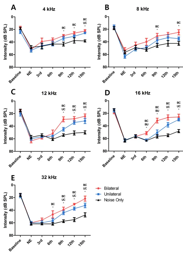
ABRs were measured before noise exposure to determine the baseline hearing threshold. Mean values (SDs) were 18.61 (5.37), 16.11 (5.57), 16.94 (6.67), 16.11 (5.3), and 16.39 (6.14) at frequencies of 4, 8, 12, 16, and 32 kHz, respectively (Fig. 2). At 24 h after noise exposure, ABRs were measured again to confirm the degree of hearing loss. Hearing thresholds were increased markedly after noise exposure. Mean values (SD) were 51.11 (6.08), 57.78 (8.44), 60.28 (6.96), 63.06 (4.79), and 60.56 (4.82) at frequencies of 4, 8, 12, 16, and 32 kHz, respectively (Fig. 2). Thus, these results indicate that overstimulation with a stimulus of 115 dB SPL can cause PTS.
Laser therapy improved hearing recovery in the bilateral and unilateral treated groups
After the sixth laser irradiation, there was a significant difference in the hearing threshold at 16 and 32 kHz between the noise-only and the bilateral laser-treated groups (p=0.001 at
16 kHz and 0.046 at 32 kHz;Figs. 2Dand2E). After the ninth laser irradiation, significant
differences existed at all test frequencies between the noise-only and the bilateral
laser-treated groups (p=0.009 at 4 kHz, 0.04 at 8 kHz, <0.001 at 12 kHz, 0.001 at 16 kHz,
and <0.001 at 32 kHz)(Fig. 2). The response of the unilateral laser-treated group was
showed difference at all frequencies except 8 kHz (Figs. 2Cand2D). Finally, after the 15th laser irradiation, the hearing threshold at all test frequencies was significant different in the noise-only compared with the bilateral laser-treated group (p<0.001 at 4 kHz, 0.005
at 8 kHz, <0.001 at 12 kHz, <0.001 at 16 kHz, and <0.001 at 32 kHz), and the difference
between the unilateral group and noise-only group increased to 4 kHz (Fig. 2). This result
showed that both bilateral and unilateral laser therapy could reduce the hearing threshold in the SD rat model after noise overstimulation. However, complete recovery of the hearing threshold (to the baseline level) was not achieved.
Bilateral laser therapy resulted in faster hearing threshold recovery than did unilateral laser therapy
A significant difference in the threshold between the bilateral group and the noise-only group was observed from the point of the sixth laser irradiation (at 16 kHz and 32 kHz)
(Figs. 2Dand2E). In contrast, significant differences between the unilateral group and the
noise-only group were observed from the points of the ninth and twelfth laser irradiations
(at 32 kHz and 16 kHz; Figs. 2D and2E). Furthermore, compared with the hearing
threshold recovery in the bilateral group at 4 kHz, 8 kHz, and 12 kHz after the ninth laser irradiation, hearing threshold recovery in the unilateral group at these frequencies (at
4 kHz and 12 kHz) was observed after the twelfth and 15th laser irradiations (Figs. 2Aand
2C), respectively. At 8 kHz, there was no significant difference between the unilateral group
and the noise-only group at any time point. This result indicated that despite the absence of differences in the extent of hearing recovery between the unilateral and bilateral laser therapy groups, the bilateral simultaneous application of laser therapy induced faster (up to 3 days) recovery of the hearing threshold after noise-induced hearing loss compared to the unilateral laser therapy group.
Laser-treated group showed better outer hair cell (OHC) preservation in the basal turn
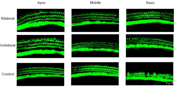
A confocal image of three representative areas is presented inFig. 3. At the apex and the
middle area, the averages of OHCs were similar across the three experiment groups (73.67, 72, and 70.33 at the apex, and 71, 72.67, and 73 at the middle, in the bilateral, unilateral, and
noise-only groups, respectively;Fig. 4). However, averages of OHCs at the basal turn were
found to be different among each group (72.67, 67.5, and 59 in the bilateral, unilateral, and noise-only groups, respectively), and both the bilateral and unilateral laser groups
showed larger number of OHCs than did the noise-only group (p=0.0052 and 0.0006,
respectively;Fig. 4).
DISCUSSION
Cochlear damage can be variable, and a hearing threshold shift can occur abruptly or progressively, depending on the intensity and duration of noise overstimulation (Clark, 1991). In the results of the present study, we found permanent threshold shifts in almost every frequency region examined. These results are consistent with our previous study
Figure 3 Representative confocal images of hair cells at three different locations (apex, middle, and base) in each experimental group.Missing hair cells were observed only at the base part of the cochlea in the noise-only group.
Figure 4 Numbers of OHCs in three parts of the basilar membrane in each group.The bilateral and unilateral laser groups showed significantly larger numbers of OHCs at the base part of the basilar mem-brane (**p<0.01, ***p<0.001).
rat model. We observed slight improvements in the hearing threshold at low-frequency regions (4 and 8 kHz) with no treatment, which could be explained as a TTS, because it
was not the main target frequency (Clark,1991) of the acoustic overstimulation applied
in the current study. Increases in hearing threshold after noise exposure as both PTS and TTS could be a result of loss or dysfunction of OHC electromotility, which contributes to
hearing sensitivity by amplifying the incoming stimulus (Liberman et al.,2002). However,
is some other mechanism of TTS or PTS that may be involved, such as the dispersal of presynaptic ribbons and postsynaptic receptors, which connect the inner hair cells and
spiral ganglion (Furman, Kujawa & Liberman,2013).
Application of laser therapy, after noise overstimulation, induced recovery of hearing
function similar to our previous study (Rhee et al.,2012). This protection mechanism is
considered to be related to the inhibition of iNOS and caspase 3 expression (Tamura et al., 2015), but the details of the underlying mechanism remain unclear. Also, another theory is that this effect may be explained by the balance of free radicals and antioxidants. Before hair cell death, ROS levels increase as a result of noise overexposure. Movement of electrons in hair cells releases energy for converting adenosine diphosphate (ADP) to adenosine triphosphate (ATP) by phosphorylation. During this process, superoxide is generated as an intermediary. When the use of oxygen is increased due to noise exposure, the generation rate of superoxide is also increased by the activity of the mitochondria (Evans & Halliwell, 1999). During noise exposure, mitochondria are strongly stimulated and produce excessive superoxide as a byproduct. Superoxide can react with other molecules in cochlear hair cells, resulting in subcellular molecular damage. Decreased cochlear blood flow due to noise exposure can also contribute to a deficiency of oxygen in the cochlea. Increased ROS can damage DNA, lipids, and proteins, leading to hair cell death (Evans & Halliwell,1999).
Despite the low penetration level in the contralateral ear, we found faster hearing recovery in the bilateral laser therapy group compared to the unilateral laser therapy group. Additional laser energy may improve the speed of hearing recovery by prompting the endo-organs of the contralateral cochlea. We hypothesize that the laser energy penetrating directly can affect the contralateral cochlea as an activator of cell metabolism. Additionally, the amount of laser energy in the middle of the head, which would be more than that reaching the contralateral cochlea, could have sufficient influence to activate the cochlear nerve or auditory pathway in the midbrain. Moreover, protective actions of cochlear efferent feedback pathway through olivocochlear bundle may also increase recovery. Several studies suggest that the protective effect of the olivocochlear bundle during hearing is allowed to suppress hyper activations of auditory nerve fibers and IHCs (Gifford & Guinan,1987;Guinan & Gifford,1988). A damaged olivocochlear bundle by surgical lesion can cause alteration of ABR response, resulting in the increase of hair cell vulnerability
related to noise exposure (Maison, Usubuchi & Liberman,2013). Depositing more laser
energy into the olivocochelar bundle in the bilateral laser group may increase this protective effect, resulting in a faster recovery compared to unilateral laser group. Additionally, the bilateral laser therapy group did not show improved recovery of the hearing threshold compared to the unilateral laser therapy group after the 15th laser irradiation. This limited effect can be explained by the destruction of the most vulnerable auditory pathways after
noise exposure, such as synaptic ribbons (Kujawa & Liberman,2009). Relatively normal
The faster effect of bilateral laser therapy versus unilateral laser therapy is promising for clinical use. Most treatments after hearing loss due to different insults, require early
intervention (Ward,1960). There are critical periods of time that can increase the success
of a treatment outcome, resulting in a more favorable prognoses (Chen et al.,2007). With
bilateral laser therapy, a shorter time frame was required to achieve a desirable outcome; thus, there is a higher chance of staying within the ‘‘golden time’’ for the treatment of hearing loss. Transcanal laser therapy treatments can lead to middle ear complications, such as acute inflammation and perforation of the tympanic membrane (Moon et al., 2016). Applying bilateral laser therapy may reduce the possibility of complications while increasing the effect because laser energy is delivered from two different sites, similar to the protocol for transcranial laser therapy. Multiple site laser irradiation has been used for transcranial laser therapy by several groups (Barrett & Gonzalez-Lima,2013;Schiffer et al., 2009). These studies reported improvements in cognitive and emotional functions in the brain, with no side effects due to laser irradiation, using lower laser power and irradiating from multiple positions. As such, if estimating the exact location of the cochlea is possible, we may be able to deliver energy to the cochlea from multiple sites transcranially. However, no methodology for transcranial aiming toward the cochlea has yet to be established or developed.
To apply bilateral laser therapy in the clinic, some practical issues must be considered. Because of anatomical differences between humans and rodents, the effects of laser energy on the contralateral side would be different in humans compared to rodents. The larger distance from one ear to the other may limit the delivery of laser energy; however, the beneficial effect of bilateral laser therapy would be expected to remain if the mechanism involves targeting the brainstem. Increasing the power of the laser may be another approach to deliver energy to the other ear, but this could cause unwanted side effects, resulting in local burning and tympanic perforation. Thus, increasing the power of transcanal laser irradiation should be carefully considered before translation to clinical application.
CONCLUSIONS
The present study showed positive effects of bilateral laser therapy after noise-induced hearing loss in an animal model. The results suggest that the use of bilateral laser therapy in a clinical setting may improve the therapeutic effects on hearing while minimizing side effects.
ADDITIONAL INFORMATION AND DECLARATIONS
Funding
Grant Disclosures
The following grant information was disclosed by the authors:
Ministry of Science, ICT and Future Planning grant: NRF-2012K1A4A3053142.
Competing Interests
The authors declare there are no competing interests.
Author Contributions
• Jae-Hun Lee conceived and designed the experiments, performed the experiments, wrote
the paper, prepared figures and/or tables.
• So-Young Chang performed the experiments, analyzed the data, contributed
reagents/materials/analysis tools.
• Wesley J. Moy and Min Young Lee wrote the paper, reviewed drafts of the paper.
• Connie Oh reviewed drafts of the paper.
• Se-Hyung Kim, Jin-Chul Ahn and Phil-Sang Chung analyzed the data.
• Chung-Ku Rhee and Jae Yun Jung conceived and designed the experiments.
Animal Ethics
The following information was supplied relating to ethical approvals (i.e., approving body and any reference numbers):
All animals were treated in accordance with the Guide for Care and Use of Laboratory Animals (7th edition, 1996), as formulated by the Institute of Laboratory Animal Resources of the Commission on Life Sciences. All procedures were approved by the Institutional Animal Care and Use Committee for the Dankook University (DKU-15-048).
Data Availability
The following information was supplied regarding data availability:
The raw data has been supplied asSupplemental Dataset.
Supplemental Information
Supplemental information for this article can be found online athttp://dx.doi.org/10.7717/
peerj.2252#supplemental-information.
REFERENCES
Anneroth G, Hall G, Ryden H, Zetterqvist L. 1988.The effect of low-energy infra-red
laser radiation on wound healing in rats.British Journal of Oral and Maxillofacial
Surgery26:12–17DOI 10.1016/0266-4356(88)90144-1.
Barrett D, Gonzalez-Lima F. 2013.Transcranial infrared laser stimulation produces
beneficial cognitive and emotional effects in humans.Neuroscience230:13–23
DOI 10.1016/j.neuroscience.2012.11.016.
Boschi ES, Leite CE, Saciura VC, Caberlon E, Lunardelli A, Bitencourt S, Melo DA, Oliveira JR. 2008.Anti-inflammatory effects of low-level laser therapy (660 nm) in
the early phase in carrageenan-induced pleurisy in rat.Lasers in Surgery and Medicine
Buck K. 1981.Influence of different presentation patterns of a given noise dose on
hearing in guinea-pig.Scandinavian Audiology Supplementum16:83–87.
Chen C-J, Dai Y-T, Sun Y-M, Lin Y-C, Juang Y-J. 2007.Evaluation of auditory fatigue
in combined noise, heat and workload exposure.Industrial Health45:527–534
DOI 10.2486/indhealth.45.527.
Clark WW. 1991.Recent studies of temporary threshold shift (TTS) and permanent
threshold shift (PTS) in animals.The Journal of the Acoustical Society of America
90:155–163DOI 10.1121/1.401309.
Evans P, Halliwell B. 1999.Free radicals and hearing: cause, consequence, and criteria.
Annals of the New York Academy of Sciences884:19–40
DOI 10.1111/j.1749-6632.1999.tb08633.x.
Ferreira D, Zangaro R, Villaverde AB, Cury Y, Frigo L, Picolo G, Longo I, Bar-bosa D. 2005.Analgesic effect of He-Ne (632.8 nm) low-level laser therapy
on acute inflammatory pain.Photomedicine and Laser Surgery23:177–181
DOI 10.1089/pho.2005.23.177.
Furman AC, Kujawa SG, Liberman MC. 2013.Noise-induced cochlear neuropathy
is selective for fibers with low spontaneous rates.Journal of Neurophysiology
110:577–586DOI 10.1152/jn.00164.2013.
Gifford ML, Guinan JJ. 1987.Effects of electrical stimulation of medial olivocochlear
neurons on ipsilateral and contralateral cochlear responses.Hearing Research
29:179–194DOI 10.1016/0378-5955(87)90166-3.
Grossman N, Schneid N, Reuveni H, Halevy S, Lubart R. 1998.780 nm low power diode laser irradiation stimulates proliferation of keratinocyte cultures:
involve-ment of reactive oxygen species.Lasers in Surgery and Medicine22:212–218
DOI 10.1002/(SICI)1096-9101(1998)22:4<212::AID-LSM5>3.0.CO;2-S.
Guinan JJ, Gifford ML. 1988.Effects of electrical stimulation of efferent olivocochlear
neurons on cat auditory-nerve fibers. I. Rate-level functions.Hearing Research
33:97–113 DOI 10.1016/0378-5955(88)90023-8.
Hu BH, Guo W, Wang PY, Henderson D, Jiang SC. 2000.Intense noise-induced
apoptosis in hair cells of guinea pig cochleae.Acta Oto-laryngologica120:19–24
DOI 10.1080/000164800760370774.
Hu BH, Henderson D, Nicotera TM. 2006.Extremely rapid induction of outer hair cell
apoptosis in the chinchilla cochlea following exposure to impulse noise.Hearing
Research211:16–25DOI 10.1016/j.heares.2005.08.006.
Kana JS, Hutschenreiter G. 1981.Effect of low—power density laser radiation
on healing of open skin wounds in rats.Archives of Surgery116:293–296
DOI 10.1001/archsurg.1981.01380150021005.
Kujawa SG, Liberman MC. 2009.Adding insult to injury: cochlear nerve
degenera-tion after ‘‘temporary’’ noise-induced hearing loss.The Journal of Neuroscience
29:14077–14085DOI 10.1523/JNEUROSCI.2845-09.2009.
Liberman MC, Gao J, He DZ, Wu X, Jia S, Zuo J. 2002.Prestin is required for
electro-motility of the outer hair cell and for the cochlear amplifier.Nature419:300–304
Littlefield PD, Vujanovic I, Mundi J, Matic AI, Richter CP. 2010.Laser
stim-ulation of single auditory nerve fibers.The Laryngoscope120:2071–2082
DOI 10.1002/lary.21102.
Maison SF, Usubuchi H, Liberman MC. 2013.Efferent feedback minimizes cochlear
neuropathy from moderate noise exposure.The Journal of Neuroscience33:5542–5552
DOI 10.1523/JNEUROSCI.5027-12.2013.
Medalha CC, Gangi GCD, Barbosa CB, Fernandes M, Aguiar O, Faloppa F, Leite VM, Renno ACM. 2012.Low-level laser therapy improves repair following com-plete resection of the sciatic nerve in rats.Lasers in Medical Science27:629–635
DOI 10.1007/s10103-011-1008-9.
Miloro M, Halkias LE, Mallery S, Travers S, Rashid RG. 2002.Low-level laser effect on
neural regeneration in Gore-Tex tubes.Oral Surgery, Oral Medicine, Oral Pathology,
Oral Radiology, and Endodontology93:27–34DOI 10.1067/moe.2002.119518. Mohammed IF, Kaka LN. 2007.Promotion of regenerative processes in injured
pe-ripheral nerve induced by low-level laser therapy.Photomedicine and Laser Surgery
25:107–111DOI 10.1089/pho.2006.1090.
Moon T-H, Lee MY, Jung JY, Ahn J-C, Chang S-Y, Chung P-S, Rhee C-K, Kim Y-H, Suh M-W. 2016.Safety assessment of transtympanic photobiomodulation.Lasers in Medical Science31(2):323–333.
Ohinata Y, Miller JM, Altschuler RA, Schacht J. 2000.Intense noise induces formation
of vasoactive lipid peroxidation products in the cochlea.Brain Research878:163–173
DOI 10.1016/S0006-8993(00)02733-5.
Park YM, Na WS, Park IY, Suh M-W, Rhee C-K, Chung P-S, Jung JY. 2013.Trans-canal laser irradiation reduces tinnitus perception of salicylate treated rat.Neuroscience Letters544:131–135DOI 10.1016/j.neulet.2013.03.058.
Rhee C-K, Bahk CW, Kim SH, Ahn J-C, Jung JY, Chung P-S, Suh M-W. 2012.Effect of low-level laser treatment on cochlea hair-cell recovery after acute acoustic trauma.
Journal of Biomedical Optics17:0680021–0680026.
Schiffer F, Johnston AL, Ravichandran C, Polcari A, Teicher MH, Webb RH, Hamblin MR. 2009.Psychological benefits 2 and 4 weeks after a single treatment with near infrared light to the forehead: a pilot study of 10 patients with major depression and
anxiety.Behavioral and Brain Functions5:46DOI 10.1186/1744-9081-5-1.
Tamura A, Matsunobu T, Mizutari K, Niwa K, Kurioka T, Kawauchi S, Satoh S, Hiroi S, Satoh Y, Nibuya M. 2015.Low-level laser therapy for
preven-tion of noise-induced hearing loss in rats.Neuroscience Letters595:81–86
DOI 10.1016/j.neulet.2015.03.031.
Viberg A, Canlon B. 2004.The guide to plotting a cochleogram.Hearing Research197(1– 2):1–10.
Ward WD. 1960.Recovery from high values of temporary threshold shift.The Journal of the Acoustical Society of America32:497–500DOI 10.1121/1.1908111.
Yamane H, Nakai Y, Takayama M, Konishi K, Iguchi H, Nakagawa T, Shibata S, Kato A, Sunami K, Kawakatsu C. 1995.The emergence of free radicals after acoustic


