Experimental Characterization of
Cis
-Acting
Elements Important for Translation
and Transcription in Halophilic Archaea
Mariam Brenneis[, Oliver Hering[, Christian Lange, Jo¨rg Soppa*
Institute for Molecular Biosciences, Goethe-University, Frankfurt, Germany
The basal transcription apparatus of archaea is well characterized. However, much less is known about the mechanisms of transcription termination and translation initation. Recently, experimental determination of the 59-ends of ten transcripts fromPyrobaculum aerophilumrevealed that these are devoid of a 59-UTR. Bioinformatic analysis indicated that many transcripts of other archaeal species might also be leaderless. The 59-ends and 39-ends of 40 transcripts of two haloarchaeal species,Halobacterium salinarumandHaloferax volcanii,have been determined. They were used to characterize the lengths of 59-UTRs and 39-UTRs and to deduce consensus sequence-elements for transcription and translation. The experimental approach was complemented with a bioinformatics analysis of theH. salinarumgenome sequence. Furthermore, the influence of selected 59-UTRs and 39-UTRs on transcript stability and translational efficiency in vivo was characterized using a newly established reporter gene system, gene fusions, and real-time PCR. Consensus sequences for basal promoter elements could be refined and a novel element was discovered. A consensus motif probably important for transcriptional termination was established. All 40 haloarchaeal transcripts analyzed had a 39-UTR (average size 57 nt), and their 39-ends were not posttranscriptionally modified. Experimental data and genome analyses revealed that the majority of haloarchaeal transcripts are leaderless, indicating that this is the predominant mode for translation initiation in haloarchaea. Surprisingly, the 59-UTRs of most leadered transcripts did not contain a Shine-Dalgarno (SD) sequence. A genome analysis indicated that less than 10% of all genes are preceded by a SD sequence and even most proximal genes in operons lack a SD sequence. Seven different leadered transcripts devoid of a SD sequence were efficiently translated in vivo, including artificial 59-UTRs of random sequences. Thus, an interaction of the 59-UTRs of these leadered transcripts with the 16S rRNA could be excluded. Taken together, either a scanning mechanism similar to the mechanism of translation initiation operating in eukaryotes or a novel mechanism must operate on most leadered haloarchaeal transcripts.
Citation: Brenneis M, Hering O, Lange C, Soppa J (2007) Experimental characterization ofcis-acting elements important for translation and transcription in halophilic archaea. PLoS Genet 3(12): e229. doi:10.1371/journal.pgen.0030229
Introduction
Determination of the 59- and 39-ends of transcripts is used to identify the points of transcription initiation and termination in order to gain important information about both processes. In fact, determination of transcript 59-ends led to the conclusion that archaea have basal promoter elements that differ from bacterial promoters and resemble eukaryotic polymerase II promoters, thus underscoring the view that archaea are a third domain of life [1]. A survey of all experimentally determined 59-ends led to the conclusion that all archaeal genes share the existence of a TATA box and a newly discovered transcription factor B recognition element (BRE), but that the respective consensus sequences are not identical in different groups of archaea [2].
While the mechanism of transcription initiation is well-characterized [3–6], transcription termination in Archaea is far from being understood. Early results indicated that extended oligo-U or oligo-pyrimidine stretches are involved in termination [7–9]. Recently, a thorough study using an in vitro termination system has been performed that corrobo-rated the importance of oligo-U stretches. However, it has also been shown that transcription termination in archaea is more complicated than anticipated and a coherent picture could not be generated [10].
Experimental determination of 59- and 39-ends also enables the calculation of the lengths of 59- and 39-UTRs of transcripts. For a long time it was thought that, with very few exceptions, mRNAs of protein-coding genes in all organisms consist of a 59-UTR, a coding region, and a 39-UTR. UTRs can have various functions. They have been shown to influence transcript stability, polyadenylation, differential translational control, and intracellular localization [11–16]. Mutations in 39-UTRs have been associated with human diseases [17]. Sequences regulating translation initiation are located in the 59-UTRs, and two different mechanisms for determining the
Editor:William F. Burkholder, Stanford University, United States of America
ReceivedAugust 8, 2007;AcceptedNovember 8, 2007;PublishedDecember 21, 2007
A previous version of this article appeared as an Early Online Release on November 8, 2007 (doi:10.1371/journal.pgen.0030229.eor).
Copyright:Ó2007 Brenneis et al. This is an open-access article distributed under the terms of the Creative Commons Attribution License, which permits unrestricted use, distribution, and reproduction in any medium, provided the original author and source are credited.
Abbreviations: BRE, B recognition element; qRT-PCR, quantitative reverse transcriptase PCR; RT, real-time; SD, Shine-Dalgarno
* To whom correspondence should be addressed. E-mail: [email protected]
codon at which translation initiates have been identified in prokaryotes and eukaryotes. The major mechanism of prokaryotes relies on the interaction of a Shine-Dalgarno (SD) sequence (located a few nucleotides upstream of the coding region) with the 39-end of the 16S rRNA, while eukaryotes use a scanning mechanism starting from the RNA 59-end. The first sequences of archaeal transcript 59-ends revealed that they did not have a 59-UTR and thus were leaderless [18–20]. However, because‘‘SD sequences’’ down-stream of the start codon were proposed and subsequently characterized archaeal transcripts were found to have 59 -UTRs; this did not lead to a change of concept. The first proposition that two different mechanisms of translation initiation might operate in archaea was based on a bioinformatic analysis of 144 Sulfolobus solfataricus genes which indicated that distal genes in operons were preceded by a SD sequence but single genes and proximal operon genes were leaderless and thus have to use an alternative mechanism [21]. Supporting this view, experimental deter-mination of 59-ends of tenPyrobaculum aerophilumtranscripts showed that all of them were leaderless, and a concomitant genome analysis led to the suggestion that most, if not all transcripts, of single genes and proximal genes in operons are leaderless [22,23]. Bioinformatic analysis of 18 archaeal genomes led to the proposal that P. aerophilum is not exceptional and to the prediction that nine other species also contain a high fraction of leaderless transcripts (31%– 74%) [24]. In two cases, experimental evidence for the efficient translation of leaderless transcripts in archaea were presented. Using an in vitro translation system of S. solfataricus it was shown that mutation of the native SD sequences of two genes resulted in a total lack of translation, but that deletion of the native 59-UTR restored translation to a large extent [25]. Using an in vivo system it was shown for oneHalobacterium salinarumgene that mutation of the native SD sequence decreased translation efficiency considerably, but that 59-UTR deletion increased the protein level by more than 10-fold [26]. Reinforcing the idea that two different
pathways are used, it was shown that the interaction between the transcript and the 30S ribosomal subunit differs for leadered SD-containing and leaderless transcripts [27].
The relative portions of leaderless and leadered transcripts in different archaea have been predicted in silico, but until now very few 59-ends have been determined experimentally that could verify these predictions. As even fewer 39-ends have been determined experimentally, the fraction of tran-scripts containing a 39-UTR and the 39-UTR length distribu-tion are unknown. This cannot be predicted in silico because the archaeal terminator structure is not known. In addition, transcript 39-ends might be modified posttranscriptionally, e.g., by polyadenylation,
In this work, we have determined the 59- and 39-ends of 40 transcripts from two species of halophilic archaea, H. salinarum and Haloferax volcanii. The two species belong to different genera and are dissimilar concerning cell morphol-ogy, optimal salt concentration, movement and chemotaxis, and energy metabolism. This collection of archaeal 59- and 39 -ends was used to determine 59- and 39-UTR lengths and to gain information on the mechanisms of transcription initiation and termination and translation initiation. A reporter gene system and tag sequence fusions were used to experimentally characterize the in vivo function of selected 59-UTRs and 39-UTRs. In addition, the translational efficien-cies of artificial 59-UTRs were characterized. The experimen-tal results were complemented by bioinformatic analyses of theH. salinarumgenome.
Results
Determination of 59- and 39-Ends of Haloarchaeal Transcripts
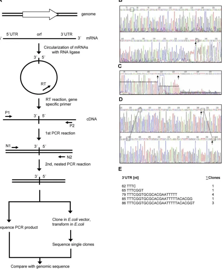
A recently developed method [28] (Figure 1A) was used to determine 59- and 39-ends of 40 transcripts from the two haloarchaeal species H. salinarum and H. volcanii. In short, cellular RNA was isolated and circularized using T4 RNA ligase. The RNA of every gene of interest was transformed into cDNA using a gene-specific primer and reverse tran-scriptase. The DNA was amplified in two consecutive nested PCR reactions with four gene-specific primers, yielding a PCR product comprising the known 59- and 39-regions of the open reading frame and the unknown 59- and 39-UTRs of the transcript. Sequencing of the PCR product and comparison with the genomic sequence allowed the determination of 59 -end and 39-end. An example is shown in Figure 1B. In several cases the signal strength of the sequence varied over the whole length of the PCR product, and one or several steps of decreasing signal intensity were observed, indicating that the 59-end, the 39-end, or both were not uniform (an example is shown in Figure 1C). In these cases the PCR product was cloned into anEscherichia colivector and the sequences of ten clones were determined. It turned out that in all cases the 59 -ends of the clones were identical, whereas the 39-ends varied. One representative example is illustrated in Figure 1D and 1E. The results revealed that transcription is initiated faithfully at only one specific nucleotide. The 39-end variability of some transcripts might either be caused by termination at several points within a region of about 30 nucleotides or by differential degradation from the 39-end.
The method was used to analyze 40 transcripts from H. salinarum or H. volcanii, thus generating by far the largest
Author Summary
Figure 1.Determination of 59- and 39-Ends of Haloarchaeal Transcripts
database of 59- and 39-ends of archaeal transcripts available. The genes were selected in order to exemplify many different functional categories (e.g., energy metabolism, anabolic metabolism, gene regulation, translation, stress response, proteins of unknown function), expression levels (e.g., ribosomal protein, transcriptional regulator), and cellular localizations (cytoplasmic, membrane-bound, extracellular). The results for the 40 genes are summarized in Tables 1 and 2. The EMBL nucleotide sequence database (http://www.ebi.ac. uk/embl/) and the NCBI literature database (http://www. pubmed.de/data/nlm.link.html) were searched for previously determined 59- and 39-ends of transcripts from both species and all publicly available examples were also included (Tables 1 and 2).
Promoter Structure in Haloarchaea
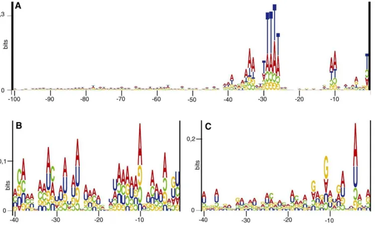
In order to characterize haloarchaeal basal promoter elements, the sequences from130 nt toþ20 nt (relative to the experimentally determined 59-ends) were extracted from the genome database ofH. salinarum(http://www.halolex.mpg. de) and partial genome database ofH. volcanii(http://www.tigr. org). Sequence logos [29,30] were generated to identify potentially functional DNA motifs present at a fixed distance from the transcription initiation point. Three sequence logos were generated, two species-specific logos for all genes ofH. salinarumandH. volcaniiwith known 59-ends (Figure 2A and 2B, respectively), and one that combined the data for both species (Figure 2C). Clearly, the most prominent promoter element is the TATA box, centered at position27/28, with the consensus sequence‘‘TTWT.’’
The‘‘transcription factor B recognition element’’(BRE) is conserved upstream of the TATA box (Figure 2). Its consensus sequence‘‘CGAAA’’is extended compared to the‘‘AA’’motif discovered in two previous studies [2,22] and to the bioinformatic analysis of theH. salinarumgenome (see below). The promoter analysis led to the detection of a ‘‘WW’’ element at positions10 and11 (Figure 2). The conserved spacing to the TATA box, the BRE, and the transcription start site strongly indicates that this novel element is a bona fide basal promoter element. It remains to be discovered which protein makes contact to these two bases. Likely candidates are TFB and the RNA polymerase.
It is known that the basal promoter elements have an optimal spacing to the transcriptional start site, e.g., the TATA box is centered at27/28, although a small degree of variation seems to be possible. Shifting of the TATA box by one or two nucleotides was reported to enhance the consensus sequence in a previous study [2]. Therefore, the upstream sequences of the genes summarized in Table 1 were inspected individually, and it was investigated whether sequence shifting would improve the alignment. However, this was not the case (unpublished data).
Transcription Termination in Haloarchaea
To identify sequence motifs important for termination of transcription, the last 80 nt of the transcribed DNA and the 50 nt downstream of the termination site were retrieved for all genes with experimentally determined 39-ends and a sequence logo was generated (Figure 3A). A motif of five Us close to the 39-end is strongly conserved. Apart from that, no conserved sequence motif was found either upstream or downstream of the termination site. To identify structural conservations, all 39-UTRs were subjected to in silico folding using three different programs (see Materials and Methods). More than 80% of the transcripts were found to contain a putative stemloop structure at their 39-ends, preceeding the penta-U motif or involving some of the Us (Table 1: transcripts 1–23 with the exception of 4 and 15; Table 2: transcripts 1–17 with the exception of 2, 5, 8, and 11). Three typical examples are shown in Figure 3B, indicating that the structure of the putative stemloops is not uniform but might include one or two bulges. Taken together, the observations suggest that transcriptional termination in haloarchaea occurs at one or several sites located downstream of a pentaU motif that is often preceded by a putative stemloop unconserved in sequence and structure.
Analysis of the 39-ends of the 40 haloarchaeal transcripts revealed that they are identical to the genome sequence and in neither species extra nucleotides were added after tran-scription. This observation corroborates a recent study that did not find any evidence for polyadenylation using bulk RNA of H. volcanii [31] and extends the analysis to a second haloarchaeal species,H. salinarum.
39-UTRs: Lengths, Putative Structural Elements, and Possible Roles
All 40 haloarchaeal transcripts were found to have a 39 -UTR (Tables 1 and 2). The GC content of the 39-UTRs was the same as that of the coding regions and no differences between the two species were detected. The lengths vary from 13 to 154 nt with an average length of 57 nt (median 49 nt). 80% of the 39-UTRs have a length between 20 and 80 nt, and only seven 39-UTRs are longer than 100 nt.
The program MEME [32] was used to search for conserved sequence motifs in different subsets of 39-UTRs (e.g., in the UTRs of all genes, of theH. salinarum, and of theH. volcanii genes). In each case, the oligo-U motif was involved in transcription termination (Figure 3A) but no additional sequence motif was retrieved. Three different programs (see Materials and Methods) were used to predict the secondary structure of the 39-UTRs either on its own or in the context of the whole mRNA. However, no conserved structural motifs apart from the stemloop structures preceding the penta-U motif (see above) could be detected.
(B) Sequence of a PCR product representing a transcript with only one specific 39-end (number 12 in Table 2). The stop and start codons of the gene are boxed, and the ligation point of the 59-end and the 39-end are denoted by an arrow.
(C) Sequence of a PCR product representing a transcript with several 39-ends (number 7 in Table 2). Different signal intensities are indicated by lines and the ligation point of the 59-end and two different 39-ends are denoted by arrows.
(D) Sequences of two clones after cloning the PCR product shown in C (number 7 in Table 2). The results of two sequencing reactions of independent clones are shown. In both cases, the stop codon and start codon of the gene are boxed, and the ligation point of the 59-end and the 39-end are denoted by an arrow.
(E) Results after sequencing ten clones. The sequences of five different 39-ends and their number of occurrence are shown. The 59-end was found to be identical in all ten cases.
Leaderless versus Leadered Transcripts and the Mechanism of Translation Initiation
Leaderless transcripts contain none or only a few nucleo-tides upstream of the translational start codon while the 59 -UTR of leadered transcripts needs to be long enough to contain a Shine-Dalgarno (SD) sequence. It has been shown that there must be at least four nucleotides between the 39 -end of the SD sequence and the start codon inH. volcanii[26], and that a SD sequence consists of at least five nucleotides (see below). Therefore we categorized all transcripts with a 59 -UTR of 10 nt or more as‘‘leadered,’’and all transcripts with fewer nucleotides as‘‘leaderless.’’In both species, about two
thirds of all transcripts are leaderless, i.e., 26 of 37 H. salinarumtranscripts (Table 1,blpwas counted only once) and 15 of 24H. volcaniitranscripts (Table 2). On average, the 59 -UTRs of leadered transcripts are longer inH. salinarumthan in H. volcanii (median 33 versus 19 nt). It remains to be discovered whether these differences are caused by the small sets of investigated genes of both species or if they reflect differences of the translation initiation process. The 59-UTRs in both species are shorter than those in eukaryotes, which have an average length of about 100 nt [13].
Translation initiation on leadered haloarchaeal transcripts was thought to depend on the‘‘conventional’’pathway, and
Figure 2.Basal Promoter Elements Identified Upstream of 63 Haloarchaeal Genes
The sequences from130 toþ20 around the experimentally determined 59-ends of haloarchaeal transcripts (Tables 1 and 2) were retrieved from databases. Sequence logos were constructed with the program‘‘RNA structure logo’’[29]. For all logos, the height of the upright letters corresponds to the degree of conservation of that nucleotide at the respective site, measured in bits. The upside down letters indicate that the occurrence of the respective nucleotide is lower than expected in random sequences of the same GC content, and the height corresponds to the degree of underrepresentation.
thus the 59-UTRs were expected to contain SD sequences. For reasons described below a SD sequence was defined to consist of at least five consecutive nucleotides able to hybridize to the 39-end of the 16S rRNA and to have a distance between three and seven nucleotides between its 39-edge and the start codon. Surprisingly, most haloarchaeal 59-UTRs were devoid of a SD sequence, and only two out of 19 leadered transcripts harbored a predicted SD sequence (Figure 4). A more relaxed criterion of four consecutive nucleotides and a distance of zero to nine nucleotides led to the inclusion of just one further gene, but four nucleotides are very unlikely to function as a SD sequence in haloarchaea (see Discussion).
It is unclear how translation is initiated on SD-less leadered transcripts, because the‘‘leaderless pathway’’cannot be used by leadered transcripts, and leadered transcripts are thought to need the interaction of a SD sequence with the 39-end of the 16S rRNA. Therefore, it seemed important to
character-ize experimentally whether or not SD-less leadered tran-scripts are translated efficiently in vivo.
Experimental Characterization of In Vivo Functions of Selected 59-UTRs and 39-UTRs Using a Reporter Gene System and Gene Fusions
To address translational efficiencies experimentally, the steady state levels of transcripts and their encoded gene products need to be quantitated. However, no quantitative assays for most of the gene products summarized in Tables 1 and 2 were available. Two different approaches were used to characterize the in vivo function of UTRs inH. volcaniiandH. salinarum.
A reporter gene system was established forH. volcanii. The H. volcanii dhfrgene was chosen as a well-established assay that has been successfully used as a reporter gene for in vivo promoter activity [33]. The dhfr transcript is leaderless. A Table 1.59-UTRs and 39-UTRs of Transcripts ofH. salinarum
Transcripts Number Identifiera Function 59-UTR [nt] 39-UTR [nt] Start Siteb Selection
Characterized in this study
1 OE3538R hypothetical protein 41 30 orf p
2 OE4187R probable DNA binding Protein 3 26 ms
3 OE2082F conserved hypothetical protein 0 50 orf p
4 OE2024F conserved hypothetical protein 1 80 ms
5 OE1982R conserved protein 0 34 orf p
6 OE3090R conserved hypothetical protein 33 13 msa
7 OE1147R protein-L-isoaspartate O-methyltransferase,pimT1 0 30 orf p
8 OE1405R conserved protein 0 27 orf p
9 OE3100F bacterioopsin-linked protein,blp 32 42 ms
10 OE3049R hypothetical protein 0 24 ms
11 OE3470F translation initiation factor,eif1a2 33 58 ms
12 OE1765R probable proteasome regulatory chain,pan1 1 29 msa
13 OE4136R translation initiation factor,eif1a1 0 132 msa
14 OE3934R citrate (si)-synthase,citZ 0 145 msa
15 OE2186R sec-independent protein translocase componenttatA 5 13 ms
16 OE4583F ferredoxin (2Fe-2S),fer5 0 41 ms
17 OE4674F alcohol dehydrogenase,adh2 0 26 ms
18 OE2935R fumarate hydratase,fumC 0 34 ms
19 OE3817R ribosomal protein S19.eR,rps19R 1 50 ms
20 OE4412R threonine synthase,thrC1 2 52 orf p
21 OE1972F methylmalonyl-CoA mutase,mut1B 2 60 ms
22 OE3717F NADH dehydrogenase 32K chain,no1A 1 77 ms
23 OE2906R superoxide dismutase,sod2 15 26 msa
Characterized in previous studies
24 L22007 bacterioopsin-linked protein,blp 32 — ms
25 J02767 cell surface glycoprotein precursor,csg 111 — ms
26 M19883 flagellin A1 und A2 (flagellin precursor),flgA1/A2 39 — ms 27 M19884 flagellin B1,B2,B3 (flagellin precursor),flg B1/B2/B3 49 — ms
28 X80931 arginine deiminase,arcA 7 — ms
29 X80931 Ornithine carbamoyltransferase,arcB 0 — ms
30 X80931 carbamate kinase,arcC 4 — ms
31 X68103 ferredoxin (2Fe-2S),fdx 1 — ms
32 D78200 adenylate kinase,adk 1 — ms
33 X57144 ribosomal protein S12,rps12 0 — ms
34 X16527 gas-vesicle proteingvpA1(p-gvpA) 20 — msa
35 X16527 gas-vesicle proteingvpA2(c-gvpA) 20 — ms
36 X94688 gas-vesicle proteingvpD2(c-gvpD) 116 120 ms
37 X01081 bop gene expression regulator,brp 0 — ms
38 V00474 bacteriorhodopsin precursor,bop 3 — —
a
The identifier of the genes characterized in this study is the gene designation of the genome project (http://www.halolex.mpg.de); the identifier of genes used in previous studies is the accession number http://www.ebi.ac.uk/embl/)..
b
variant of the gene without its native 39-UTR was generated and used as a control. Two genes encoding a ‘‘HoxA-like regulator’’(hlr) and a ‘‘conserved hypothetical protein’’(hp) were chosen for the investigation of the in vivo roles of UTRs (numbers 2 and 3 in Table 2). The native transcripts of both genes contain a 59-UTR lacking a SD sequence. Four plasmids were generated that encode transcripts containing either the 59-UTR or the 39-UTR of the two genes fused to thedhfropen reading frame (Figure 5A).H. volcaniicultures containing the respective constructs were grown to the exponential growth phase (23108cells/ml) and the DHFR activities as well as the dhfrtranscript levels were determined quantitatively (Figure 5A).
The determination of the average transcript level showed that the fusion of 59-UTRs to the control transcript did not alter the stability of the transcript. In contrast, fusion of the 39-UTRs to the control transcript in both cases enhanced transcript abundance by a factor of 1.4 and 2, respectively (numbers 3 and 5 in Figure 5A). Different transcription initiation rates can be excluded as the 59-parts of both transcripts are identical to the control transcript, therefore it can be inferred that the transcripts have different stabilities. Apart from the discovery that 39-UTRs are involved in selenocysteine incorporation in some methanogenic archaea [34], this enhancement of transcript stability is the second experimentally proven biological function of 39-UTRs in archaea, and this role is probably of much more general relevance.
The DHFR activities and transcript levels were used to calculate translational efficiencies of the five transcripts under investigation (Figure 5A). The results were normalized
to the translational efficiency of the control transcript and are summarized in Figure 5B. In both cases, the fusion of a 59 -UTR to the leaderless transcript led to a decrease in translational efficiency by a factor of about two (Figure 5B, numbers 2 and 4). This underscores that leaderless transcripts do not only form the majority of transcripts but that they are very efficiently translated in haloarchaea, in contrast to Escherichia coli [35]. For the first time the results prove experimentally that transcripts with a SD-less 59-UTR are translated in haloarchaea.
In one case fusion of 39-UTRs to thedhfrtranscript had no effect on translational efficiency (Figure 5B, number 3); in the second example it led to a slight decrease (Figure 5B, number 5).
In order to determine the in vivo function of UTRs in H. salinarum, a different strategy was chosen, i.e., the addition of a hexahistidine (his6) tag to the respective proteins; thus
enabling us to quantify the levels of different proteins using the same commercial anti-his-tag antibody. Two genes were chosen for the analysis, one gene encoding a SD-less leadered transcript (OE3100F, number 9 in Table 1) and one gene encoding a leaderless transcript with a long 39-UTR (OE2082F, number 3 in Table 1). His-tagged control versions as well as his-tagged versions carrying a deletion of the 59 -UTR or a deletion within the 39-UTR were integrated into the chromosome ofH. salinarum(Figure 6). Exponentially growing cultures (23108cells/ml) were used to determine transcript
levels by quantitative reverse transcriptase PCR (qRT-PCR) and protein levels with an anti-his4-tag antibody.
Unexpect-edly, the transcript level of the deletion variant was elevated about 2-fold suggesting that the 59-UTR destabilizes the Table 2.59UTRs and 39UTRs of Transcripts ofH. volcanii
Transcripts Number Identifiera Function 5’ UTR [nt] 3’ UTR [nt]
Characterized in this study 1 FS 451E8 ribonuclease HIIrnhB 0 95
2 FS 437B8 hoxA-like transcriptional regulator 14 30
3 FS 448H7 conserved hypothetical protein 22 45
4 FS 452E1 ferrichrome-binding protein 0 22
5 FS 436G6 rieske 2Fe-2S family protein 1 78
6 FS 433H6 transcription regulatorarcR 1 73
7 FS 459C6 glucose 1-dehydrogenase 0 79
8 FS431G8 cdc cell division proteinftsH 1 76
9 FS 458C1 transcription factor B (not identical with No.18) 5 53 10 FS 431F1 bacterial extracellular solute-binding protein 5 47
11 FS 451C1 dihydrofolate reductasefolA 1 101
12 FS 436F5 mandelate racemase/muconate lactonizing enzyme 0 153
13 FS 434E1 signal peptide peptidase 16 34
14 FS 441B9 conserved hypothetical protein 1 122
15 FS 456E3 cold shock transcription regulator 3 49
16 FS 456E4 conserved domain protein 30 55
17 FS 456E5 conserved hypothetical protein 14 33
Characterized in previous studies 18 AF143693 transcription factor B (not identical with No. 9) 79 n.d.
19 M97486 superoxide dismutase 1sod1 15 140
20 M97487 superoxide dismutase 2sod2 19 18
21 X58924 ribosomal protein L1e 63 41–55
22 X58924 ribosomal protein L11e 0–1 24–66
23 AF010470 heat shock proteincct1 4–5 n.d.
24 AF010469 heat shock proteincct2 5 n.d.
a
The identifier of the genes characterized in this study is the gene designation of a library [61]; the identifier of genes used in previous studies is the accesssion number (http://www.ebi.ac. uk/embl/).
transcript or that the transcription rate is enhanced in the mutant. However, it turned out that the transcript carrying a SD-less leader sequence, as well as the transcript lacking its native 59-UTR, was translated with very similar or identical efficiency (Figure 6). Deletion of more than half of the 39 -UTR of the OE2082F transcript had no effect on transcript stability or translation efficiency.
Taken together, the results demonstrate that leaderless transcripts, as well as leadered transcripts without a SD sequence, are efficiently translated in vivo in both haloarch-aeal species.
Translational Efficiencies of Transcripts with Artificial 59 -UTRs of Random Sequence
Bioinformatic analysis of SD-less 59-UTRs did not identify consensus sequences or conserved structural elements possi-bly involved in translation initiation (unpublished data). Therefore we tested whether artificial 59-UTRs of random sequence have the ability to initiate translation in vivo. Four random sequences of 20 nt (equivalent to the average length of natural 59-UTRs inH. volcanii) were generated and fused to the dhfr reporter gene. Very unexpectedly, translational
Figure 3.Transcription Termination in Halophilic Archaea
(A) The sequences from80 toþ50 around the experimentally determined 39-ends of haloarchaeal transcripts (Tables 1 and 2) were retrieved from databases. A sequence logo was constructed with the program‘‘RNA structure logo’’[29].
(B) Three typical examples of secondary structures predicted to be formed at the 39-end of transcripts. Note that the oligo-U motif is partially involved in basepairing in the stemloop in all three cases. From left to right, the examples show the 39-ends of the transcripts of the genes numbers 4, 15, and 16 in Table 2.
efficiencies of all four transcripts were at least as high as of the leaderless control transcript and the efficiencies of two transcripts were even 4-fold to fold higher (Figure 7). The 8-fold difference in translational efficiencies indicates that the random sequences contain as yet unrecognized sequence or structural information that influences translation initiation. Nevertheless, the results of all four artificial 59-UTRs of random sequence underscore that translation was efficiently initiated at leadered transcripts devoid of a SD sequence.
Genome-Wide Bioinformatic Analysis of Translation Initiation
Experimental characterization of a limited number of genes revealed that the majority of transcripts were found to be leaderless and that leaderless as well as leadered tran-scripts lacking a SD sequence were efficiently translated in vivo. To address the question whether these results can be generalized, a bioinformatic analysis of the H. salinarum genome was performed (the H. volcanii genome is not yet annotated and thus could not be analyzed). TheH. salinarum R1 genome guarantees a very high quality of translational start point annotation, because many proteomic data and gene homology data have been integrated (Pfeiffer et al., unpublished data; http://www.halolex.mpg.de). First, a ge-nome-wide operon prediction was performed. Two adjacent genes were regarded as co-transcribed if they had the same orientation and their intergenic distance was smaller than 40 nt. This value was chosen because the distance between two monocistronic adjacent genes has to contain the termination signal of the first gene (i.e., at least a pentaU motif, Figure 3) as well as the promoter of the second gene (i.e., BREþTATA box at35 to27, Figure 2). 2,055 genes were predicted to be monocistronic or proximal genes of operons and the regions upstream of their start codons were searched for conserved DNA or RNA elements. Figure 8A shows that no evidence for the existence of a SD consensus sequence was found in the region between1 and12. Instead, all three basal promoter elements were retrieved. Remarkably, the distance between the consensus sequences in Figure 8A and the translational start point is identical to the known distance between the promoter elements and the transcriptional start point, strongly indicating that the genes contributing to the consensus sequence have leaderless transcripts without any extra nucleotides upstream of the translational start codon. It should be pointed out that the information content is only 0.3 bits and thus this group contains also other transcripts, i.e., leadered transcripts and leaderless transcripts including one or a few upstream nucleotides. Nevertheless leaderless transcripts beginning at the translational start codon show the highest occurrence in this group of genes (Figure 8A).
Next, all predicted distal operon genes were analyzed. Initial attempts revealed that further subdivision was advis-able. 109 genes had a distance of 26–40 nt to the adjacent gene. Unexpectedly, the generation of a sequence logo revealed that consensus sequences for the three basal promoter elements were present, but a termination signal from the preceding gene was missing (unpublished data). Therefore, this group of genes was seemingly transcribed into a bicistronic or polycistronic mRNA from the promoter of the proximal gene of the operon, and in addition, from a second promoter that is localized in the small intergenic region. This group is highly enriched in ‘‘hypothetical
Figure 4.59-UTRs of Leadered Transcripts ofH. salinarumandH. volcani
proteins’’ and ‘‘conserved hypothetical proteins’’ (67%, in comparison to the average value of 45%). 83 genes had a distance of 10 to 25 nt from the preceding genes. Generation of a sequence logo did not reveal the presence of any conserved element (Figure 8B).
A large group of genes (618) showed a distance of less than ten nucleotides to the preceding gene or had an overlap of up to ten nucleotides. Very faint overlapping signals with similarity to a SD sequence were detected, i.e.,14 GAGGU-GA8 and12 AGGUGA7 (Figure 8C). To reveal whether the signal from 14 to 7 in Figure 8C was due to SD sequences, 100 sequences were visually inspected and convincing SD sequences were indeed detected. The program ‘‘PatSearch’’ [36] was used for a systematic search for the occurrence of possible SD sequences in this group of 618 genes. As the minimal length of a functional SD sequence in archaea is not known, consecutive as well as non-consecutive matches of different length were retrieved, and the results are shown in Table 3. The spacing was set to a distance of 3–7 nt (the optimal distance determined for E. coli þ/ 2 nt). Changing the spacing to a distance from 0–9 nt led only to a slight change of results (unpublished data). Consecutive matches of seven or eight nucleotides occurred nearly exclusively at the right distance and can be expected to be
functional SD sequences. In contrast, four consecutive or five non-consecutive matches were found mostly in the wrong spacing and thus they do not seem to be sufficient to determine the translation initiation point. When the minmal requirement for a haloarchaeal SD sequence is set to$5 or $6 consecutive nucleotides that can hybridize with the 16S rRNA then 27% or 11%, respectively, of this group of genes is preceded by a SD sequence. Therefore the exact values vary with the definition of a SD sequence. Nevertheless, the analysis revealed that SD sequences do exist, but that only a minority of genes is preceded by a SD sequence even in this group of distal genes of operons. Due to the small intergene distances in this group the putative SD motifs overlap with the preceding open reading frame.
The analysis was also performed with the group of 2,055 single genes and first operon genes and the results are shown in Table 4. In this case, 100 nt upstream of the start codon were used. Again, the data indicate that five non-consecutive matches do not function as a SD sequence because the vast majority is found at a wrong distance. The fraction of genes with five or six consecutive matches in the right distance is 3.6% and 1.6%, respectively, values that are considerably lower than in the group of distal operon genes. Taken together, the genome analysis revealed that a SD sequence
Figure 5.Characterization of the In Vivo Function of UTRs inH. volcaniiUsing a Reporter Gene
(A) Thedhfrreporter gene and the gene fusions containing 59-UTRs and 39-UTRs of genes number 2 and 3 of Table 2 are shown schematically. The genes encode a‘‘HoxA-like transcriptional regulator’’(hlr, number 2 in Table 2) and a‘‘conserved hypothetical protein’’(hp, number 3 in Table 2). The DHFR enzymatic activity, thedhfrtranscript level, and the translational efficiency are tabulated. Three biological replicates were perfomed and average values were calculated. The numbers in parentheses are standard deviations. The translational efficiencies were calculated by dividing the DHFR activity by thedhfrtranscript level.
plays a very minor role for translation initiation in haloarchaea, thus reinforcing the experimental results obtained with a limited set of genes (Figures 4–7).
Discussion
Determination of 59- and 39-Ends of Transcripts
We have determined the 59- and 39-ends of 40 haloarchaeal mRNAs using a recently developed method. In the original description of the method, Kuhn and Binder [28] came to the conclusion that only processed 59-ends could be determined, but no primary 59-ends. The experimental indication was that they found several different 59-ends belonging to the single transcript analyzed. The theoretical explanation was that primary 59-ends carry triphosphates which presumably cannot be ligated by RNA ligase. Therefore, for the determination of primary 59-ends the triphosphates would need to be first converted to monophosphates using a pyrophosphatase [37]. However, this turned out to be unnecessary for haloarchaeal transcripts. In a proof of principle experiment the 59-end of the blp transcript was determined, and the result was identical to an earlier study using primer extension (compare numbers 9 and 24 in Table 1). The results of the whole gene set yielded the following additional proofs that primary 59-ends have been deter-mined: (1) in all 40 cases a single 59-end was discovered, and (2) the basal promoter elements (TATA box and BRE) had exactly the known distance to the experimentally analyzed 59 -ends, which can only occur when primary 59-ends were determined.
The most conceivable explanation as to why the method worked so well with haloarchaeal transcripts is that a fraction of transcripts was dephosphorylated from the triphosphate to the monophosphate form. In E. coli it was shown that conversion of the 59-triphosphate to a 59-monophosphate is the first step in mRNA degradation. The fractions of
5-monophosphorylated cellular transcripts were quantified for two genes and found to be‘‘18%–35%’’and‘‘up to half’’[38]. If that would also be true for haloarchaea, it would explain the success of the method. However, it should be noted that for unknown reasons the method was not successful with some genes. Possibly, in these cases the treatment with a pyrophosphatase might have helped. However, either in the simple version applied here or in the version including a pyrophosphatase step [37], the method is very powerful and is expected to be extremely helpful for the determination of transcriptional and translational signals in a variety of archaeal, bacterial, and eukaryotic species.
Promoter Structure of Haloarchaea
The regions upstream of experimentally determined start sites yielded consensus sequences for three basal promoter elements, i.e., the TATA box, the BRE, and a novel element at 10/11. The same basal promoter elements were retrieved in a genome-wide analysis of the more than 2,000 genes, which are monocistronic or first genes in predicted operons.
The conserved region of the TATA box comprises only four (or five) nucleotides (Figures 2 and 8A). This is considerably shorter than the TATA box length of eight nucleotides determined in structures of TBP/DNA complexes from different species [39–41] and which is thought to be universally conserved in eukaryotes and archaea. However, two previous studies already reported that the consensus sequence for a haloarchaeal TATA box is shorter than eight nucleotides. A comparison of the regions upstream of experimentally determined 59-ends available in 1999 revealed that the conserved region in haloarchaea was shorter than those in methanogenic archaea and in thermophilic archaea [2]. The in vivo selection of TATA boxes out of a random library of more than 106 clones yielded a strong consensus sequence of six nucleotides [33]. The most probable explan-ation is based on the fact that haloarchaea have multiple
Figure 6.Characterization of the In Vivo Function of UTRs inH. salinarumUsing Gene Fusions
(A) The constructs that were integrated into the genome ofH. salinarumare shown schematically. Protein and transcript levels were determined using a anti-his6-tag antibody and qRT-PCR, respectively. The translational efficiencies were calculated by dividing the protein levels by the transcript levels. Three biological replicates were performed and the average values and standard deviations (in parentheses) were calculated.
TBPs, in contrast to all other species [42]. The TATA box consensus sequence of four nucleotides might comprise the common core that is detected by all haloarchaeal TBPs, while the remaining four nucleotides might be differentially recognized by individual TBPs. Thereby haloarchaeal TBPs could act as global regulators of transcription initiation, similar to bacterial sigma factors [42,43].
Transcription Termination in Archaea
The results indicate that both a pentaU-motif at the very 39-end of the transcripts and possibly a small stemloop near the 39-end are involved in transcription termination. It should be pointed out that the putative stemloops are neither conserved in sequence nor in structure, therefore it is unclear how they could be involved in a general mechanism for transcription termination. Clearly, experimental data are needed to clarify whether the predicted stemloops are indeed involved in transcription termination.
A comparison with previous results is difficult since termination has not been thoroughly studied in archaea. The only report for a haloarchaeon revealed that the sequence ‘‘UUAUUCUUU’’ functioned as a terminator in
vivo, while the mutated sequence‘‘UUAUUCGGU’’did not, confirming the importance of Us [43]. Some earlier studies with stable RNA genes from thermophilic archaea showed that termination occurred after extended U or oligo-pyrimidine stretches of more than ten nucleotides [7–9,44]. This might indicate that the oligoU-stretch required for termination is shorter in halophilic archaea, which have very GC-rich genomes, than in thermophilic crenarchaea. Re-cently one very thorough study of transcriptional termination in Methanothermobacter thermoautotrophicus was reported that used an in vitro termination system [10]. Natural and synthetic U-tracts were found to have the capacity and to be essential for termination while stemloops influenced termination in a way that could not be rationalized. Surprisingly, termination was also found to be influenced both by the topology of the substrate and by sequences up to 200 nt upstream of the termination site. Taken together, it appears that transcription termination in archaea does not have a uniform mechanism and that it is far from being understood. Determination of 40 haloarchaeal 39-ends has identified a much higher number of in vivo functional
Figure 7.Translational Efficiencies of Artificial UTRs of Random Sequences inH. volcanii
Four 59-UTRs of random sequence were generated and cloned upstream of the reporter genedhfr.
(A) The transcripts are shown schematically. The DHFR activities, the transcript levels and the translational efficiencies are tabulated. Three biological replicates were performed and average values and standard deviations (in parentheses) were calculated.
(B) The translational efficiencies are shown after normalization to the control transcript devoid of a 59-UTR.
(C) The sequences of the four random 59-UTRs are tabulated including the start codon (bold) are shown. On top, the sequence of eight nucleotides that are complementary to the 39-end of the 16S rRNA is shown. The optimal spacing between SD sequence and start codon in archaea is unknown, therefore the optimal spacing determined withE. colihas been chosen.
terminators than in any other archaeal species and the results will guide further experiments.
39-UTRs and Their Putative Biological Roles
Experimental determination of 40 39-ends, a much higher number than in previous studies, revealed that all analyzed genes have a 39-UTR with an average length of 57 nt. In
eukaryotes, 39-UTRs are longer and their length increased in evolution, e.g., 39-UTRs of yeast and humans have an average length of 200 nt and 500 nt, respectively [13]. Eukaryotic 39 -UTRs are involved in a variety of different functions [12,13,16]. In archaea, the only function characterized previously was selenocysteine incorporation at stop codons within a few genes in some methanogenic archaea [34]. However, haloarchaea do not contain selenoproteins, and thus 39-UTRs must have other functions. The reporter gene system showed that the addition of 39-UTRs stabilized thedhfr transcript, and thus transcript stabilization is the second experimentally proven function of 39-UTRs in archaea, which might be of very general relevance.
The 39-UTRs are collinear with the genome sequence and posttranscriptional modification was not found to occur in any of the haloarchaeal transcripts analyzed. It has been shown that in exosome-containing archaea the 39-ends are tailed post-transcriptionally with A-rich sequences important for RNA degradation [31,45]. However, Haloarchaea are devoid of an exosome and thus they have to use a different, as yet unidentified, RNA degradation pathway.
Translation Initiation at Leaderless and Leadered mRNAs The results of the experimental determination of 59-ends of transcripts from two haloarchaea species as well as the bioinformatic genome analysis ofH. salinarumrevealed that
Figure 8.Bioinformatic Analysis of theH. salinarumGenome
(A) Sequence logo of the upstream regions of 2,055 genes that are predicted to be either monocistronic or proximal genes in operons with an intergenic distance to the adjacent gene of at least 40 nt.
(B) Sequence logo of the region upstream of 83 genes that are predicted to be distal genes in operons at a distance between 10 nt and 25 nt to the adjacent gene.
(C) Sequence logo of the region upstream of 618 genes that are predicted to be distal genes in operons but less than 10 nt apart from the adjacent gene.
doi:10.1371/journal.pgen.0030229.g008
Table 3.Occurrence of Strings Matching the SD-Sequence
‘‘GGAGGUGA’’Upstream of 618 Distal Operon Genes ofH. salinarum
Matches Number of Matches
Distance from Translation Start 3–7 nt
Distance from Translation Start 0–2 nt ; 8 to$32 nt
Consecutive 8 18 1
7 31 2
6 19 8
5 101 59
4 98 272
Non-consecutive 7 55 10
6 88 81
5 82 400
the majority of transcripts are leaderless. Thus, this is regarded as the default pathway of translation initiation at least in haloarchaea. The characterization of a transcript as ‘‘leaderless’’ as well as the elucidation whether a leader contains a SD sequence (see below) critically depends on the correct annotation of translational start sites. ForH. salinarum a high quality annotation of the genome was used, which integrates numerous proteomics results and cross-species comparisons (http://www.halolex.mpg.de; Pfeiffer et al., un-published data). The translation initiation site of more than half of the leaderedH. salinarumtranscripts used in this study had been experimentally determined by peptide mass finger-printing, and in the remaining cases it was verified that also possible start codons further downstream were not associated with SD sequences. For H. volcanii, codon preference plots were used to enhance the quality of correct start site selection [46].
Translation initiation on leaderless transcripts is brought about by a different mechanism than on leadered transcripts. In two archaea it has been shown that mutation of a SD sequence inhibited translation, while total deletion of the natural 59-leader restored translation capacity [25,26]. In bacteria it has been shown that leaderless transcripts differ from leadered transcripts with regard to the requirement of initiator tRNA and translation initiation factors, the inter-action with ribosomal subunits and the ribosome, and the resistance to the antibiotic kasugamycin. While translation on leadered transcripts initiates via an interaction with the small ribosomal subunit, bacterial and eukaryotic leaderless tran-scripts bind undissociated ribosomes and initiator tRNA to initiate translation [47–52]. Archaeal, bacterial, and eukary-otic in vitro translation systems are capable of translating leaderless transcripts, and therefore it has been proposed that this pathway is universally conserved and ancient [47,51]. As leadered transcripts are the minority at least in haloarchaea, their 59-UTRs might have one or more bio-logical functions in addition to translation initiation. The biological role of 59-UTRs might be to repress translational efficiency compared to leaderless transcripts. This would allow fine-tuning of the protein amount produced per mRNA and it would enable the usage of differential translational control if positive regulators would enhance translational
efficiency under certain conditions. 59-UTRs could also be involved in (differential) regulation of transcript half-lives. Experiments to characterize the biological roles of 59-UTRs in haloarchaea are underway.
It is currently believed that two major pathways of translation initiation exist, which are different in prokaryotes and eukaryotes. Prokaryotic transcripts require the inter-action of a SD sequence with the 16S rRNA 39-end, while in eukaryotes a scanning mechanism is used for translation initiation (in addition, exceptions exist like leaderless tran-scripts, internal ribosomal entry sites [53], and short up-stream ORFs). The data presented here show that this does not hold true at least for haloarchaea, which use SD-mediated translational initiation only for a small subset of their genes. Evidences are (1) that most of the 19 leadered haloarcheal transcripts reported here do not contain a SD sequence, (2) that the seven experimentally characterized 59-UTRs without a SD sequence mediate efficient translation in vivo (Figures 5–7), and (3) that the bioinformatic genome analysis indicates that SD sequences are not typical even for distal genes in operons.
The definition of a SD sequence is critical for the decision whether or not SD sequences are present in 59-UTRs. InE. coli it is part or all of the sequence TAAGGAGGT that can hybridize to the 39-end of the 16S rRNA. A bioinformatic analysis of all E. coli genes has revealed the consensus sequence AAGGA [54]. In most experimental studies, four to six consecutive nucleotides are used (e.g., [55] and references therein). The spacing is 5 ntþ/ 2 nt between the 39-edge of the aligned SD sequence and the start codon [54,55]. In contrast to the decades of research with bacteria only two experimental studies on archaeal SD sequences are available. Using aS. solfataricusin vitro translation system it was revealed that mutation of the native SD sequence
GAGGUGA to the sequence GACGUCA (nt with
comple-mentarity to the 16S rRNA are shown in bold, mutated nt are underlined) led to a complete loss of translation competence [25]. Note that the inactive mutant sequence retained five nt that could hybridize to the 16S rRNA. Using aH. volcaniiin vivo system it was shown that mutation of the natural SD sequence GGAGGUCA to UUAGGUCA decreased transla-tional efficiency by more than 90% although the mutant retained five SD nucleotides [26]. These results are in excellent agreement with the bioinformatic analysis of the H. salinarumgenome. Four consecutive and five non-consec-utive matches are found predominantly at wrong distances to the translational start point, and this is even true for five consecutive matches if the analyzed region is 100 nt (Tables 3 and 4). Taken together, bioinformatic as well as experimental results indicate that archaeal SD sequences might need more extensive base-pairing to the 16S rRNA for determining the translational start site than E. coli SD sequences. Even if a rather relaxed definition of ‘‘five consecutive matches’’ is applied, less than 10% of all H. salinarumgenes use the SD pathway for translation initiation and more than 90% are either leaderless or use a novel mechanism devoid of a SD sequence.
It might turn out that a low occurrence of SD sequences and the usage of a non-SD pathway for translation initation is not confined to haloarchaea. A large-scale bioinformatic genome analysis predicted that 50% of all archaeal and all bacterial genes are not preceded by a SD sequence [56]. If that Table 4.Occurrence of Strings Matching the SD-Sequence
‘‘GGAGGUGA’’Upstream of the 2,055 Proximal Operon Genes or Monocistronic Genes ofH. salinarum
Matches Number of Matches
Distance from Translation Start 3–7 nt
Distance from Translation Start 0–2 nt ; 8 to$32 nt
Consecutive 8 4 5
7 13 27
6 15 42
5 43 279
Non-consecutive 7 11 80
6 60 730
5 175 3,598
would be true, a high fraction of prokaryotic transcripts must either be leaderless or carry 59-UTRs devoid of a SD sequence, in contrast to the current belief. It will be interesting to reveal (1) how widespread the occurrence of leaderless transcripts and of leadered SD-lacking transcripts is in different groups of prokaryotes, (2) whether translation initiation on leadered SD-lacking transcripts uses a scanning or a novel mechanism, and (3) how initiation on SD-lacking transcripts works mechanistically and to uncover the nature of the molecules involved.
Materials and Methods
Microorganisms, media, and growth conditions. H. salinarumwas
provided from the German culture collection (DSMZ, Braunschweig,
Germany, strain number DSM 670),H. volcaniiWR 340 was obtained
from Moshe Mevarech (Tel Aviv University, Tel Aviv, Israel), andE.
coliXL1 Blue MRF’ was purchased from Stratagene (Amsterdam, The
Netherlands).H. salinarumwas grown aerobically in complex medium
containing 4.3 M NaCl, 80 mM MgSO4, 10 mM Na3-Citrate, 27 mM
KCl, 1% (w/v) peptone (pH 7.2) at 428C [57].H. volcaniiwas grown
aerobically in complex medium containing 2.9 M NaCl, 150 mM
MgSO4, 60 mM KCl, 4 mM CaCl2, 0.275% (w/v) yeast extract, 0.45%
(wt/vol) tryptone, and 50 mM Tris-HCl (pH 7.2) at 428C [58].E. coli
XL1 Blue MRF’ was grown in SOB Medium at 378C [59].
Total RNA preparation and determination of 59-ends and 39-ends.
RNA was isolated from exponentially growing cultures as described by Chomczynski and Sacchi [60] or by using the RNeasy system (Qiagen) with DNase treatment according to the manufacturer’s instructions. The quantity and quality of the extracted total RNA were determined by UV spectroscopy (at 260, 280, and 230 nm) and denaturing formaldehyde agarose gel electrophoresis [59,61]. RNA circularization, reverse transcription, and PCR amplification were carried out as described [28]. After denaturation at 658C for 10 min,
total RNA was self-ligated by incubating 5–10lg RNA with 40 U T4
RNA ligase (New England BioLabs), 10 U RNase Inhibitor (Promega), and 13T4 ligase buffer in a reaction volume of 25ll at 378C for 1 h.
After adjusting the total volume to 500ll, proteins were removed by
phenol chloroform extraction following standard protocols [59]. Self-ligated RNA was denatured and hybridized with 0.5 pmol of gene
specific primer RT (see Tables S1 and S2) at 658C for 10 min. cDNA
synthesis was carried out with M-MLV Reverse Transcriptase RNase
H Minus point mutant (Promega) on 5–10lg ligated RNA according
to the manufacturer’s instructions. The cDNA of the 59-39ligated
RNA was then amplified with gene-specific primer pairs PCR1/PCR2 followed by a second PCR with nested primer pairs NES1/NES2 (see Tables S1 and S2). The second, nested PCR enhances the specificity of amplification considerably and eliminates any possible false-positive fragments of the first PCR reaction. Nested PCR products of the 59-39
ligated RNA were subsequently analyzed by sequencing.
To analyze variable transcription stop sites, amplified nested PCR products were blunt-end ligated into the pBluescript vector pSKII (Stratagene) according to standard procedures [59]. Ten different cDNA clones were then analyzed by sequencing.
Identification of conserved DNA and RNA sequence motifs.The
program‘‘RNA structure logo’’[29,30] allows us to identify and to
visualize conserved sequence motifs that have a fixed distance to a point used for the alignment of a set of sequences, e.g., the transcriptional start site and the termination site. To create a logo, sequences with a defined length upstream and downstream of the alignment site were extracted from sequence databases, and the sequence logos were generated using the website: http://www.cbs.dtu. dk/;gorodkin/appl/slogo.html.
The program MEME [32] allows us to identify conserved sequence motifs in a set of sequences. The motifs can have variable local-izations in different sequences, can occur in all or a subset of sequences, can occur 1- or several-fold in a sequence, and can be present in either strand. The website http://meme.sdsc.edu./meme/ meme.html was used for the analysis.
Prediction of secondary structures of complete RNAs, 59-UTRs,
and 39-UTRs.Three different programs were used for the prediction
of the possible secondary structures of complete transcripts, 59-UTRs
and 39-UTRs. The program ‘‘Mfold 3.2’’ [62,63] was used at the
website http://www.bioinfo.rpi.edu/applications/mfold/rna/form1.cgi
and the program ‘‘RNAshapes’’ [64,65] was used at the website
http://bibiserv.techfak.uni-bielefeld.de/rnashapes. Dirk Metzler
(Bio-informatics, University of Frankfurt) was so kind to fold the RNAs using a self-developed program that can take pseudoknots into account (unpublished data).
Construction of plasmids containing 59-UTR and 39-UTR reporter
gene fusions.The shuttle vector pSD1/M2–18 [33] was used for the
generation of a reporter system for the in vivo function of 59-UTRs
and 39-UTRs. It contains replication origins and resistance genes for
E. coliandH. volcaniias well as thedhfrgene under the control of a constitutive promoter of medium strength, which allows detection of both up- and downregulation of expression levels.
First, a control plasmid was constructed. The promoter region and
the open reading frame of thedhfrwere amplified separately by PCR
reactions (the oligonucleotides for the construction of all plasmids are summarized in Table S3). The two PCR fragments were fused into one by a subsequent third PCR reaction, and the resultant fragment was cloned into the basal vector using ApaI and KpnI. The newly generated plasmid, pMB1, is very similar to the basic vector pSD1/
M2–18, but the native 39-UTR of thedhfrand nonnative nucleotides
at the 59-UTR (used for cloning in previous studies) were removed.
Joining of promoter region and ORF by fusion PCR instead of ligation has the advantage that not a single nonnative nucleotide has to be introduced.
The plasmids pMB3 and pMB6 (Figure 5, numbers 2 and 4) were constructed in a similar way. The promoter fragments and the ORF were amplified as separate PCR fragments and joined by fusion PCR.
The 59-UTRs were part of the primers (see Table S3). The plasmid
pMB4 (Figure 5, number 3) was also constructed by the same method.
The 39-UTR and a KpnI site were part of the downstream primer for
amplification of the ORF fragment. For construction of plasmid pMB7 (Figure 5, number 5) three PCR fragments were generated
containing the promoter region, the ORF, and the 39-UTR,
respectively. The three fragments were joined into one fragment by two consecutive fusion PCRs. In all five cases, the final fragments were cloned into the shuttle vector pSD1/M2–18 using single ApaI and KpnI sites. The plasmids pMB14–17 (Figure 7, numbers 10–13)
containing artificial 59-UTRs of random sequence upstream of the
dhfrreporter gene were constructed as described above for pMB2 and
pMB3. Primer sequences are included in Table S3. The newly generated regions of all plasmids were verified by sequencing. Then
they were used to transformH. volcaniias described previously [58].
Determination of dhfr transcript levels. RNA was isolated from
exponentially growing cultures (2 3108 cells/ml) as described by
Chomczynski and Sacchi [60]. DNA was removed from the samples by
incubating 6 lg RNA with RNase-free RQ1-DNase (Promega)
according to the manufacturer’s instructions. The RNA was purified using the RNeasy Mini Kit (clean up protocol; Qiagen). For reverse
transcription, 1lg RNA was denatured and was incubated with 0.6lg
of random hexamers (0.2 mM dATP and dTTP, 0.3 mM dCTP and dGTP) for 10 min at 658C in 38ll 1x buffer (Promega). After cooling
on ice for 2 min, cDNA synthesis was started with 2ll M-MLV Reverse
Transcriptase RNase H Minus (Promega) at 42 8C. After 1 h,
additional reverse transcriptase (1ll) was added. The reaction was
stopped after 2 h by heat inactivation at 808C for 5 min.
Each real-time reaction contained 1ll or 1.5ll of template, 0.8lM
of forward primer and reverse primer and 12,5ll DyNAmo SYBR
Green qPCR Master Mix (Finnzymes OY) in a final volume of 25ll.
dhfr transcript levels were determined with the upstream primer
Dhfr-RT_f, and the downstream primer Dhfr-RT_r. As an
unregulated control, the ribL10 transcript levels were determined
with the primer pair RibL-RT_f and RibL-RT_r.
The real-time PCR included 10 min initial denaturation at 948C,
60 cycles of 30 s 948C, 45 s 658C, 60 s 728C, and a final extension of 5 min at 728C. At last, samples were heated to 998C, cooled to 608C in
0.58C steps and the melting point of each sample was determined.
The real-time PCRs were performed in the ‘‘Rotor Gene 3000’’
(Corbett Research). Data analysis was performed using the software
‘‘Rotor Gene 6.35’’ (Corbett Research). For each sample the
intersection to a threshold line in the early exponential phase of the reaction was determined (Ct value). Data analysis was performed according to Livak and Schmittgen [66]. The Ct levels of the control
transcript ribL10 were used to normalize the Ct levels of thedhfr
transcripts. The dhfr level of the chromosomal gene copy was
determined using a strain carrying a plasmid without a dhfr gene
(pNP10) [67]. It was less than one-third compared with the strains
carrying a plasmid with a dhfr gene. The value was subtracted to
quantitate the transcript level of the plasmid-encodeddhfrgene. The
dhfrtransript level of the strain containing pMB1, which encodes a
dhfrwithout 59-UTR and 39-UTR, was set to 1.
Determination of DHFR activities and of translational efficiencies.
Novobiocin. 10 ml of cultures in exponential growth phase (23108 cells/ml) were harvested by centrifugation (2,800g, 48C, 20 min). The cells were washed once in basal salt solution (2.9 M NaCl, 150 mM
MgSO4, 60 mM KCl, and 4 mM CaCl2) and were then resuspended in 2
ml of basal salt solution. The cells were broken by sonication (2330 s, output control: 5, duty cycle: 50%; Branson sonifier 250). The extract
was then centrifuged at 18,000gand 48C for 30 min, and the pellet
was discarded.
Dihydrofolate reductase activity was measured in 1-ml volume
containing 100ll of cytoplasmic extract, 700ll buffer (3 M KCl, 25
mM potassium phosphate, 25 mM citrate [pH 6.0]) 0.05 mM dihydrofolic acid (Sigma), and 0.1 mM NADPH (AppliChem). The
oxidation of NADPH was determined at 340 nm and 258C with a
Specord S600 photometer (Analytik Jena). The enzymatic activity was
calculated using an extinction coefficient of 6.22 mM1
cm1
. Protein contents were determined using the BCA Assay Kit (Pierce) according to the manufacturer’s instructions, with BSA as standard, and used to calculate specific enzyme activities (nkat/mg protein). The DHFR level
encoded by the chromosomaldhfrcopy was determined using a strain
carrying a plasmid without adhfrgene (pNP10) [67]. It was less than
one-third compared with the plasmid-encoded DHFR values. Never-theless, it was subtracted to quantitate the plasmid-encoded DHFR level (Figures 5 and 7).
The translational efficiencies were calculated by dividing the specific enzyme activities with the transcript levels. Three independ-ent experimindepend-ents were performed, and average values and standard deviations were calculated.
Construction of his-tagged fusion genes and determination of
translational efficiencies inH. salinarum.The fusion genes encoding
the respective protein with a C-terminal hexahistidine tag were generated by the production of two overlapping PCR fragments, both containing the histidine codons introduced by the PCR primers, and their consecutive fusion into one fragment using‘‘ligation PCRs.’’All primers are summarized in Table S3.
For the construction of the gene shown as number 1 in Figure 7, one PCR fragment contained 476 nt upstream sequences of the gene
OE3100F, the 59-UTR, and the open reading frame. The second PCR
fragment contained 560 nt downstream sequences. Both fragments were fused by PCR.
For the construction of the gene shown as number 2 in Figure 7,
the 59-UTR was deleted. The deletion was performed by generating
two overlapping PCR fragments, which contained the 476 nt upstream region and the open reading frame, respectively, but were
devoid of the 59-UTR. They were fused by a PCR reaction, and the
resulting fragment was fused to the 560 nt downstream region by a further PCR reaction.
For the construction of the gene shown as number 3 in Figure 7, one PCR fragment was generated that contained 430 nt upstream of gene OE2082F and the open reading frame, and a second PCR fragment that contained 502 nt of the downstream region including
the full-length 39-UTR. Again, both contained the primer-encoded
histidine codons and were fused by a subsequent PCR reaction. For the construction of the gene shown as number 4 in Figure 7,
the first 27 nt of the 39-UTR were omitted from the downstream
fragment. The shorter downstream fragment (475 nt) was fused to the identical fragment containing the upstream region and the open reading frame described above.
In all four cases, the resulting PCR fragments were cut with BamHI and KpnI (the sites were introduced with the PCR primers) and
ligated with a BamHI-KpnI fragment generated from vector pSD1/
R1–6 [33]. This fragment contained theE. colivector pSKII and a gene
conferring Novobiocin resistance to haloarchaea. The resulting four
plasmids were used to transform H. salinarum as described [58].
Novobiocin (0.4lg/ml) was used to select for clones that had stably
integrated the plasmids into the chromosome by homologous recombination.
The transcript levels were quantitated by qRT-PCR as described
above for thedhfrgene ofH. volcanii. The qRT-PCR protocol differed
in two details, i.e., annealing was performed at 65 8C and the
elongation phase was 60 s. The primers are included in Table S4. The
39-regions of the reverse primers are complementary to the histidine
codons. Thereby it is guaranteed that only transcripts are quantitated that give rise to the his6-including fusion protein. As an unregulated
control, the transcript level of thehstAgene was used.
To prepare cell extracts for immunoblotting, 15 ml of cultures in
exponential growth phase (2 3108 cells/ml) were harvested by
centrifugation (3,200g, 20 min, 218C).
The pellet was suspended in 1 ml a. bid. to lyse the cells. Proteins were then precipitated according to the method of Wessel and Flu¨gge [68], and the precipitate was dissolved in 25 mM Tris-HCl (pH 8,6).
Protein concentrations were determined with the BCA protein assay reagent (Pierce). Solutions of bovine serum albumin (BSA) were used as standard. Before electrophoretic separation, one-quarter volume
of 43SDS sample buffer was added. The samples were incubated for 5
min at 958C and 80lg total protein/lane was separated on standard
sodium dodecyl sulphate (SDS)-polyacrylamide gels. Subsequently, the proteins were transferred onto nitrocellulose membranes (Protran BA 83; Whatman, Schleicher and Schu¨ll) by semi-dry blotting and probed with a specific Tetra-His antibody (Quiagen) according to the manufacturer’s instructions. Horseradish perox-idase-conjugated goat anti-mouse antibody (Sigma) was used at concentrations recommended by the manufacturer. Immunoreactive bands were visualized by chemiluminescence (ECL substrate). For quantitation the films were scanned and the pictures were analyzed with the software‘‘ImageJ’’(http://rsb.info.nih.gov/ij/index.html). The background was determined locally for each band and subtracted from the signal.
Genome-wide in silico analyses. The identifiers, positions of the
start and stop codons, and the direction were retrieved from the genome database (http://www.halolex.mpg.de). Microsoft Excel was used to identify groups of genes that have the same direction of transcription and have intergene distances of at least 40 nt, 26 nt–40 nt, 10 nt–15 nt, and below 10 nt. For the first group of genes, the
upstream regions from1 to100 were retrieved from the genome
database, for the other groups the upstream regions from1 to40.
For data analysis the programs ‘‘RNA structure logo’’ [29,30] and
MEME [32] were used as described above. The program‘‘PatSearch’’
[36] was used for the identification of putative SD sequences in the four groups of genes. The search pattern was varied, e.g., both consecutive as well as nonconsecutive matches to the SD sequence
‘‘GGAGGUGA’’were retrieved and the length of the search string was
changed. The searches were performed (1) in the 40 nt upstream of the 618 genes that are distal genes of operons and have a distance of less than 10 nt to the preceding gene and (2) in the 100 nt upstream of the 2,055 single genes or proximal genes of operons. The results tables were sorted to derive the number of genes with matches in a distance of 3–7 nt from the translation start and the number of genes with other distances. From the results tables that contained the
number of genes with‘‘at least x matches’’the number of genes were
calculated that contain‘‘exactly x matches,’’with‘‘x’’being an integer between 4 and 8 as shown in Tables 3 and 4.
Supporting Information
Table S1. Primer Used for 59- and 39-End Determination of
Transcripts ofH. salinarum
Found at doi:10.1371/journal.pgen.0030229.st001 (50 KB DOC).
Table S2. Primer Used for 59- and 39-End Determination of
Transcripts ofH. volcanii
Found at doi:10.1371/journal.pgen.0030229.st002 (46 KB DOC).
Table S3.Primer Used for Reporter Plasmid Construction and
Real-Time PCR
Found at doi:10.1371/journal.pgen.0030229.st003 (77 KB DOC).
Table S4.Primer Used for Construction of his-Tagged Fusion Genes
and Real-Time PCR
Found at doi:10.1371/journal.pgen.0030229.st004 (37 KB DOC).
Acknowledgments
Dirk Metzler (Bioinformatics, University of Frankfurt) is thanked for
predicting possible secondary structures of 59-UTRs, 39-UTRs, and
complete RNAs using a self-developed program that takes pseudo-knots into account, in contrast to other programs. Kathrin Klee (MPI for Biochemistry, Martinsried, Department of Dieter Oesterhelt) is thanked for the retrieval of upstream sequences from the genome
database of H. salinarum. Jo¨rg Simon (University of Frankfurt,
Germany) is thanked for reading the manuscript. The reviewers are thanked for the speed of review and for valuable comments and the editor is thanked for informing us about some important references and the program‘‘PatSearch.’’
Author contributions.MB, OH, and JS conceived and designed the
experiments. MB and OH performed the experiments. MB, OH, CL, and JS analyzed the data. MB, OH, and JS wrote the paper.