Extracellular Histones Induce Chemokine
Production in Whole Blood
Ex Vivo
and
Leukocyte Recruitment
In Vivo
Johannes Westman1*, Praveen Papareddy1, Madelene W. Dahlgren2,
Bhavya Chakrakodi3, Anna Norrby-Teglund3, Emanuel Smeds1, Adam Linder1, Matthias Mörgelin1, Bengt Johansson-Lindbom2, Arne Egesten4, Heiko Herwald1
1Department of Clinical Sciences, Division of Infection Medicine, Biomedical Center, Lund, Sweden,
2Department of Experimental Medical Science, Adaptive Immunity, Biomedical Center, Lund, Sweden,
3Department of Medicine, Center for Infectious Medicine, Karolinska Institutet, Karolinska University Hospital Huddinge, Stockholm, Sweden,4Department of Clinical Sciences, Respiratory Medicine & Allergy, Biomedical Center, Lund, Sweden
Abstract
The innate immune system relies to a great deal on the interaction of pattern recognition receptors with pathogen- or damage-associated molecular pattern molecules. Extracellular histones belong to the latter group and their release has been described to contribute to the induction of systemic inflammatory reactions. However, little is known about their functions in the early immune response to an invading pathogen. Here we show that extracellular his-tones specifically target monocytes in human blood and this evokes the mobilization of the chemotactic chemokines CXCL9 and CXCL10 from these cells. The chemokine induction involves the toll-like receptor 4/myeloid differentiation factor 2 complex on monocytes, and is under the control of interferon-γ. Consequently, subcutaneous challenge with extracellu-lar histones results in elevated levels of CXCL10 in a murine air pouch model and an influx of leukocytes to the site of injection in a TLR4 dependent manner. When analyzing tissue biopsies from patients with necrotizing fasciitis caused byStreptococcus pyogenes, extra-cellular histone H4 and CXCL10 are immunostained in necrotic, but not healthy tissue. Col-lectively, these results show for the first time that extracellular histones have an important function as chemoattractants as their local release triggers the recruitment of immune cells to the site of infection.
Author Summary
The detrimental effects of extracellular histones under pathological conditions have lately attracted considerable attention. However, little is known about their functions as dam-age-associated molecular pattern molecules. Our study shows for the first time that extra-cellular histones trigger the induction of chemotactic chemokines from monocytes. As this interaction is dependent on a pattern recognition receptor, namely toll-like receptor 4, our
OPEN ACCESS
Citation:Westman J, Papareddy P, Dahlgren MW, Chakrakodi B, Norrby-Teglund A, Smeds E, et al. (2015) Extracellular Histones Induce Chemokine Production in Whole BloodEx Vivoand Leukocyte RecruitmentIn Vivo. PLoS Pathog 11(12): e1005319. doi:10.1371/journal.ppat.1005319
Editor:Renée M. Tsolis, University of California, Davis, UNITED STATES
Received:August 31, 2015
Accepted:November 11, 2015
Published:December 8, 2015
Copyright:© 2015 Westman et al. This is an open access article distributed under the terms of the
Creative Commons Attribution License, which permits unrestricted use, distribution, and reproduction in any medium, provided the original author and source are credited.
Data Availability Statement:All relevant data are within the paper and its Supporting Information files.
http://www.stiftelsen-data indeed point to an important role of extracellular histones in danger signaling. Nota-bly, CXCL9 and CXCL10 are chemoattractants, and the recruitment of immune cells to the site of histone injection in a subcutaneous mouse model supports the concept that low levels of extracellular histones constitute a part of the host response.
Introduction
The rapid response to infections or tissue injury is one of the main features of the innate immune system. To this end pattern recognition receptors (PRRs) play an important role in these processes. PRRs can be divided into different groups or classes such as toll-like receptors (TLRs), C-type lectin receptors, scavenger receptors, and complement receptors [1–3]. Many PRRs are exposed on the host immune cells and they are evolutionary developed to target con-served non-self patterns (pathogen-associated molecular patterns, PAMPs). However, apart from sensing microbial signatures, PRRs can also bind and respond to host derived danger sig-nals [1–3], also referred to as damage-associated molecular patterns (DAMPs). Upon binding to PRRs, DAMPs can trigger several signaling pathways which in turn can lead to a production of pro-inflammatory mediators, including cytokines, chemokines, and vasoactive peptides [4–
6]. DAMPs thereby act as adjuvants that further increase the host response in addition to the initial cause of inflammation. One important group of DAMPs consists of the extracellular his-tones. Histones are ubiquitous proteins that are mainly involved in organizing DNA into nucleosomes and chromatin. Therefore they normally have only an intracellular function and are not actively released into the extracellular environment. However, during inflammatory and/or necrotic conditions histones can be mobilized from stressed, damaged, or dying cells. Such complications are for instance seen in patients with malignant tumors, trauma associated lung injuries, malaria, or severe infectious diseases [7–10].
Several studies have reported that intravenous injection of high histone doses (10–50 mg/ kg) causes severe thrombocytopenia and tissue damage in mice and at even higher concentra-tions (75 mg/kg) death within minutes [11,12]. Subsequentin vitroandin vivoexperiments have shown that at such concentrations, extracellular histones can evoke an aggregation of platelets, a formation of thrombi, exposure of phosphatidylserine on erythrocytes, and cell necrosis [11–18]. (For a review [19]). By employing knockout animals (TLR2, TLR4, TLR9, and MyD88), extracellular histones have also been found to induce release of pro-inflamma-tory cytokines in mice (interleukin-6 (IL-6), IL-8, and tumor necrosis factor-α(TNF-α)).
Nota-bly, extracellular histones can be found in complex with DNA which has been reported to enhance their immunostimulatory and immunogenic properties [20,21]. Though these results clearly point to the involvement of toll-like receptors, a characterization of the interaction between histones and TLRs at protein chemical level has not been described. Further, it has not been reported whether histones are able to induce the release of other mediators (for instance substances with chemotactic activities).
The present study was undertaken to analyze a potential role of extracellular histones as sen-tinels in innate immunity. We show that histone H4 binds directly to the TLR4/myeloid differ-entiation factor 2 (MD-2) complex and that extracellular histones specifically target monocytes in human peripheral blood. As a consequence, monocytes release the non-ELR CXC chemo-kines CXCL9 and CXCL10, respectively. In addition,in vivoexperiments show that this leads to a recruitment of leukocytes. Together our findings implicate an important role of extracellu-lar histones in evoking the innate immune system by sensing danger and damage signals with-out causing harmful effects for the host.
Extracellular Histones Induce Leukocyte Recruitment
sojansons-livsverk.se), the Royal Physiographic Society in Lund (http://www.fysiografen.se/sv/), the Medical Faculty at Lund University (http://www.med. lu.se), the Swedish Foundation for Strategic Research (http://www.stratresearch.se, K2014-56X-13413-15-3) and the Swedish Research Council (http://www.vr.se, SB12-0019). The funders had no role in study design, data collection and analysis, decision to publish, or preparation of the manuscript.
Results
Extracellular histones induce the release of chemokines CXCL9 and
CXCL10 but not CXCL11
In the first series of experiments, we wished to study the role of extracellular histones as potential DAMPs and their ability to induce inflammatory reactions. To this end, calf thymus histones (CTHs) were incubated with human heparinized blood and cytokine levels were determined semi-quantitatively with a multi-cytokine membrane array. A densitometric evaluation of the secreted cytokine pattern revealed that CTH stimulation triggered an increase in the levels of IL-6, IL-8, TNF-α, and IFN-γwhen compared to blood incubated with buffer (PBS) alone (Fig 1A).
These findings are in line with reports from Xu et al. who studied the release of these mediators in a murine model of inflammation [17]. We also found increased levels of the IFN-γinducible
chemokines, CXCL9 and CXCL10, but not CXCL11, and noted an up-regulation of the chemo-kines CCL2, CCL3, CCL7, and CCL20, respectively (Fig 1A, boxes).
CXCL9, CXCL10, and CXCL11 belong to the family of non-ELR CXC chemokines that exert their chemotactic activities by binding to CXCR3, a G protein-coupled receptor expressed on monocytes, macrophages, neutrophils, eosinophils, activated T-lymphocytes and NK-cells [22–26]. As the induction of these chemokines by extracellular histones has not been described, the focus was put on these three proteins and in particular on CXCL10 throughout this study. Thus, we investigated the production of the three chemokines over time. Blood from healthy volunteers was treated with CTHs for 12h and the chemokine response was recorded. Plasma levels of CXCL9 and CXCL10 increased significantly (Fig 2A and 2B) while no protein eleva-tion for CXCL11 was observed (Fig 2C). We also found elevated levels of the other three che-mokines, CCL3, CCL20, and CCL7 in these samples, suggesting that extracellular histones induce a broad immune response (Fig 2D–2F).
Further analysis of the non-ELR CXC chemokines revealed that the CXCL10 concentration reached a plateau after an incubation time of 10h, while the CXCL9 levels continued to increase even up to 12h. This was in contrast to the levels of CXCL11 which remained constant low over time (S1A Fig). The release of CXCL10 in blood was dose-dependent (S1B Fig) and did not cause cell damage as measured by the release of LDH (S1C Fig). However, at histone concentrations exceeding 50μg/ml LDH release from PBMCs was recorded (S1D Fig). Together the data show
that extracellular histones can trigger the mobilization of chemokines in human blood.
Histone-induced CXCL10 production is dependent on TLR4 and IFN-γ
Cytokine (IL-6, IL-8, and TNF-α) induction evoked by stimulation with extracellular histonesFig 1. Cytokine imprint of blood stimulated with CTHs.(A) Human heparinized blood was stimulated with CTHs (60μg/ml) or PBS. After a 12-hour incubation time, samples were centrifuged and the resulting plasma was analyzed using a multi-cytokine membrane array. Mean pixel densities were
The mobilization of CXCL10 is regulated by interferons [27] and incubation of whole blood with extracellular histones can induce the release of IFN-γ(Fig 1). We therefore tested the role
of IFN-γin our experimental model. To this end, human blood was incubated with CTHs in
the absence or presence of an inhibitory monoclonal antibody against human IFN-γor an
iso-type control. As seen inFig 3C, CXCL10 production was down-regulated in the presence of the IFN-γantibody, but not when the control antibody was applied. Together our data suggest that
the release of CXCL10 is induced by a chain of events involving an activation of TLR4 that sub-sequently leads to activation of the IFN-γreceptor, resulting in the expression of chemokines.
Histone H4 binds to TLR4/MD-2 on monocytes
Having characterized the mechanisms that lead to an up-regulation of histone-induced CXCL10 in human blood, we next wished to identify the cell populations that are involved in these processes. We decided to focus on histone H4, since we and others have shown that his-tone H4 interacts with many cell types including endothelial cell, platelets, and erythrocytes [12,18]. To study the binding to human blood cells, histone H4 was added to blood and after fixation, cell-bound protein was detected with an antibody against histone H4 followed by a FITC-conjugated secondary antibody. Antibody binding was then recorded by flow cytometry
quantified by an image software and respective cytokine identity is represented as a bar graph. Boxes in different colors and shapes mark cytokines that were found up-regulated (n = 1). (B) Mean pixel densities of cytokine signals were quantified by image software and represented as a bar graph.
doi:10.1371/journal.ppat.1005319.g001
Fig 2. Determination of cytokine levels by ELISA.Human heparinized blood from different healthy donors was incubated with CTHs (60μg/ml) at 37°C. After a 12-hour incubation the content of CXCL9 (n = 7) (A), CXCL10 (n = 9) (B), CXCL11 (n = 6) (C), CCL3 (n = 4) (D), CCL20 (n = 4) (E), and CCL7 (n = 4) (F) was measured by ELISA.
Fig 3. Histone-induced CXCL10 production is dependent on TLR4 and IFN-γ.(A) CTHs (60μg/ml) were
added to heparinized blood in presence of antibodies against human TLR2 (25μg/ml), human TLR4 (25μg/ ml) or isotype control (25μg/ml). After a 12-hour incubation at 37°C, the release of CXCL10 was measured. Blood stimulated with CTHs in the absence of an antibody served as control. (B) CTHs (60μg/ml) were mixed with heparinized blood in the presence or absence of the TLR4 antagonist CLI-095 (1.25μg/ml). After a 12-hour incubation at 37°C, the release of CXCL10 was measured. Blood stimulated with CTHs in the absence of antagonist served as control. (C) CTHs (60μg/ml) were incubated with heparinized blood in presence of antibodies against human IFN-γ(25μg/ml) or an isotype control (25μg/ml). After a 12-hour incubation at 37°C, the release of CXCL10 was measured. Blood stimulated with CTHs in the absence of an antibody served as control.
doi:10.1371/journal.ppat.1005319.g003
analysis. Histone H4 interacts with monocytes and platelets, while no binding to neutrophils or the lymphocyte population was observed (Fig 4A). To investigate whether the interaction of histones and TLR4 takes place at the plasma membrane of monocytes, histone H4 was incu-bated with THP-1 monocytes. Cells were double immunostained with gold-labeled antibodies against histone H4 and human TLR4 respectively, and analyzed by transmission electron microscopy. TLR4 and exogenous histone H4 were found in close proximity (Fig 4B), suggest-ing that this interaction is responsible for the activation of monocytes. Additional surface plas-mon resonance experiments were conducted to confirm the binding of TLR4 to histone H4. A sensorchip was immobilized with histone H4 and a flow of TLR4 in complex with its co-factor MD-2 was applied (Fig 4C). An antibody against human TLR4 was then used to confirm that the receptor and not only MD-2 had bound to histone H4 (Fig 4D). Together our results show that histone H4 binds TLR4/MD-2 on monocytes.
Histones induce CXCL10 production in monocytes
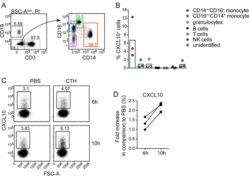
To exclude involvement of other cell types than monocytes for CXCL10 production, human PBMCs were separated from neutrophils using Polymorphprep. Isolated PBMCs were then stimulated with CTHs for 6 and 10h and analyzed for intracellular CXCL10. A gating strategy for cellular subsets was defined (Fig 5A), where CXCL10 production was found in CD14 +-+CD16+monocytes, while little or no CXCL10 was produced in response to CTHs in the other
cell populations (Fig 5B). The production of CXCL10 started to appear after 6h and was clearly induced at 10h (Fig 5C and 5D) which is consistent with other results (S1A Fig). Beyond this, no CXCL10 was produced in neutrophils (S1E Fig). Thus, these findings clearly demonstrate that monocytes are crucial for histone-induced CXCL10 production.
Mapping of the cell-binding site in histone H4
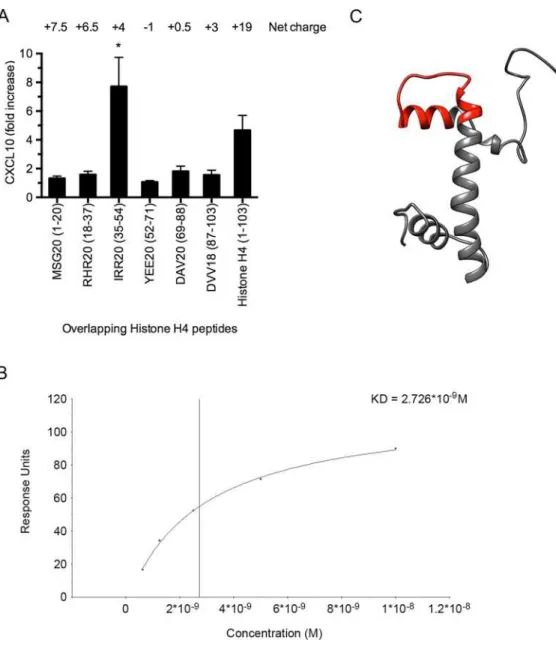
Next, we wished to map the cell-binding site in histone H4. Therefore, a panel of overlapping histone H4-derived peptides was incubated with heparinized blood and the induction of CXCL10 was monitored. These results revealed that the cell-binding site is located at amino acids 35–54 (IRR20) towards the amino terminal portion of the protein (Fig 6A). To measure the affinity of the interaction between peptide IRR20 and TLR4/MD-2, surface plasmon reso-nance experiments were performed. IRR20 was immobilized to a CM5 sensorchip and TLR4/ MD-2 was applied in a flow over the surface. Steady-state affinity analysis showed that IRR20 binds to TLR4/MD-2 with high affinity (KD = 2.726 nM) (Fig 6B). Additional modeling sug-gests that the binding segment forms a hairpin loop surrounded byα-helices (Fig 6C).
CXCL9 and CXCL10 are elevated in the circulation of mice injected with
CTHs
Histones induce recruitment of leukocytes
in vivo
CXCL9 and CXCL10 have been reported to act as chemoattractants for cells expressing the CXCR3 receptor, such as monocytes, macrophages, neutrophils, eosinophils, activated T-lym-phocytes and NK-cells [22–26]. To test whether the local injection of histones into Balb/c mice Fig 4. Histone H4 interacts with TLR4/MD-2 on monocytes.(A) Human blood was incubated with histone H4 (red curves) or PBS (blue curves) for 30 min at 37°C. Cells were fixed and stained with a primary antibody against histone H4 followed by a FITC-conjugated secondary antibody. Cells were subjected to flow cytometry analysis and gated for platelets (upper left), monocytes (upper middle), lymphocytes (upper right) and neutrophils (lower left). Histone stimulated unstained cells are shown in black curves. (B) THP-1 cells were incubated with histone H4 for 30 min at 37°C. Cells were then fixed and prepared for transmission electron microscopy. Samples were first labeled with rabbit anti-histone H4 and rat anti-TLR4 followed by gold-conjugated goat anti-rabbit (5 nm) and goat anti-rat (15 nm). Arrows indicate co-localization of histone H4 (5 nm gold particles) and TLR4 (15 nm gold particles). Scale bar: 500 nm. (C) Histone H4 was immobilized on a CM5 sensor chip and TLR4/MD-2 complex was applied in a flow. (D) The TLR4/MD-2 complex was injected and after a 580 s dissociation time, an antibody against TLR4 (black) or an isotype control antibody (grey) was applied.
doi:10.1371/journal.ppat.1005319.g004
Fig 5. Histones induce CXCL10 production by CD14++CD16+monocytes.Human PBMCs was stimulated with CTHs (10μg/ml), or PBS for 6h or 10h respectively, with Brefeldin A being present during the last 4h of stimulation. Cells were collected and analyzed for intracellular CXCL10. (A) Gating strategy used to identify indicated peripheral blood cellular subsets after exclusion of granulocytes based on high SSC signal (SSC-Ahigh). Cells with a CD16+CD14 -phenotype were confirmed to be NK cells based on CD56 expression. (B) Percentage CXCL10+cells within indicated subsets after 10h of CTH-stimulation. Background (PBS control) is subtracted. Data from three separate experiments are shown. (C and D) CXCL10 expression by CD14++CD16+monocytes after 6h and 10h stimulation with CTHs or PBS. Representative FACS plots (C) and pooled results, from three separate experiments showing CXCL10 expression in CTH-stimulated cultures relative to PBS control cultures for indicated time-points (D).
leads to a recruitment of such immune cells, we employed a CTH air pouch model (see Mate-rial and Methods). Mice receiving a PBS injection instead of CTHs served as control. Sixteen hours after injection, fluids from the air pouch were collected and analyzed in the presence of counting beads by flow cytometry.Fig 8Adepicts the increased cellular influx when animals were treated with CTHs. Further flow cytometry analysis revealed that different populations of leukocytes recruited into the air pouch are monocytes, eosinophils, NK-cells, and neutrophils, respectively (Fig 8B). As these cells have been reported to express the CXCR3 receptor [22–26] we next tested whether the recruitment of these cells correlates with the CXCL10 levels in the Fig 6. Histone H4 binds to TLR4/MD-2 on THP-1 cells.(A) Overlapping peptides from histone H4 (100μM) were incubated with heparinized blood for 12h at 37°C and CXCL10 release was measured by ELISA. (B) IRR20 was immobilized on a CM5 sensorchip and serial dilutions (200 nM–12.5 nM) of the TLR4/MD-2 complex were applied in a flow. Steady-state affinity analysis was used to calculate the KD value
(BIAevaluation software, GE Healthcare, Uppsala, Sweden). (C) The predicted structure of histone H4 was modeled using the UCSF Chimera software (http://www.cgl.ucsf.edu/chimera) with a hairpin loop
corresponding to the TLR4-binding 20-mer peptide IRR20 marked in red.
doi:10.1371/journal.ppat.1005319.g006
air pouch. For this purpose C57BL/6 wild-type mice received a subcutaneous injection of CTHs and the CXCL10 levels in the air pouch were measured after 16h of incubation. TLR4 and MyD88 knockout animals were used to confirm that the histone-induced release of CXCL10 is dependent on TLR4 receptor signaling. As shown inFig 8C, increased levels of the chemokine were recovered from the air pouch of wild-type, but not of TLR4 and MyD88 knockout mice. Consequently, also the influx of leukocytes into the air pouch was significantly reduced in the knockout animals when compared to wild-type animals (Fig 8D). To exclude a LPS contamina-tion in the CTH preparacontamina-tion, the synthetic histone H4-derived peptide IRR20 was injected in wild-type and TLR4 knockout mice. As seen for CTHs, IRR20 was able to trigger the induction of CXCL10 (Fig 8E) and recruitment of leukocytes (Fig 8F) in wild-type but not TLR4 knockout mice. Together these experiments show that subcutaneous injection of extracellular histones leads to the local release of CXCL10 and recruitment of leukocytes to the side of injection.
CXCL10 production in
S
.
pyogenes
infection is dependent on
endogenous extracellular histones
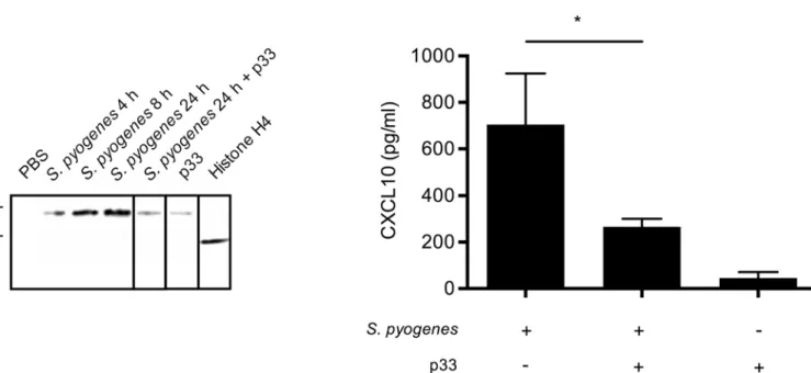
To confirm that extracellular histones are important for chemokine production duringS. pyo-genesinfection, we inoculated C57BL/6 wild-type mice withS.pyogenesby subcutaneous injec-tion. Accumulation of extracellular histone H4 in the air pouch was detected by Western blot analysis andFig 9Adepicts that increasing concentrations were measured 4h, 8h, and 24h after infection, respectively. We recently reported that p33, also known as gC1q receptor is a specific inhibitor of extracellular histones [12]. To test whether p33 can interfere with the chemotactic activity of the extracellular histones, the protein was injected into the air pouch 8h after infec-tion. Subsequent Western blot analysis revealed that this treatment let to a reduction of his-tones in the air pouch which was in a similar low range as seen in animals that were treated with p33 for 24h in the absence of bacteria (Fig 9A). No histones were detected in air pouches of mice that were injected with PBS only. Purified recombinantly produced histone H4 was used to show the specificity of the antibody.
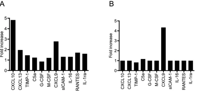
Fig 7. Intravenous injection of CTHs triggers the systemic release of CLXC9 and CXCL10.CTHs (25 mg/kg body weight) or PBS was injected intravenously into Balb/c mice (5 mice per group). After 4h (A) and 24h (B), mice were euthanized and blood samples were recovered by cardiac puncture followed centrifugation. The resulting plasma samples from each group were pooled and analyzed using a mouse multi-cytokine array (n = 1). Mean pixel densities were quantified by Image Lab 4.1 (Bio-Rad Laboratories, Berkeley, CA, USA).
Fig 8. Subcutaneous injection of CTHs induces leukocyte recruitment and local production of CXCL10.(A) Balb/c mice (CTHs (n = 7) and PBS (n = 7)) were challenged with 100μl of a 10μg/ml CTHs solution and the number of leukocytes that infiltrated the air pouch were measured. (B) Identification of cellular subsets recruited into the mice air pouch was done as described in Material and Methods. Monocytes (CD11b+Ly6C+CD64+), neutrophils (CD11b+Ly6G+), eosinophils (CD11b-CD64-Ly6CintSSC-Ahigh) and NK cells (CD11b-Ly6G-NK1.1+) were identified after gating on PI-CD3-B220-events. (C) Mice received an injection with 100μl of a 10μg/ml CTH solution and the air pouch supernatants from C57BL/6 wild-type (n = 12), TLR4 KO (n = 7), and MyD88 KO (n = 7) mice were used to measure the local levels of CXCL10. (D) Mice received an injection with 100μl of a 10μg/ml CTH solution and the number of leukocytes that infiltrated the air pouch, are depicted for wild-type (n = 7), TLR4 KO (n = 7), and MyD88 KO (n = 7) and mice. (E) Mice received an injection with 100μl of a 100μM IRR20 solution and the air pouch supernatants from wild-type (n = 6) and TLR4 knockout (n = 6) mice were used to measure
Having shown that extracellular histones are generated during infection, we next investi-gated whether this leads to an induction of CXCL10 in the air pouch.Fig 9Bshows that ele-vated concentrations of the chemokine are detected in mice infected for 24h withS.pyogenes. However, when the mice were treated with p33, the levels of CXCL10 were significantly decreased and only minor CXCL10 concentrations were found in non-infected animals that received a p33 injection (Fig 9B). Together these findings suggest that endogenous extracellular histones were responsible for the CXCL10 production in the infected mice.
Histone H4 and CXCL10 are mobilized in patients under infectious
disease conditions
Our previous results show that histone H4 is released into the tissue during severe deep tissue infections such as necrotizing fasciitis and cellulitis [12]. Thus, we wished to study whether the release of extracellular histones co-localizes with the induction of CXCL10 in patients suffering fromS.pyogenestissue infections. Immunofluorescence microscopy was used to analyze tissue biopsies from patients with severe deep tissue infections, i.e. severe cellulitis or necrotizing fas-ciitis, evoked byS.pyogenes.Fig 10A(upper panel) illustrates that the infection caused necrotic tissue leading to severe cell damage. Further immunostaining analysis revealed the mobiliza-tion of extracellular histone H4 and CXCL10 and at some locamobiliza-tions co-localizamobiliza-tion of the two factors was noted (Fig 10A, arrows, yellow). In the healthy tissue, only intact cells and
the concentration of CXCL10. (F) Mice received an injection with 100μl of a 100μM IRR20 solution and the number of leukocytes that infiltrated the air pouch, was determined in wild-type (n = 6) and TLR4 knockout (n = 6) mice.
doi:10.1371/journal.ppat.1005319.g008
Fig 9. Histone H4 and CXCL10 expression afterS.pyogenesinfection.(A) Histone H4 was immunostained by Western blot analysis in the supernatants from air pouch of C57BL/6 wild-type mice inoculated withS.pyogenes(AP1 strain) for 4h, 8h, and 24h. p33 treatment was administrated 8h after infection. Note that the shift in the molecular weight of histone H4 recovered from the air pouch compared to recombinant histone H4 is due to histone acetylation and/ or methylation [28]. (B) CXCL10 was measured by ELISA in the supernatant from air pouch of C57BL/6 wild-type mice inoculated withS.pyogenes(AP1 strain, n = 3), infected mice treated with p33 8h after infection (n = 3), and mice injected with p33 only (n = 3).
consequently also only intracellular DNA was seen. We were therefore not able to co-localize histone H4 or CXCL10 in these tissue samples (Fig 10A, lower panel).
To investigate if CXCL10 is systemically up-regulated during infection, the levels of the che-mokine were measured in plasma samples from patients admitted at the intensive care unit (ICU) suffering from septic shock or non-septic critical illness. For comparison samples from patients with milder infections (upper respiratory tract viral infections and gastroenteritis) or from healthy individuals were used. The highest levels of CXCL10 were found in plasma sam-ples from ICU patients (Fig 10B). Though CXCL10 concentrations in plasma samsam-ples from patients suffering from mild infection were lower than in ICU patients, their levels were still significantly increased compared to those measured in samples from healthy donors (Fig 10B). Taken together, the patient data may support ourin vitroandin vivofindings that the release of histones induces the induction of CXCL10 and that patients with severe disease conditions have a systemic increase of the chemokine.
Discussion
The role of extracellular histones in the pathology of severe diseases such as sepsis has lately attracted considerable attention [10,11,15–17,19,29,30]. Extracellular histones are released into the surrounding tissue or circulation from NETotic/necrotic cells and at high concentra-tions they can trigger toxic responses and evoke systemic inflammatory reacconcentra-tions. This in turn may lead to life-threatening complications including disturbed hemostatic functions and acute organ injury [19]. Animal studies have shown that mice receiving sub-lethal doses of histones develop symptoms that are also often seen in patients suffering from severe infectious diseases [11,15,17,19]. These findings point to an important role for extracellular histones in disease progression. However, little is known whether the release of lower histone levels is less devas-tating and perhaps even could contribute to host reactions that are involved in immune defense or wound healing.
In the present study we sought to address this issue by studying if extracellular histones can act as danger signals by triggering local host responses. Our data indeed supported this notion, as histones induced the production of the IFN-γinducible chemokines, CXCL9 and CXCL10,
respectively. Both chemokines are important chemoattractants [23,31] and our results show that the subcutaneous injection of extracellular histones evokes an influx of inflammatory cells to the challenged sites. Further characterization revealed that histones specifically target mono-cytes by approaching a pattern recognition receptor, namely TLR4 andin vivoexperiment with TLR4 and MyD88 knockout mice confirmed the involvement of the receptor. Additional examination of tissue biopsies from patients with tissue infections depicted co-localization of extracellular histones and CXCL10, suggesting that the chemokine is released at the site of infection under disease conditions. When analyzing plasma samples from patients suffering from infectious diseases, we found that the increase of CXCL10 in these samples correlated well with the severity state of the disease. However, unlike described for other inflammatory mediators such as IL-6 and TNF-α[17] CXCL10 concentrations did not reach pathological
Fig 10. Detection of extracellular histone H4 and CXCL10 in patient material.(A) Immunostaining of snap-frozen tissue biopsies from a patient withS.pyogenesdeep tissue infection (upper level) and a healthy donor (lower level) are depicted. Histone H4 staining is shown in red, CXCL10 in green, and DNA (DAPI) in blue. The boxed areas provide a larger magnification (right side). Co-localization of histone H4 and CXCL10 is shown in yellow and indicated by arrows. The image shows staining of one representative patient biopsy out of three analyzed. (B) CXCL10 was measured in the blood plasma from patients admitted at the ICU suffering from septic shock or non-septic critical illness (n = 15), patients with milder infections (upper respiratory tract viral infections or gastroenteritis) (n = 15) and healthy individuals (n = 10).
levels within the microgram range (cytokine storm). We can therefore conclude that extracellu-lar histones are able to cause local and systemic release of IFN-γinducible chemokines,
how-ever, under systemic conditions it seems that these chemokines do not contribute as much to disease progression as other cytokines.
It is noteworthy to mention that analyzing the cytokine release pattern of murine blood samples revealed that CXCL9 and CXCL10 are the most abundant inflammatory mediators detected when mice were injected with CTHs. Notably, both chemokines have in addition to their chemotactic activities other features. For instance, CXCL9 and CXCL10 have been reported to carry antimicrobial activity [32,33] and are pro-apoptotic [34]. The two effects have in common that they help prevent an induction of systemic inflammatory complications. This further strengthens our concept that the two chemokines are released in response to a danger signal and are part of a local immune response. Taken together we here provide for the first time evidence that histones can act as DAMPs/alarmins through inducing leukocyte migration and trigger the release of the IFN-γinducible chemokines, CXCL9 and CXCL10.
These findings demonstrate a dual role of extracellular histones in local and systemic disease conditions. It will be important to understand the details of this dual role in future develop-ment of therapies targeting extracellular histones.
Materials and Methods
Ethics statement
The study was approved by the Institutional review board (IRB) at the Lund University Hospi-tal (Protocol #790/2005). Plasma samples and tissue biopsies (Lund University HospiHospi-tal, Swe-den and University of Toronto, Canada) were taken with a written informed consent, and the study included patients above 18 years of age. Animal use protocols (#M108-10, #M326-12 and #M327-12) were approved by the local Institutional Animal Care and Use Committee
(IACUC) of Malmö/Lund, Sweden. All animals were handled according to the Swedish Animal Welfare Act (SFS 1988:534). The Institutional Review Boards (IRBs) of the University of Toronto (Toronto, ON) and Karolinska University Hospital (Stockholm, Sweden) approved the studies involving humans, and written informed consent was obtained from the patients and the volunteers.
Animals
Female and male Balb/c, C57BL/6 wild-type and knockout mice were purchased from Charles River (Sulzfeld, Germany) and were at least 8 weeks old at the initiation of the experiments.
Proteins and antibodies
Bovine calf thymus histones (CTHs) were purchased from Roche (Basel, Switzerland). Over-lapping Histone H4-derived peptides were synthesized at Biopeptide Co. (San Diego, CA, USA). CTHs have been used by researchers for bothin vivoandin vitrostudies of the function of extracellular histones [11,12,15–18,20]. LPS-free CTHs and LPS-free histone H4-derived peptides were dissolved in LPS-free water. Recombinant histone H1, H2A, H2B, H3.1 and H4 were purchased from New England Biolabs (Ipswich, MA, USA). Recombinant human TLR4/ MD-2 complexes were obtained from R&D Systems (Minneapolis, MN, USA) Peroxidase-con-jugated goat anti-rabbit and goat anti-mice immunoglobulin G was from Bio-Rad Laboratories (Berkeley, CA, USA). Antibodies against histone H4 (polyclonal chip-grade anti-histone H4) were from Abcam (Cambridge, UK). Rat polyclonal antibodies to human TLR4, TLR2 and iso-type control were purchased from Invivogen (San Diego, CA, USA). Mouse anti-human
interferon-γ(clone B27) and isotype control were purchased from BioLegend (San Diego, CA,
USA). Pam3CSK was obtained from Invivogen (San Diego, CA, USA) and LPS from Salmo-nella entericaserotype typhimurium from Sigma-Aldrich (St. Louis, MO, USA).
Patient samples
Plasma samples from 30 patients enrolled at Lund University Hospital, Sweden were analyzed. Fifteen patients treated in the ICU due to septic shock (n = 8) and non-septic critical illness (n = 7) and another 15 patient with milder infections such as upper respiratory tract viral infec-tions (n = 10) and gastroenteritis (n = 5) were enrolled. Blood samples for the analyses were collected at enrollment in 5 ml plastic vacutainer tubes containing 0.5 mL of 0.129 mol/l sodium citrate.
Blood collection
Peripheral venous blood samples were collected from healthy human donors into 6.0 ml sodium heparin tubes (102 IU) or 2.7 ml 0.109 M buffered sodium citrate (Becton Dickinson, Franklin Lakes, NJ, USA). Blood samples were immediately used for experiments upon collection.
LPS measurement in histones
The presence of LPS was measured in all histone subclasses, histone H4-peptides and CTHs using the Pierce LAL Chromogenic Endotoxin Quantitation Kit (ThermoFisher Scientific, MA, USA) according to manufacturer’s protocols. Histone H2A, H2B and H3.1 contained signifi-cant levels of LPS (S3A Fig), while no LPS was found in histone H4, CTHs or histone H4-derived synthetic peptides (S3B Fig). Therefore, only CTHs, histone H4 and histone H4-derived peptides were used throughout this work.
Cytokine arrays
Human XL Cytokine Array Kit and Mouse Cytokine Array Panel A were purchased from R&D Systems (Minneapolis, MN, USA) and experiments were performed according to the manufac-turer’s protocols. Human heparin blood was stimulated with CTHs (60μg/ml, 12 h)
centri-fuged (2000 x g for 10 min) and the resulting plasma was used for the Human XL Cytokine Array Kit. Mice were injected with CTHs (25 mg/kg body weight, 4 h) and euthanized with CO2inhalation followed by cervical dislocation. Blood was drawn by cardiac puncture using a syringe filled with 70μl of buffered sodium citrate and then transferred into K2 Microtainer
MAP tubes pre-coated with 1 mg EDTA (Becton Dickinson, NJ, USA). Blood was centrifuged and the mouse plasma was used for the Mouse Cytokine Array Panel A.
Surface plasmon resonance
Analyses were performed with a BIAcore X100 instrument (GE Healthcare, Uppsala, Sweden) using Sensor Chip CM5 technology at 25°C in a HBS-EP buffer (10 mM HEPES, 150 mM NaCl, 3 mM EDTA, 0.05% (v/v) Surfactant P20, pH 7.4). Histone H4 or IRR20 was diluted in sodium acetate (10 mM, pH 5.5) and immobilized via amine coupling to flow cell 2. Flow cell 1 was subjected to the coupling reaction but without protein, and was used as a control in each experiment. TLR4/MD-2 was injected over the coated surface at 30μl/min in running buffer.
Cytokine production in whole blood
CTHs (60μg/ml), Pam3CSK4 (200 ng/ml) or LPS (100 ng/ml) were incubated with
heparin-ized blood at 37°C on rotation. At different time points, aliquots were removed and centrifuged at 2000 x g for 10 min. All plasma samples were stored at -20°C for further experiments. For inhibition experiments, the TLR4 signaling inhibitor CLI-095 (1.25μg/ml) or antibodies with
blocking activity against human IFN-γ(25μg/ml), human TLR2 (25μg/ml), human TLR4
(25μg/ml) or polyclonal control antibodies (25μg/ml) were added simultaneously with 60μg/
ml CTHs, Pam3CSK4 or LPS. Histone H4-derived synthetic peptides were incubated with hep-arinized blood at 37°C on rotation for 12 h. After centrifugation, CXCL10 was measured in the plasma using Human CXCL10/IP-10 Quantikine ELISA kit from R&D Systems (Minneapolis, MN, USA).
Cytokine production in peripheral blood mononuclear cells (PBMCs)
PBMCs were isolated from heparinized human blood using Polymorphprep (Axis-Shield, Dundee, Scotland) according to manufacturer’s protocols. PBMCs (8x10⁵cells/ml) were stimu-lated with different concentrations of CTHs for 6h, 10h or 12h at 37°C, 5% CO2.Cytokine ELISA
Cytokine ELISA kits for human CXCL10, mouse CXCL10, human CXCL9 and human CXCL11 were purchased from R&D Systems (Minneapolis, MN, USA) and performed accord-ing to manufacturer’s instructions. ELISA kits from R&D Systems were not optimized for plasma samples from citrated blood. Therefore heparinized blood was used for these experi-ments. Cytokine ELISA kits for human CCL3, CCL20, CCL7 were purchased from Sigma-Aldrich (St. Louis, MO, USA).
Cytotoxicity assay
Histone-induced cell toxicity was measured in heparinized blood or PBMCs using the Tox-7 kit (Sigma Aldrich, St. Louis, MO, USA). Cells were stimulated with CTHs (0–100μg/ml) for
12h at 37°C, 5% CO2. Cells were pelleted and the supernatants were used for measurement of lactate dehydrogenase (LDH) according to the manufacturers’protocol. Histone-induced LDH release was calculated as percentage of Tox-7 lysis-induced LDH release.
Histone-white blood cell interactions
Citrated blood (20μl) was diluted with 40μl Cell Staining Buffer (BioLegend, San Diego, CA,
USA). CTHs (final concentration 200μg/ml) were added and samples were incubated for 30
min, 37°C, 5% CO2. Cells were washed in 900μl cell staining buffer, centrifuged (500 x g for 15
min, 4°C) and the pellet was resuspended in 200μl buffer. Histone H4 bound to cells was
stained with 2μg/ml of a primary rabbit anti-human histone H4 (Abcam, Cambridge, UK) for
30 min on ice. Erythrocytes were lysed first with 200μl Lysis buffer A (10 min, room
tempera-ture, dark) followed by 1 ml Lysis buffer B (10 min, room temperatempera-ture, dark). Supernatant con-taining lysed erythrocytes were removed and remaining white blood cells were resuspended in 1 ml cold cell staining buffer. A secondary FITC-conjugated goat anti-rabbit antibody (Abcam, Cambridge, UK) was added (2μg/ml) and samples were incubated for 30 min on ice in dark.
Two more wash steps were performed and each sample was analyzed in flow cytometry.
Intracellular cytokine staining in human PBMCs
Total PBMCs were cultured with or without CTHs (final concentration 10μg/ml) in RPMI
1640 for 6h or 10h with 1X Brefeldin A (eBioscience, San Diego, CA, USA) added during the last 4 h. Stimulated cells were collected in cold FACS buffer (PBS supplemented with 2% FCS and 2 mM EDTA) and unspecific binding was blocked with 10% mouse serum. Surface stain was done with the following antibodies; anti-CD19 (HIB19), anti-CD16 (3G8) (BioLegend, San Diego, CA, USA), anti-CD4 (RPAT4) (BD Biosciences, Franklin Lakes, NJ, USA), CD3 (UCHT1), anti-CD8 (RPAT8), anti-CD14 (61D3) and anti-CD56 (CMSSB) (eBioscience, San Diego, CA, USA) conjugated to allophycocyanin-Cy7, FITC, Alexa700, eFluor450, PE-Cy7, allophycocyanin or biotin revealed by streptavidin-PECF594 (BD Biosciences, Franklin Lakes, NJ, USA). Dead cells were excluded with Live/Dead fixable aqua dead cell stain kit (Life Tech-nologies). Intracellular staining was done with the FoxP3 Fixation/Permeabilization kit (eBioscience, San Diego, CA, USA) using anti-IP10 (J034D6) conjugated to PE (BioLegend). Data were collected using a LSRII (BD Biosciences, Franklin Lakes, NJ, USA) and analyzed with the FlowJo software (Tree star, Ashland, OR, USA).
In vivo
cell migration
CTHs (100μl, 10μg/ml,), IRR20 (100μM) or PBS were injected subcutaneously into an air pouch
of male and female wild-type (C57BL/6), TLR4 knockout (C57BL/6), MyD88 knockout (C57BL/ 6), or wild-type Balb/c mice. After 16h the air pouch was rinsed with 1 ml PBS and the rinsing solution was fixed in 2% paraform aldehyde for 30 min on ice. CountBright Counting Beads (Ther-moFisher Scientific, MA, USA) were diluted 1:10 into the fixed cells and the samples were counted with a FACSVerse Flow Cytometer (Becton Dickinson, NJ, USA). Cells and counting beads were gated with side scatter and forward scatter and analyzed with FlowJo 9.3.1 Software. Identification of cellular subsets was done by flow cytometry in conjunction with mAb against CD64 (X54-5/ 7.1), CD3 (145-2C11) (BD Biosciences, Franklin Lakes, NJ, USA), NK1.1 (PK136), Ly6G (1A8), Ly6C (HK1.4), CD11c (N418), CD11b (M1/70) (BioLegend), B220 (RA3-6B2) and CD4 (GK1.5) (eBioscience, San Diego, CA, USA). Unspecific binding was blocked using anti-FcR mAb (2.462) and dead cells were excluded with propidium iodide (PI). Data were collected using a LSRII (BD Biosciences, Franklin Lakes, NJ, USA) and analyzed with FlowJo (Tree star). Monocytes (CD11b +Ly6C+CD64+), neutrophils (CD11b+Ly6G+), eosinophils (CD11b-CD64-Ly6CintSSC-Ahigh) and NK cells (CD11b-Ly6G-NK1.1+) were identified after gating on PI-CD3-B220-events.
Animal model of
S
.
pyogenes
infection
Wild-type C57BL/6 mice were subjected to a subcutaneous infection ofS.pyogenes(AP1 strain) as previously described [35]. Briefly, mice were inoculated with 108CFU/ml log-phase bacteria, and euthanized at different time points. To remove endogenous histones released afterS. pyo-genesinfection, mice were treated by administration of p33 (50μg/ml) 8h after initial infection.
After 4 h, 8h or 24 h, the air pouch was rinsed with 1 ml PBS and cells and bacteria were pelleted. Samples were subjected to Western blot analysis in the cell supernatant by pooling samples from mice in the same group. Histone H4 was detected using mouse anti-histone H4 antibody (ab31830, Abcam, Cambridge, UK) and secondary goat anti-mouse HRP (1:1000, Bio-Rad Labo-ratories Berkeley, CA, USA). CXCL10 levels in the air pouch were measured by ELISA.
Co-localization Histone H4 and CXCL10
(kindly provided by Prof. Donald E Low, Mount Sinai Hospital, Toronto, Canada). Also included were biopsies of healthy skin tissue obtained at plastic surgery at Karolinska Univer-sity Hospital. The biopsies were cryosectioned to 8μm, fixed in ice-cold acetone and
immuno-fluorescent stainings of serial sections were conducted as described previously [36]. The following antibodies were used: rabbit polyclonal histone H4 and mouse monoclonal anti-IP10 (clone 6D4) (both from Abcam, Cambridge, UK), Alexa 546 conjugated donkey anti-rab-bit IgG and Alexa 488 conjugated donkey anti-mouse IgG (both from Molecular Probes, Eugene, OR, USA). Slides were mounted using DAPI supplemented mounting media (Molecu-lar Probes, Eugene, OR, USA). Single stainings were performed to assure specificity of staining patterns. For image evaluation, a Nikon A1R confocal microscope was used (Nikon Instru-ments, Amstelveen, the Netherlands).
Immunoelectron microscopy
Monocyte THP-1 cells were incubated with histone H4 (50μg/ml) for 30 min at 37°C. For
immunohistochemistry and transmission electron microscopy, samples were embedded in Epon resin, sectioned and subjected to antigen retrieval with metaperiodate as recently described in detail [37]. Sections were labeled with rabbit anti-histone H4 and rat anti-TLR4 followed by gold-conjugated goat-anti rabbit (5 nm) and goat-anti rat (10 nm) antibodies. Samples were observed in a Philips/FEI CM 100 transmission electron microscope at the Core Facility for Integrated Microscopy, Panum Institute, University of Copenhagen.
Statistical analysis
Data were analyzed using GraphPad Prism 6 (GraphPad Software, San Diego, CA, USA). Sta-tistical significance was determined by using the non-parametric Mann-Whitney U test for comparison between two groups. The level of statistical significance was defined as a two-tailed p-value of<0.05 (),<0.005 () and<0.0005 ().
ID numbers
CXCL9, Q07325 CXCL10, P02778 CXCL11, O14625 CXCR3, P49682 IFN-γ, P01579TLR4, O00206 TLR2, O60603 p33, Q07021 Histone H4, P62805
Supporting Information
S1 Fig. Dose dependency of Histone-induced CXCL10 release and LDH release.(A) Human heparinized blood was incubated with CTHs (60μg/ml) at 37°C. At different time points, the
content of CXCL9, CXCL10 and CXCL11 was measured by ELISA. The image shows one rep-resentative time curve out of three. (B) Human heparinized blood was incubated with different concentrations of CTHs for 12h at 37°C. CXCL10 levels were measured by ELISA. (C) Human heparinized blood were incubated with different concentrations of CTHs for 12h at 37°C. LDH release was measured in the supernatant. (D) Human PBMCs was incubated with different concentrations of CTHs for 12h at 37°C. LDH release was measured in the plasma supernatant.
(E) Human neutrophils were incubated with different concentrations of CTHs for 12h at 37°C. CXCL10 levels were measured by ELISA.
(TIFF)
S2 Fig. Dose response curves of CXCL10 induction by different histone subclasses. Pam3CSK4 (200 ng/ml) or LPS (100 ng/ml) was added to heparinized blood in presence of antibodies against human TLR2 (25μg/ml), human TLR4 (25μg/ml), or an isotype control
(25μg/ml). After a 12-hour incubation at 37°C, the release of IL-6 was measured. Blood
stimu-lated with CTHs in the absence of an antibody served as control. (TIFF)
S3 Fig. LPS levels in recombinant histones.Traces of LPS were measured in different histone subclasses (A) and histone H4-derived synthetic peptides (B) using Pierce LAL Chromogenic Endotoxin Quantitation Kit.
(TIFF)
S1 Appendix. Manuscript raw data.Raw data from the manuscript has been added as an appendix.
(XLSX)
Acknowledgments
We wish to thank Pia Andersson and Maria Baumgarten for excellent technical assistance.
Author Contributions
Conceived and designed the experiments: JW BJL AE HH. Performed the experiments: JW PP MWD BC ANT ES MM. Analyzed the data: JW MWD AL ANT MM. Contributed reagents/ materials/analysis tools: AL. Wrote the paper: JW PP MWD BC ANT ES AL MM BJL AE HH.
References
1. Beutler BA. TLRs and innate immunity. Blood. 2009; 113(7):1399–407. doi:
10.1182/Blood-2008-07-019307PMID:WOS:000263316400004.
2. Ward PA. Seeking a heart salve. Nat Med. 2009; 15(5):497–9. doi:10.1038/Nm0509-497PMID:
WOS:000265889300027.
3. Zhang X, Mosser DM. Macrophage activation by endogenous danger signals. J Pathol. 2008; 214 (2):161–78. doi:10.1002/Path.2284PMID:WOS:000252597000005.
4. Chen GY, Tang J, Zheng P, Liu Y. CD24 and Siglec-10 Selectively Repress Tissue Damage-Induced Immune Responses. Science. 2009; 323(5922):1722–5. doi:10.1126/Science.1168988PMID:
WOS:000264559800038.
5. Kawai T, Akira S. The role of pattern-recognition receptors in innate immunity: update on Toll-like receptors. Nat Immunol. 2010; 11(5):373–84. doi:10.1038/Ni.1863PMID:WOS:000276852200006.
6. Medzhitov R. Origin and physiological roles of inflammation. Nature. 2008; 454(7203):428–35. doi:10.
1038/Nature07201PMID:WOS:000257860300037.
7. Holdenrieder S, Stieber P, Bodenmuller H, Busch M, Fertig G, Furst H, et al. Nucleosomes in serum of patients with benign and malignant diseases. (vol 95, pg 114, 2001). Int J Cancer. 2001; 95(6):398–. PMID:ISI:000171870200012.
8. Holdenrieder S, Stieber P, Bodenmuller H, Fertig G, Furst H, Schmeller N, et al. Nucleosomes in serum as a marker for cell death. Clin Chem Lab Med. 2001; 39(7):596–605. doi:10.1515/Cclm.2001.095 PMID:ISI:000170779000003.
9. Zeerleder S, Zwart B, Wuillemin WA, Aarden LA, Groeneveld ABJ, Caliezi C, et al. Elevated nucleo-some levels in systemic inflammation and sepsis. Crit Care Med. 2003; 31(7):1947–51. doi:10.1097/
10. Abrams ST, Zhang N, Manson J, Liu TT, Dart C, Baluwa F, et al. Circulating Histones Are Mediators of Trauma-associated Lung Injury. Am J Resp Crit Care. 2013; 187(2):160–9. doi:10.1164/Rccm.
201206-1037ocPMID:ISI:000313924600012.
11. Fuchs TA, Bhandari AA, Wagner DD. Histones induce rapid and profound thrombocytopenia in mice. Blood. 2011; 118(13):3708–14. doi:10.1182/blood-2011-01-332676PMID:21700775; PubMed Cen-tral PMCID: PMC3186342.
12. Westman J, Smeds E, Johansson L, Mörgelin M, Olin AI, Malmström E, et al. Treatment with p33 cur-tails morbidity and mortality in a histone-induced murine shock model. Journal of innate immunity. 2014; 6(6):819–30. doi:10.1159/000363348PMID:24942226.
13. Kleine TJ, Gladfelter A, Lewis PN, Lewis SA. Histone-Induced Damage of a Mammalian Epithelium— the Conductive Effect. Am J Physiol-Cell Ph. 1995; 268(5):C1114–C25. PMID:ISI:
A1995QW58000005.
14. Kleine TJ, Lewis PN, Lewis SA. Histone-induced damage of a mammalian epithelium: the role of pro-tein and membrane structure. Am J Physiol-Cell Ph. 1997; 273(6):C1925–C36. PMID:
ISI:000071106400018.
15. Xu J, Zhang X, Pelayo R, Monestier M, Ammollo CT, Semeraro F, et al. Extracellular histones are major mediators of death in sepsis. Nat Med. 2009; 15(11):1318–21. doi:10.1038/nm.2053PMID:
19855397; PubMed Central PMCID: PMC2783754.
16. Semeraro F, Ammollo CT, Morrissey JH, Dale GL, Friese P, Esmon NL, et al. Extracellular histones promote thrombin generation through platelet-dependent mechanisms: involvement of platelet TLR2 and TLR4. Blood. 2011; 118(7):1952–61. doi:10.1182/Blood-2011-03-343061PMID:
ISI:000294011500035.
17. Xu J, Zhang XM, Monestier M, Esmon NL, Esmon CT. Extracellular Histones Are Mediators of Death through TLR2 and TLR4 in Mouse Fatal Liver Injury. J Immunol. 2011; 187(5):2626–31. doi:10.4049/
Jimmunol.1003930PMID:WOS:000294059500066.
18. Semeraro F, Ammollo CT, Esmon NL, Esmon CT. Histones induce phosphatidylserine exposure and a procoagulant phenotype in human red blood cells. Journal of thrombosis and haemostasis: JTH. 2014; 12(10):1697–702. doi:10.1111/jth.12677PMID:25069624; PubMed Central PMCID: PMC4194154.
19. Allam R, Kumar SV, Darisipudi MN, Anders HJ. Extracellular histones in tissue injury and inflammation. Journal of molecular medicine. 2014; 92(5):465–72. doi:10.1007/s00109-014-1148-zPMID:
24706102.
20. Huang H, Evankovich J, Yan W, Nace G, Zhang LM, Ross M, et al. Endogenous Histones Function as Alarmins in Sterile Inflammatory Liver Injury Through Toll-like Receptor 9 in Mice. Hepatology. 2011; 54 (3):999–1008. doi:10.1002/hep.24501PMID:WOS:000294738300028.
21. Ishii KJ, Akira S. Innate immune recognition of, and regulation by, DNA. Trends Immunol. 2006; 27 (11):525–32. doi:10.1016/j.it.2006.09.002PMID:WOS:000241937400006.
22. Booth V, Keizer DW, Kamphuis MB, Clark-Lewis I, Sykes BD. The CXCR3 binding chemokine IP-10/ CXCL10: structure and receptor interactions. Biochemistry. 2002; 41(33):10418–25. PMID:12173928.
23. Dufour JH, Dziejman M, Liu MT, Leung JH, Lane TE, Luster AD. IFN-gamma-inducible protein 10 (IP-10; CXCL10)-deficient mice reveal a role for IP-10 in effector T cell generation and trafficking. J Immu-nol. 2002; 168(7):3195–204. PMID:11907072.
24. Ichikawa A, Kuba K, Morita M, Chida S, Tezuka H, Hara H, et al. CXCL10-CXCR3 Enhances the Devel-opment of Neutrophil-mediated Fulminant Lung Injury of Viral and Nonviral Origin. Am J Resp Crit Care. 2013; 187(1):65–77. doi:10.1164/rccm.201203-0508OCPMID:WOS:000313606700013.
25. Zhou J, Tang PCY, Qin LF, Gayed PM, Li W, Skokos EA, et al. CXCR3-dependent accumulation and activation of perivascular macrophages is necessary for homeostatic arterial remodeling to hemody-namic stresses. J Exp Med. 2010; 207(9):1951–66. doi:10.1084/jem.20100098PMID:
WOS:000282650700014.
26. Katoh S, Fukushima K, Matsumoto N, Ehara N, Matsumoto K, Yamauchi A, et al. Accumulation of CXCR3-expressing eosinophils and increased concentration of its ligands (IP10 and mig) in bronchoal-veolar lavage fluid of patients with chronic eosinophilic pneumonia. Int Arch Allergy Imm. 2005; 137 (3):229–35. doi:10.1159/000086335PMID:WOS:000230478400007.
27. Luster AD, Unkeless JC, Ravetch JV. Gamma-interferon transcriptionally regulates an early-response gene containing homology to platelet proteins. Nature. 1985; 315(6021):672–6. PMID:3925348.
28. Georgieva EI, Sendra R. Mobility of acetylated histones in sodium dodecyl sulfate-polyacrylamide gel electrophoresis. Anal Biochem. 1999; 269(2):399–402. doi:10.1006/abio.1999.4050PMID:
WOS:000080059300024.
29. Gillrie MR, Lee K, Gowda DC, Davis SP, Monestier M, Cui L, et al. Plasmodium falciparum histones induce endothelial proinflammatory response and barrier dysfunction. The American journal of
pathology. 2012; 180(3):1028–39. doi:10.1016/j.ajpath.2011.11.037PMID:22260922; PubMed Cen-tral PMCID: PMC3448071.
30. Saffarzadeh M, Juenemann C, Queisser MA, Lochnit G, Barreto G, Galuska SP, et al. Neutrophil Extra-cellular Traps Directly Induce Epithelial and Endothelial Cell Death: A Predominant Role of Histones. Plos One. 2012; 7(2). doi: ARTN e32366 doi:10.1371/journal.pone.0032366PMID:
ISI:000302999600034.
31. Tensen CP, Flier J, Van Der Raaij-Helmer EM, Sampat-Sardjoepersad S, Van Der Schors RC, Leurs R, et al. Human IP-9: A keratinocyte-derived high affinity CXC-chemokine ligand for the IP-10/Mig receptor (CXCR3). The Journal of investigative dermatology. 1999; 112(5):716–22. doi:10.1046/j.
1523-1747.1999.00581.xPMID:10233762.
32. Cole AM, Ganz T, Liese AM, Burdick MD, Liu L, Strieter RM. Cutting edge: IFN-inducible ELR- CXC chemokines display defensin-like antimicrobial activity. J Immunol. 2001; 167(2):623–7. PMID:
11441062.
33. Jovic S, Shikhagaie M, Mörgelin M, Kjellström S, Erjefält J, Olin AI, et al. Expression of MIG/CXCL9 in cystic fibrosis and modulation of its activities by elastase of Pseudomonas aeruginosa. Journal of innate immunity. 2014; 6(6):846–59. doi:10.1159/000365399PMID:25115612.
34. Liu M, Guo S, Hibbert JM, Jain V, Singh N, Wilson NO, et al. CXCL10/IP-10 in infectious diseases path-ogenesis and potential therapeutic implications. Cytokine & growth factor reviews. 2011; 22(3):121–30. doi:10.1016/j.cytogfr.2011.06.001PMID:21802343; PubMed Central PMCID: PMC3203691.
35. Shannon O, Rydengård V, Schmidtchen A, Mörgelin M, Alm P, Sörensen OE, et al. Histidine-rich
glyco-protein promotes bacterial entrapment in clots and decreases mortality in a mouse model of sepsis. Blood. 2010; 116(13):2365–72. doi:10.1182/blood-2010-02-271858PMID:WOS:000282369700025.
36. Thulin P, Johansson L, Low DE, Gan BS, Kotb M, McGeer A, et al. Viable group A streptococci in mac-rophages during acute soft tissue infection. PLoS medicine. 2006; 3(3):e53. doi:10.1371/journal.pmed. 0030053PMID:16401174; PubMed Central PMCID: PMC1326258.