Acta Cir. Bras. 2018;33(12):1052-1060 DOI: http://dx.doi.org/10.1590/s0102-865020180120000002
Qiao-Hong ZhaoI, Xi-Shan ZhangII, Kun WuIII, Jie ZhangIII, Tian-Fang XiaIII, Jian ChenIII, Zhen-Shen QinIV,
Li-Qun PangIV
Preparation of Zoledronate liposome and its impact on
apoptosis of Kupffer cells in rat liver
1Abstract
Purpose: To establish a method for the preparation of zoledronate liposome and to observe its effect on inducing the apoptosis of rat liver Kupffer cells.
Methods: Zoledronate was encapsulated in liposomes, and then the entrapment rate was detected on a spectrophotometer. The prepared Zoledronate liposome (0.01 mg/mL) was injected into the tail vein of SD rats. Three days later, the number of Kupffer cells (CD68 positive) in rat liver tissue was detected by immunohistochemistry. Flow cytometry was used to detect the apoptosis rate of the isolated liver Kupffer cell cultured in vitro.
Results: The entrapment rate of Zoledronate was 43.4±7.8%. Immunohistochemistry revealed that the number of Kupffer cells was 19.3±2.1 in PBS group and 5.5±1.7 in Zoledronate liposome group, with a significant difference (P<0.05). The apoptosis rate of Kupffer cells was 4.1±0.8% in PBS group, while it was 9±2.2% and 23.3±5.9% in Zoledronate liposomes groups with different concentrations of Zoledronate liposome (P<0.05).
Conclusions: Zoledronate liposomes can effectively induce the apoptosis of Kupffer cells in vivo and in vitro, and the apoptosis rate is related to the concentration of Zoledronate liposome. To establish a rat liver Kupffer cell apoptosis model can provide a new means for further study on Kupffer cell function.
Key words: Kupffer Cells. Zoledronate. Liposomes. Liver. Rats.
IBachelor of Medical Science, Department of Nursing, Jiangsu College of Nursing, Huai’an, China. Conception of the
study, acquisition and interpretation of data.
IIBachelor of Medical Science, Department of General Surgery, Lian’shui County People’s Hospital, Lian’shui, China.
Analysis and interpretation of data.
IIIFellow Master degree, Postgraduate Program in Surgical Science, Department of General Surgery, The Affiliated Huaian
No.1 People’s Hospital of Nanjing Medical University, Huai’an, China. Immunohistochemical and flow cytometryanalysis.
IVPhD, Associate Professor, Department of General Surgery, The Affiliated Huaian No.1 People’s Hospital of Nanjing
in the tumor was induced changes, which then was enhanced the antitumor efficacy by the use of liposomal11.
For this reason, Zoledronate was used in this study to prepare liposome-encapsulated Zoledronate for intravenous injection to selectively induce apoptosis of Kupffer cells (KCs) in rat liver, which laid the foundation for further study of its function.
■
Methods
All experimental operations were approved by the Ethics Committee of Jiangsu University. This study was carried out in strict accordance with the recommendations in the Guide for the Care and Use of Laboratory Animals of the National Institutes of Health. The animal use protocol has been reviewed and approved by the Institutional Animal Care and Use Committee (IACUC) of Nanjing Medical University. The approval number was KY-P-2016-007-01.
Specific pathogen free (SPF) male Sprague Dawley (SD) rats (license number: SCXK: (Su) 2009-0002), aged 10-12 weeks and weighing 180-280 g, were provided by the Animal Center of Jiangsu University, China. Animals were fed in cages with controlled temperature and humidity at the Animal Center of Jiangsu University, China. Animals were free for foods and drinking.
Preparation of liposomes
We used the methods reported by Van Rooijen et al.12 and made some improvement. In brief, 8 mg cholesterol was dissolved in 10 mL chloroform. Then 0.86 mL of the stock solution of phosphatidylcholine was added to remove the chloroform, followed by elution with 0.3 M phosphate buffered Zoledronate (20 mg Zoledronate dissolved in 10mL phosphate buffer) to diffuse the phospholipid membrane, forming a milky suspension. After stored in Nitrogen for 2h, the suspension was shocked
■
Introduction
The first closed bilayer phospholipid systems, called liposomes, is described in 1965 and soon were proposed as drug delivery systems that can reduce the toxic effects of drugs and mainly be phagocytized by the mononuclear macrophage system of liver and spleen in the body1. The pioneering work of
countless liposome researchers over almost 50 years led to the development of important technical advances such as long-circulating (PEGylated) liposomes, triggered release liposomes, liposomes containing nucleic acid polymers, ligand-targeted liposomes and liposomes containing combinations of drugs2.
Zoledronate (ZOD), a nitro diphosphonate, was the third generation of bisphosphonates, which was more potent and safe to induce osteoclast apoptosis to cure bone tumors in clinics, while the mechanism is the formation of toxic analogues of ATP in osteoclasts (macrophages residing in the bone) after they phagocytize the bisphosphonate bound to hydroxyapatite crystals leads to the intracellular energy metabolism disorder and causes osteoclast apoptosis, thereby reducing osteoclast absorption of bone3,4. In addition to
inhibition of cell multiplication and initiation of apoptosis in cultured cancer cells5, ZOD was widely used in giant cell tumor of bone, multiple myeloma, metastatic bone cancer and not only suppress the progression to bone metastatic lesions but also prevented growth of primary hepatocellular carcinoma6-8.
for 3 min in a water bath sonicator and then stored at room temperature for 2h to expand liposomes. Then liposomes were washed 2-3 times in sterile phosphate buffer saline (PBS) to remove the uncoated Zoledronate. Finally, the precipitated liposomes were suspended in 4mL PBS to form a milky colloidal solution.
Determination of entrapment rate
Drug solution at 0.1 mg/mL was diluted into 0.01 mg/mL. Appropriate amount of diluted drug solution, empty liposome, Zoledronate lipidsome were scanned in full wavelength including UV and visible light to determine its maximum absorption wavelength. PBS solution served as a blank control.Zoledronate was separated by dissolving the liposomes in chloroform/methanol mixture (concentration ratio 9:1) since with nitric acid zoledronate would form a colored complex with copper ions. The concentration of Zoledronate was determined by ultraviolet spectrophotometer at 240 nm. The results showed that the encapsulation efficiency of Zoledronate was (43.4+7.8)%.
Drug loading of zoledronate liposome
4 mL Zoledronate liposome loading = entrapment rate×total dosage=(43.4±7.8) %×20(mg)=8.68(mg), so the loading dose of Zoledronate liposome is 8.68/4=2.17(mg/ml).
Animal grouping and treatment
Food fasting for 12h to avoid reflex and aspiration during anesthesia for better recover after operationand liquid fasting for 6h to all of rats before treatment. Twenty SD male rats were randomly divided into two groups: control group (Group A, treated with PBS by tail vein injection) and Zoledronate liposome group (Group B, treated with Zoledronate liposome (0.01 mg/mL) by tail vein injection). Three days later, the rats were sacrificed and the liver was
collected for paraffin embedding.
Isolation of rat liver Kupffer cells
Rat liver tissue was minced and disgested with collagenase in a water bath at 37°C for 20 min. then the tissue mince was isolated by density gradient centrifugation. KCs were purified by adherent method.
Immunohistochemical detection (IHC)
Liver sections underwent conventional dewaxing and hydration, and then applied for IHC using streptavidin-perosidase (SP) method. Sections were incubated with 3% H2O2 for 5 min, and then blocked by 10% normal goat serum for 10 min at room temperature, followed by incubation with mouse anti-rat CD68 antibody at 37°C for 2h. Subsequently, sections were incubated with biotin-labeled secondary antibody (1:200 goat anti-mouse IgG) for 30 min, followed by incubated with streptavidin labeled with streptozotocin (1:200) for 30 min. Then the sections were developed with DAB and counterstained with Hematoxylin. After mounting, sections were observed under a light microscope. Cytoplasm showed brown or brown particles were defined as positive. The IHC results were quantitatively analyzed using an image analysis software.
Flow cytometry
The rat liver cells were made into cell suspension and isolated by gradient centrifugation. After adjusting the cell concentration, cells were inoculated in 24-well plates (1 mL per 24-well) and incubated in an incubator with 5% CO2 at 37°C for 1 h. Then the plates were taken out. All non-adherent cells were washed off, and the left adherent cells were Kupffer cells. 1×10⁶ Kupffer cells were
Zoledronate liposomes (0.03 mg/mL). After cultured at 37°C and 5% CO2 for 2h, cells were metered volume with 100 μL staining buffer, and incubated with 1 μL FITC-labeled anti-CD11c antibody at 4°C for 20-30 min in dark. Then the cells were washed with PBS and resuspended in 100 μL PBS and detected on a flow cytometer.
Statistical analysis
Statistical analysis was performed using SPSS v17.0 package. Data were expressed as mean±standard deviation (SD). Comparisons between the two groups were carried out using t test. P<0.05 was considered as significant different.
■
Results
Entrapment rate of zoledronate
Zoledronate can react with copper ions and form colored compound in the presence
of nitric acid. For this reason, Zoledronate liposome was dissolved in a chloroform/ methanol mixed solution (concentration ratio of 9:1) to separate Zoledronate, so that its concentration could be detected by ultraviolet spectrophotometer at 240 nm. The results showed that the entrapment rate (%) of Zoledronate was 43.4±7.8.
Number of Kupffer cells
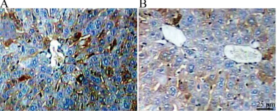
Immunohistochemical staining showed Kupffer cells settled in the hepatic sinusoid with irregular shape. Most of the cell body intruded into or completely mobilized into the hepatic sinusoid. They extended into the perisinusoidal space and directly contacted with hepatocytes (Figure 1A). After treated with PBS or Zoledronate liposomes, the numbers of Kupffer cells (positive for CD68) were (19.3±2.1) in PBS group and (5.5±1.7) in Zoledronate liposome group, with a significant difference (P<0.05, Figure 1B).
Figure 1 - CD68+ Kupffer cells in liver tissue of the two groups determined by IHC (×400). A: PBS group; B:
Zoledronate liposome group.
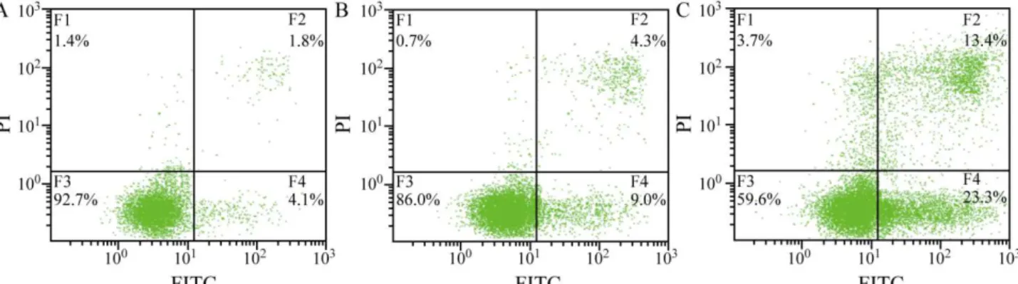
Apoptosis rate of KCs
The apoptosis rates of KCs were 4.1±0.8% in PBS group and 9.0±2.2% and
Figure 2 - The apoptosis rates of KCs determined by flow cytometry. A: PBS group; B: Zoledronate liposome
group (0.01 mg/mL); C: Zoledronate liposome group (0.03 mg/mL).
■
Discussion
Kupffer cells (KCs) locate in hepatic sinusoids and attach to the endothelial cells. They extend into the sinus space through the fenestra. The amount of KCs accounts for 35% of the non-parenchymal cells in the liver and 80%-90% of the total monocytes-macrophages system13. Studies have shown that KCs have
strong phagocytosis which can be enhanced or inhibited by a variety of drugs. Otogawa
et al.14 found that the expression of dead ligands and cytokines in KCs is increased after they phagocytize the apoptotic bodies, which aggravates the inflammation and fibrosis of liver15. KCs can secrete and synthesize a variety of bioactive substances such as tumor necrosis factor α (TNF-α), interleukin, platelet-activating factor (PAF), reactive oxygen species (OFR), nitric oxide (NO) and so on. They regulate the body›s immune response through inducing the proliferation of lymphocytes, secreting cytokines and chemokines, expressiing certain molecules and other pathways16.
Recent studies have shown that PK2/Bv8 expression of KCs is decreased in liver cancer as compared with that in normal liver17. KCs are involved in the hepatic metastasis from gastrointestinal malignant tumors promoted by carcinoembryonic antigen (CEA) and the carcinogenesis of peroxisome proliferator.
Meanwhile, KCs also participate in the carcinogenesis of hepatocarcinogen through producing inflammatory cytokines such as superoxide. In addition, KCs are also involved in tumor immunity through phagocytosis, antibody-dependent cell-mediated cytotoxicity, antigen presentation and release of cytokines.
It has been found that the expression of CD40/CD40L of KCs plays a role in chronic rejection following liver transplantation. KCs also exacerbate self-apoptosis by secreting NO, leading to acute rejection in liver transplantation. At the same time, KCs induce T cell apoptosis and differentiation through the Fas-FasL apoptotic pathway, playing a role in the tolerance of liver transplantation18,19.
In the early stage of transplantation and reperfusion, cytokines such as TNF-α, IL-1 and IL-6 released by the activated Kupffer cells and a large number of reactive oxygen species (ROS) cause direct damage to hepatocytes, while the late injury is the serious injury on hepatocytes caused by complicated inflammatory cascade induced by neutrophil infiltration, release of a large number of oxygen free radicals and protease and microcirculation disturbance of the grafted liver20-22. The activation of KCs and
anti-inflammatory self-stabilizing mechanism
in vivo and leading to the inflammatory cascade reaction, which further leads to the pathological process of IRI and grafted liver organ failure. Effective regulation of the function of KCs may become an effective way to prevent and treat the IRI in transplanted liver.
At present, the inhibition and regulation of KC function has become one of the hotspots in the researches of protective measure from IRI in transplanted liver. In the past five years, 40 researches have been retrieved on PubMed by the MESH “liver transplantation AND Kupffer word cell AND ischemia reperfusion injury”. Most of them used taurine, carbon monoxide and hemeoxygenase 1 (HO-1) to inhibit the secretion and phagocytosis of KCs, or used gadolinium chloride (GdCl3) to induce the apoptosis of KCs, in order to reduce the hepatic ischemia-reperfusion injury23-26.
Although they have achieved protective effects to some extent, the reagents have not been commercialized, and there is no related clinical application reported.
Bisphosphonates are a class of synthetic pyrophosphonic acid analogues3,4.
The osteoclasts (macrophages settled in the bone) in bone phagocytize the bisphosphonate combined with hydroxyapatite crystal and form ATP analogs with toxic effects in osteoclasts, resulting in cellular energy dysmetabolism that causes the apoptosis of osteoclast and thereby reduces the bone resorption of osteoclasts. Bisphosphonates can selectively eliminate monocytes/macrophages. In this regard, Fukushima et al.27 used Clodronate
liposome to inhibit the function of macrophages and significantly alleviate the blepharoconjunctivitis caused by immune reaction. Shifrin et al.28 used Clodronate
liposome to induce macrophage apoptosis in mice, which significantly reduced the local and systemic inflammatory response in severe acute pancreatitis, but has no effect on other
cells. Recent studies have shown that as a specific scavenger of macrophages, Clodronate has no effect on other cells such as vascular endothelial cells and smooth muscle cells. ZOD is a bisphosphonate containing nitrogen with greater effect on inducing osteoclast apoptosis and better safety, which has just entered the Chinese market29. Zoledronate is superior to
disodium chlorondronate on the induction of KC apoptosis in terms of dose- and time- effect relationship. Due to the smaller molecular weight, Zoledronate liposome has better entrapment rate30.
Liposome, one of the most common galenic pharmacy methods to reduce drug toxicity, is an ideal drug carrier. The conventional liposomes prepared with lecithin or cholesterol are phagocytized by reticuloendothelial system in vivo and act on the liver and spleen through blood circulation31. It has been widely
researched experimentally and clinically and its application has been widely used in many fields, especially in medical engineering. As a drug carrier, liposome has the characteristics of improving drug efficacy, reducing adverse drug reactions and targeting32.
In this study, a preparation method of Zoledronate liposome was established and its inductive effect on the apoptosis of Kupffer cells in rat liver was observed. There has been animal experiment showed that Zoledronate liposomes would help ZOD to concentrate on target zone, so the concentration in other sites decreased and the toxicity was less33.
different concentrations, showing a significant difference (P<0.05). The results showed that Zoledronate liposomes can effectively induce apoptosis of Kupffer cells in vitro and in vivo, and the apoptosis rate of KCs is related to the concentration of Zoledronate liposomes. To establish a rat liver Kupffer cell apoptosis model provides a new way to further study the function of Kupffer cells.
■
References
1. Pattni BS, Chupin VV, Torchilin VP. New Developments in liposomal drug delivery. Chem Rev. 2015;115(19):10938-66. doi: 10.1021/acs.chemrev.5b00046.
2. Allen TM, Cullis PR. Liposomal drug delivery systems: from concept to clinical applications. Adv Drug Deliv Rev. 2013;65(1):36-48. doi: 10.1016/j.addr.2012.09.037.
3. Frith JC, Monkkonen J, Blackburn GM, Russell RG, Rogers MJ. Clodronate and liposome-encapsulated clodronate are metabolized to a toxic ATP analog, adenosine 5’-(beta, gamma-dichloromethylene) triphosphate, by mammalian cells in vitro. J Bone Miner Res. 1997;12(9):1358-67. doi: 10.1359/ jbmr.1997.12.9.1358.
4. Naito M, Nagai H, Kawano S, Umezu H, Zhu H, Moriyama H, Yamamoto T, Takatsuka H, Takei Y. Liposome-encapsulated dichloromethylene diphosphonate induces macrophage apoptosis in vivo and in vitro. J Leukoc Biol. 1996;60(3):337-44. doi: 10.1002/jlb.60.3.337.
5. Singh T, Kaur V, Kumar M, Kaur P, Murthy RS, Rawal RK. The critical role of bisphosphonates to target bone cancer metastasis: an overview. J Drug Target. 2015;23(1):1-15. doi: 10.3109/1061186X.2014.950668. 6. Nishisho T, Hanaoka N, Miyagi R, Sakai T,
Toki S, Takahashi M, Kenji E, Yasui N, Sairyo K. Local administration of zoledronic acid for giant cell tumor of bone. Orthopedics. 2015;38(1):e25-30. doi: 10.3928/01477447-20150105-56.
7. Sanfilippo KM, Gage B, Luo S, Weilbaecher K, Tomasson M, Vij R, Colditz G, Carson K. Comparative effectiveness on survival of zoledronic acid versus pamidronate in multiple myeloma. Leuk
Lymphoma. 2015;56(3):615-21. doi: 10.3109/10428194.2014.924117.
8. Cohen PR. Zoledronic acid-associated symmetrical drug-related intertriginous and flexural exanthema (SDRIFE): report of baboon syndrome in a woman with recurrent metastatic breast cancer after receiving zoledronic acid. Dermatol Online J. 2015;21(8). PMID: 26437156.
9. Honda Y, Takahashi S, Zhang Y, Ono A, Murakami E, Shi N, Kawaoka T, Miki D, Tsuge M, Hiraga N, Abe H, Ochi H, Imamura M, Aikata H, Chayama K. Effects of bisphosphonate zoledronic acid in hepatocellular carcinoma, depending on mevalonate pathway. J Gastroenterol Hepatol. 2015;30(3):619-27. doi: 10.1111/jgh.12715.
10. Salzano G, Marra M, Porru M, Zappavigna S, Abbruzzese A, La Rotonda MI, Leonetti C, Caraglia M, De Rosa G. Self-assembly nanoparticles for the delivery of bisphosphonates into tumors. Int J Pharm. 2011;403(1-2):292-7. doi: 10.1016/j. ijpharm.2010.10.046.
11. Hattori Y, Shibuya K, Kojima K, Miatmoko A, Kawano K, Ozaki K, Yonemochi E. Zoledronic acid enhances antitumor efficacy of liposomal doxorubicin. Int J Oncol. 2015;47(1):211-9. doi: 10.3892/ ijo.2015.2991.
12. van Rooijen N, van Kesteren-Hendrikx E. ‘’In vivo’’ depletion of macrophages by liposome-mediated ‘’suicide’’. Methods Enzymol. 2003;373:3-16. PMID: 14714393. 13. Bilzer M, Roggel F, Gerbes AL. Role of
Kupffer cells in host defense and liver disease. Liver Int. 2006;26(10):1175-86. doi: 10.1111/j.1478-3231.2006.01342.x.
14. Otogawa K, Kinoshita K, Fujii H, Sakabe M, Shiga R, Nakatani K, Ikeda K, Nakajima Y, Ikura Y, Ueda M, Arakawa T, Hato F, Kawada N. Erythrophagocytosis by liver macrophages (Kupffer cells) promotes oxidative stress, inflammation, and fibrosis in a rabbit model of steatohepatitis: implications for the pathogenesis of human nonalcoholic steatohepatitis. Am J Pathol. 2007;170(3):967-80. doi: 10.2353/ ajpath.2007.060441.
expression. Hepatology. 2003;38(5):1188-98. doi: 10.1053/jhep.2003.50472.
16. Bottcher JP, Knolle PA, Stabenow D. Mechanisms balancing tolerance and immunity in the liver. Dig Dis. 2011;29(4):384-90. doi: 10.1159/000329801.
17. Monnier J, Piquet-Pellorce C, Feige JJ, Musso O, Clement B, Turlin B, Theret N, Samson M. Prokineticin 2/Bv8 is expressed in kupffer cells in liver and is down regulated in human hepatocellular carcinoma. World J Gastroenterol. 2008;14(8):1182-91. doi: 10.3748/wjg.14.1182.
18. Liu G, Ma H, Jiang L, Zhao Y. Allograft inflammatory factor-1 and its immune regulation. Autoimmunity. 2007;40(2):95-102. doi: 10.1080/08916930601083946. 19. Chen Y, Liu Z, Liang S, Luan X, Long F, Chen
J, Peng Y, Yan L, Gong J. Role of kupffer cells in the induction of tolerance of orthotopic liver transplantation in rats. Liver Transpl. 2008;14(6):823-36. doi: 10.1002/lt.21450. 20. Briceño J, Ciria R. Early graft dysfunction
after liver transplantation. Transplant Proc. 2010;42(2):631-3. doi: 10.1016/j. transproceed.2010.02.004.
21. Hanschen M, Zahler S, Krombach F, Khandoga A. Reciprocal activation between CD4+ T cells and kupffer cells during hepatic
ischemia-reperfusion. Transplantation. 2008;86(5):710-8. doi: 10.1097/ TP.0b013e3181821aa7.
22. Montalvo-Jave EE, Escalante-Tattersfield T, Ortega-Salgado JA, Piña E, Geller DA. Factors in the pathophysiology of the liver ischemia-reperfusion injury. J Surg Res. 2008;147(1):153-9. doi: 10.1016/j. jss.2007.06.015.
23. Kincius M, Liang R, Nickkholgh A, Hoffmann K, Flechtenmacher C, Ryschich E, Gutt CN, Gebhard MM, Schmidt J, Büchler MW, Schemmer P. Taurine protects from liver injury after warm ischemia in rats: the role of kupffer cells. Eur Surg Res. 2007;39(5):275-83. doi: 10.1159/000102982.
24. Tomiyama K, Ikeda A, Ueki S, Nakao A, Stolz DB, Koike Y, Afrazi A, Gandhi C, Tokita D, Geller DA, Murase N. Inhibition of Kupffer cell-mediated early proinflammatory response with carbon monoxide in transplant-induced hepatic ischemia/reperfusion injury in rats. Hepatology. 2008;48(5):1608-20. doi: 10.1002/hep.22482.
25. Zeng Z, Huang HF, Chen MQ, Song F, Zhang YJ. Heme oxygenase-1 protects donor livers from ischemia/reperfusion injury: the role of Kupffer cells. World J Gastroenterol. 2010;16(10):1285-92. doi: 10.3748/wjg. v16.i10.1285.
26. Jahnke C, Mehrabi A, Golling M, Frankenberg MV, Kashfi A, Nentwich H, Fonouni H, Nickkholgh A, Schemmer P, Gutt CN, Weitz J, Schmidt J, Gebhard MM, Büchler MW, Kraus T. Evaluation of microperfusion disturbances in the transplanted liver after Kupffer cell destruction using GdCl3: an experimental porcine study. Transplant Proc. 2006;38(5):1588-95. doi: 10.1016/j. transproceed.2006.02.067.
27. Fukushima A, Ozaki A, Ishida W, Rooijen NV, Fukata K, Ueno H. Suppression of macrophage infiltration into the conjunctiva by clodronate liposomes in experimental immune-mediated blepharoconjunctivitis. Cell Biol Int. 2005;29(4):277-86. doi: 10.1016/j.cellbi.2004.12.011.
28. Shifrin AL, Chirmule N, Zhang Y, Raper SE. Macrophage ablation attenuates adenoviral vector-induced pancreatitis. Surgery. 2005;137(5):545-51. doi: 10.1016/j. surg.2005.01.004.
29. Li F, Wang W, Li L, Chang Y, Su D, Guo G, He X, Li M. An effective therapy to painful bone metastases: cryoablation combined with zoledronic acid. Pathol Oncol Res. 2014;20(4):885-91. doi: 10.1007/s12253-014-9769-7.
30. Zlatev HP, Auriola S, Mönkkönen J, Määttä JA. Uptake of free, calcium-bound and liposomal encapsulated nitrogen containing bisphosphonates by breast cancer cells. Eur J Pharm Sci. 2016;86:58-66. doi: 10.1016/j. ejps.2016.02.016.
31. Caron WP, Lay JC, Fong AM, La-Beck NM, Kumar P, Newman SE, Zhou H, Monaco JH, Clarke-Pearson DL, Brewster WR, Van Le L, Bae-Jump VL, Gehrig PA, Zamboni WC. Translational studies of phenotypic probes for the mononuclear phagocyte system and liposomal pharmacology. J Pharmacol Exp Ther. 2013;347(3):599-606. doi: 10.1124/ jpet.113.208801.
hepatocytes, and immune cells. Pharmacol Rep. 2014;66(5):788-98. doi: 10.1016/j. pharep.2014.04.007.
33. Ho EA, Ramsay E, Ginj M, Anantha M, Bregman I, Sy J, Woo J, Osooly-Talesh M, Yapp DT, Bally MB. Characterization of
cationic liposome formulations designed to exhibit extended plasma residence times and tumor vasculature targeting properties. J Pharm Sci. 2010;99(6):2839-53. doi: 10.1002/jps.22043.
Correspondence: Li-Qun Pang
Department of General Surgery
The Affiliated Huaian No.1 People’s Hospital of Nanjing Medical University
Huai’an 223300 China Phone: +86 13515248309 Fax: +86 517 83165499 cnliqunpang@163.com
Received: Aug 10, 2018 Review: Oct 13, 2018 Accepted: Nov 16, 2018
Conflict of interest: none
Financial source: Huai’an Science and Technol-ogy Bureau International Cooperation Project (HG201112)
1Research performed at Central Laboratory,