Effect of two erosive protocols using acidic beverages on the
shear bond strength of orthodontic brackets to bovine enamel
Catielma Nascimento Santos1, Felipe de Souza Matos2, Sigmar de Mello Rode3, Paulo Francisco Cesar4, Flávia Pardo Salata Nahsan5, Luiz Renato Paranhos6
Objective: To assess the short-term effect of two in vitro erosive challenge protocols on the bond strength of metal orthodontic brackets on bo-vine enamel. Methods: Sixty bobo-vine incisors were selected and randomly divided into six groups: AS7 (artificial saliva - 7 days, Control Group); CC7 (Coca-Cola™ - 7 days); LJ7 (lime juice - 7 days); AS30 (artificial saliva - 30 days, Control Group); CC30 (Coca-Cola™ - 30 days); LJ30 (lime juice - 30 days). Microhardness testing was performed prior to the erosive challenge to verify the standardization of samples. Immersion was performed 4x/day for five minutes, for either 7 or 30 days. After immersions were concluded, the brackets were bonded and shear bond strength was assessed after 48 hours. The Adhesive Remnant Index (ARI) was also assessed. Data were analyzed by two-way ANOVA, followed by Tukey’s post-hoc and Student’s t test for paired samples, and the Kruskal-Wallis non-parametric test (α = 5%). Results: The mean and standard de-viation of microhardness testing of total samples were 281.89 ± 44.51 KHN. There was no statistically significant difference in shear bond strength for the time factor (7 or 30 days; F5.54 = 0.105; p= 0.901). However, there was a statistically significant difference for the solution factor (F5.54 = 6.671; p= 0.003). These differences occurred among solutions of Saliva x Coca-Cola™ (p= 0.003) and Coca-Cola™ x Lime Juice (p= 0.029). The as-sessment of the Adhesive Remnant Index showed no significant difference between groups. Conclusions: The immersion time used in the erosion protocols did not affect the bond strength of brackets to teeth. Coca-Cola™ induced significantly higher shear bond strength values than lime juice and artificial saliva. However, the short term effects of 7/30 days in this in vitro study may not be extrapolated for in vivo ones. Clinical studies should be conducted, substantiating the laboratory results. Keywords:Erosion. Orthodontics. Orthodontic brackets. Shear strength.
1 Universidade Federal de Sergipe, Programa de Pós-graduação em Odontologia
(Aracaju/SE, Brazil).
2 Universidade Estadual Paulista, Instituto de Ciência e Tecnologia, Programa
de Pós-Graduação em Odontologia Restauradora (São José dos Campos/SP, Brazil).
3 Universidade Estadual Paulista, Instituto de Ciência e Tecnologia,
Departamento de Materiais Dentários e Prótese (São José dos Campos/SP, Brazil).
4 Universidade de São Paulo, Faculdade de Odontologia, Departamento de
Biomateriais e Biologia Oral (São Paulo/SP, Brazil).
5 Universidade Federal de Sergipe, Departamento de Odontologia (Lagarto/SE,
Brazil).
6 Universidade Federal de Uberlândia, Faculdade de Odontologia (Uberlândia/
MG, Brazil).
DOI: https://doi.org/10.1590/2177-6709.23.6.064-072.oar
How to cite: Santos CN, Matos FS, Rode SM, Cesar PF, Nahsan FPS, Para-nhos LR. Effect of two erosive protocols using acidic beverages on the shear bond strength of orthodontic brackets to bovine enamel. Dental Press J Orthod. 2018 Nov-Dec;23(6):64-72. DOI: https://doi.org/10.1590/2177-6709.23.6.064-072.oar
Submitted: June 22, 2017 - Revised and accepted: February 17, 2018
» The authors report no commercial, proprietary or financial interest in the products or companies described in this article.
Contact address: Luiz Renato Paranhos Av. Pará, 1720, bloco 2G, sala 1, bairro Umuarama CEP: 38.405-320 – Uberlândia/MG – Brasil E-mail: [email protected]
Objetivo: avaliar o efeito de curto prazo de dois protocolos de desafio erosivo, in vitro, na resistência adesiva de braquetes ortodônticos metálicos em esmalte bovino. Métodos: Sessenta incisivos bovinos foram selecionados e divididos aleatoriamente em seis grupos: SA7 (saliva artificial - 7 dias, Grupo Controle); CC7 (Coca-Cola® - 7 dias); SL7 (suco de limão - 7 dias); SA30 (saliva artificial - 30 dias, Grupo Controle);
CC30 (Coca-Cola® - 30 dias); SL30 (suco de limão - 30 dias). Foi realizado o teste de microdureza antes do desafio erosivo, para verificar a
padronização das amostras. A imersão foi realizada quatro vezes ao dia, por cinco minutos, durante 7 ou 30 dias. Finalizadas as imersões, os bra-quetes foram colados e, após 48 horas, foi avaliada a resistência ao cisalhamento. O Índice de Adesivo Remanescente (IAR) também foi avaliado. Para análise dos dados, foram utilizados os testes ANOVA dois fatores, seguido do post-hoc de Tukey e teste t de Student para amostras pareadas, e o teste não-paramétrico de Kruskal-Wallis (α = 5%). Resultados: a média e o desvio-padrão do teste de microdureza das amostras totais foi igual a 281,89 ± 44,51 KHN. Não houve diferença estatisticamente significativa na resistência ao cisalhamento para o fator tempo (7 ou 30 dias; F5,54 = 0,105; p = 0,901). Contudo, houve diferença estatisticamente significativa para o fator solução (F5,54=6,671; p = 0,003). Essas diferenças ocorreram entre as soluções de Saliva x Coca-Cola® (p = 0,003) e Coca-Cola® x suco de limão (p = 0,029). Ao avaliar o Índice de
Adesivo Remanescente, não foi possível verificar diferença significativa entre os grupos. Conclusões: o tempo de imersão utilizado nos protocolos de erosão não afetou a resistência de união dos braquetes aos dentes. A Coca-Cola® induziu valores de resistência ao
INTRODUCTION
Dental erosion is a problem with increasing
in-cidence in the worldwide population.1 This type of
dental lesion is characterized by wear on the tooth surface caused by a chemical process involving the activity of acids, without the involvement of
bacte-ria.2 Erosion has a multifactorial etiology and is
re-lated to intrinsic and extrinsic factors. Intrinsic fac-tors are related to endogenous acids produced by the human body and commonly present in individuals with bulimia or diseases affecting the
gastrointesti-nal tract.3 The extrinsic factors are related to
exog-enous acids found in foods and beverages.2
Several commercially available acidic beverages accelerate the erosion process, such as citric
acid-based4-8 and cola-based3,4,7-14 drinks, energy drinks,15
and isotonic drinks.4 The erosive potential of these
beverages is related to their low pH and low buffering capacity. Acidic foods and beverages with pH lower than 5.5 may cause the dissolution of hydroxyapatite
and fluorapatite present in tooth enamel.5
Tooth enamel is a mineralized tissue and its mi-crostructure influences the bonding mechanism
involving this substrate and the bracket.1 A
satis-factory bond between bracket and enamel is cru-cial for the success of the orthodontic treatment, considering that the bonded bracket, apart from the fact that it will eventually be removed, should resist the orthodontic forces and the masticatory
loads occurring during the treatment.16 Oncag et
al13 found that carbonated beverages, such as
Coca-Cola™ and Sprite™, negatively affected the reten-tion force of brackets bonded to enamel previously subjected to an erosion process. On the other hand,
Khoda et al14 showed that the intake of acidic
bev-erages does not decrease the bond strength of orth-odontic brackets to tooth enamel.
Some behavioral factors such as eating habits may change bracket bond strength to enamel dur-ing orthodontic treatment. The pH of beverages, type of acid present, buffering capacity of saliva, constant acidity (pKa), and concentrations of phos-phate, calcium, fluoride and phosphorus may
in-fluence the erosion of hard dental tissues.3,10-12 Few
current studies correlate the bonding of orthodon-tic attachments in previously eroded enamel and its
potential complications.17
This study aimed to assess the effect of storage time in the erosive solution and the effect of the substance used in the erosive challenge on the bond strength of metal orthodontic brackets bonded to bovine enamel. The null hypotheses were: [1] there would be no differences on the bond strength of the brackets to enamel due to the different immersion times, and [2] the different solutions would not af-fect the bond strength of the brackets to enamel.
MATERIAL AND METHODS
This in vitro study was carried out with 60
bo-vine central incisors. Based on the study by Pasha et
al,18 who found the highest standard deviation of the
groups equal to 2.74 MPa, at 5% significance level, in order to prove that 7 elements per group are re-quired to detect a minimum difference of 2.5 MPa among groups. Predicting potential losses, the
num-ber of 10 elements13,18 per group was adopted. These
teeth were sectioned in sizes of 7 x 7 x 2 mm, at the
flattest central region of the buccal aspect in the cervical-incisal and mesiodistal directions, forming
enamel blocks,7 and poured in acrylic resin within a
polyvinyl chloride (PVC) tube. The same evaluator performed all the procedures.
The random distribution of specimens in their respective groups was performed as follows: speci-mens were numbered from 1 to 60, placed in one single recipient, and picked one by one to compose the groups. The groups were separated by time (7 and 30 days) and by the beverage used (Coca-Cola [CC], Lime Juice [LJ] and artificial saliva [AS]), arranged as follows: AS7; CC7; LJ7; AS30; CC30; LJ30 (Fig 1).
The surfaces were flattened and polished in or-der to standardize the specimens and prepare them for the dental microhardness test. Therefore, #320,
#600, and #1200 silicon carbide grit papers9,12,19-21
(Norton™, Guarulhos/SP, Brazil) were used for 30 seconds in high rotation and refrigeration in the pol-isher (Politriz Polipan™ 2, São Paulo/SP, Brazil).
Assessment of dental microhardness testing In order to verify the standardization of enamel
surface hardness19 a microhardness tester (FM 700,
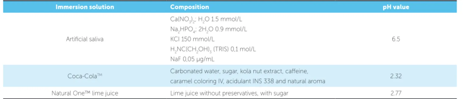
Table 1 - Description of solutions composition and their pH value.
Figure 2 - Indentation performed by Knoop microdurometer. Red and green lines delimiting the indentation size.
5 seconds on enamel (Fig 2). Three indentations were made on the same specimen according to the
following protocol19: one indentation to the right,
one in the middle, and one to the left, with distance
of 100 µm separating each indentation.5 To conclude
the test, samples were subjected to the erosive pro-cess with the selected beverages.
Measurement of pH
The pH was measured in a previously calibrated bench pH meter (Q400AS Quimis™, Diadema/SP, Brazil). Thirty mL of each compound were placed in a test tube and tested in the glass electrode of the
Figure 1 - Flowchart of the method design of the study. 60 bovine
central inci-sors
- Removal of the root - Forming enamel blocks
Poured in acrylic resin
Polishing the specimens
Assessment of dental
micro-hardness
EROSIVE CHALLENGE
DENTAL MICROHARDNESS TEST
BRACKET BONDING
SHEAR BOND STRENGTH TEST
ADHESIVE REMNANT INDEX (ARI)
7 days AS7 CC7 LJ7
30 days AS30 CC30
LJ30
Immersion solution Composition pH value
Artificial saliva
Ca(NO3)2; H2O 1.5 mmol/L Na2HPO4; 2H2O 0.9 mmol/L
KCI 150 mmol/L
H2NC(CH2OH)3 (TRIS) 0,1 mol/L
NaF 0,05 µg/mL
6.5
Coca-ColaTM Carbonated water, sugar, kola nut extract, caffeine,
caramel coloring IV, acidulant INS 338 and natural aroma 2.32 Natural One™ lime juice Lime juice without preservatives, with sugar 2.77
pH meter, and the value obtained was shown in the
ATT digital display.4 The operation was repeated
three times with a five-minute interval, to standardize and certify the values obtained in the test (Table 1).
Immersion method
Immersion cycles were performed by submerg-ing specimens in the specific solution for five
min-utes,4 four times a day (8h, 12h, 16h, and 20h),20
under agitation, for seven19 and 30 days.4 The
so-lutions composition is described in Table 1. After each immersion cycle, the specimens were washed in distilled water, dried in absorbent paper, and im-mersed in 15 mL of artificial saliva; then they were
incubated at 37oC until the immersion procedure.4
In the AS7 and AS30 groups, specimens were im-mersed in artificial saliva for the selected time.
Sa-liva was changed weekly4 for groups AS30, CC30,
Bonding procedure
The metal orthodontic brackets, Roth prescription, with 0.022-in slot (3M Unitek, São José do Rio Preto/ SP, Brazil), were bonded to bovine tooth surfaces with Transbond™ XT orthodontic adhesive system (3M Unitek, Monrovia, CA, USA). Prophylaxis was previ-ously performed with an extra-thin pumice (S.S. White, Rio de Janeiro/RJ, Brazil) and distilled water solution with a Robinson brush (Microdont, São Paulo/SP, Bra-zil) for 10 seconds in low-rotation handpiece (Kavo, Joinville/SC, Brazil), and water sprayed (manufacturer’s recommendation). Acid-etching was performed with
37% phosphoric acid (Dentsply, Petrópolis/RJ, Brazil) for 30 seconds on the dental surface, followed by water spraying and air-drying. Next, a primer was applied to the etched sample according to the manufacturer’s pro-tocol and light-cured for 15 seconds.
The adhesive was applied with a syringe (from the Transbond™ XT kit) using a sufficient amount to completely fill the base of the bracket. Then, the bracket was lightly placed on the dental surface aided by orthodontic tweezers, and pressed to remove ex-cesses. The structure composed by tooth/adhesive system/bracket was light-cured for 20 seconds.
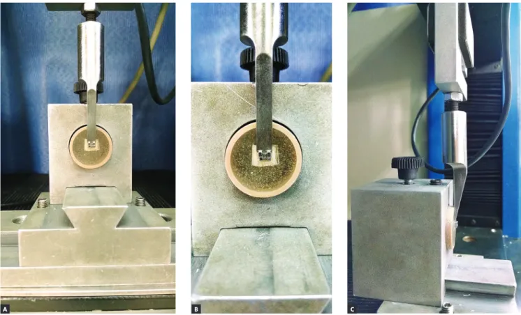
Figure 3 - Shear test at EMIC: A) front view; B) position of the chisel tip on the upper surface of the bracket; C) side view performing the test.
Shear bond strength test
Specimens were subjected to the shear bond strength test using a universal testing machine (EMIC DL-1000, São José dos Pinhais/PR, Brazil)
with 10 KN maximum capacity, 50 KgF cell load21
and 0.5 mm/min crosshead speed,5 48 hours22-23 after
bonding the orthodontic attachments.
Specimens were positioned in the testing machine so that the vertical rod of the shearing machine was perpendicular to the incisal edge of the bracket (flattest part), close to enamel surface, and parallel to the latter (Fig 3), in such a way that the force was perpendicular to
the orthodontic bracket during the test.5 The force
re-quired for detachment was obtained in Kilograms-force (KgF), then converted into Newtons (N), and finally recorded and divided by the bonding area (area of the
base of the bracket = 12.89 mm2), thus obtaining bond
strength values in MegaPascal (MPa).
Adhesive Remnant Index (ARI)
After shear test, the Adhesive Remnant Index (ARI) was assessed with a stereoscope (SteREO Discovery.
V20, Zeiss, Germany) with 10 x magnification.
Any adhesive remaining after bracket removal was
as-sessed according to the ARI.24 The ARI scale ranges from
5 to 1, where 5 indicates that no composite remained on the enamel; 4 = less than 10% of the composite remained on the tooth surface; 3 = between 10% and 90% of the composite remaining; 2 = more than 90% remained on the tooth, and 1 = all composite remained on the tooth, along with the impression of the bracket base.
Statistical analysis
The Kolmogorov-Smirnov test was used to verify sample normality. Two-way ANOVA (solution fac-tor and treatment time facfac-tor) was used for
statisti-cal analysis of data, followed by Tukey’s post-hoc and
Student’s t tests for paired samples. All analyses
con-sidered a significance level of 95% and all tests were performed in the SPSS 16.0 software (IBM).
The Kruskal-Wallis non-parametric test was used to compare the six groups, regarding the ARI score.
All statistical procedures were performed in the Sta-tistica software (StatSoft Inc., Tulsa, USA) version 13.
RESULTS
The sample showed normal distribution
accord-ing to the Kolmogorov-Smirnov test (p = 0.77).
The mean value (±standard deviation) of enamel mi-crohardness was 281.89 ± 44.51 KHN.
Two-way ANOVA followed by Tukey’s test showed
significant effect only for the solution factor (F5.54 = 6.671;
p= 0.003), while the immersion time factor (F5.54 = 1.282;
p= 0.263) and the interaction among the factors studied
were not statistically significant (F5.54 = 0.105; p= 0.901).
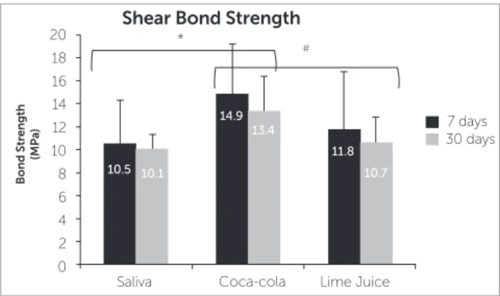
Figure 4 shows that Tukey’s test identified a statistical difference among the bond strength values of Artificial
Saliva versus Coca-Cola™ (p= 0.003) and Coca-Cola™
versus Lime Juice (p= 0.029), regardless of the immersion
time. The bond strength values obtained for the group immersed in Coca-Cola™ were significantly higher when compared to those of the groups subjected to Arti-ficial Saliva and Lime Juice.
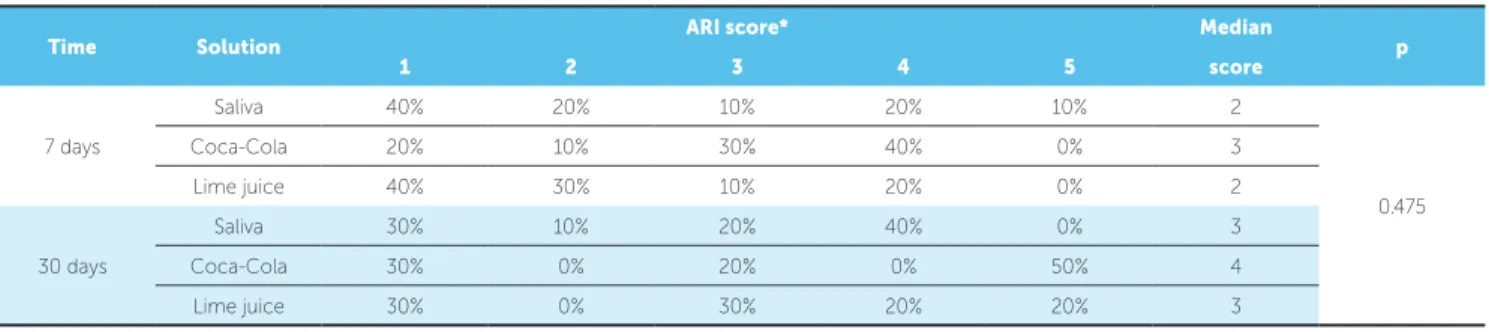
Table 2 - Rate of occurrences of ARI score, median score, and result of the comparison between the groups by the Kruskal-Wallis test.
*1 = all composite remained on tooth, 2 = more than 90% of composite remained on tooth, 3 = between 10% and 90% remained on tooth, 4 = less than 10% of composite on tooth, 5 = no composite on tooth.
Time Solution ARI score* Median p
1 2 3 4 5 score
7 days
Saliva 40% 20% 10% 20% 10% 2
0.475
Coca-Cola 20% 10% 30% 40% 0% 3
Lime juice 40% 30% 10% 20% 0% 2
30 days
Saliva 30% 10% 20% 40% 0% 3
Coca-Cola 30% 0% 20% 0% 50% 4
Figure 4 - Effect of the type of solution and immersion time on shear bond strength measurements. The values in the bars refer to the mean and stan-dard deviation. The * and the # represent statistically significant difference for the “solution” factor (F5.54 = 6.671; p = 0.003) among solutions of Saliva x Coca-Cola™ (p = 0.003) and Coca-Cola™ x Lime Juice (p = 0.029).
20
30 days
Coca-cola
Bond Strength
(MPa)
Lime Juice *
#
10.5 10.1
14.9 13.4
11.8 10.7
7 days
Saliva 16
10
4 18
12
6 14
8
2 0
Shear Bond Strength
Table 2 lists the ARI scores. In the 7-days protocol, both in Saliva and Lime Juice groups, 40% of samples presented all the adhesive on the enamel surface. On the other hand, in the Coca-Cola™ group, 40% of samples indicated score 4, that is, less than 10% of adhesive on the enamel surface. In the 30-days protocol, in the Sa-liva group, 40% of samples indicated score 4, that is, less than 10% of adhesive on the enamel surface. In the Lime Juice group, scores 1 (all adhesive on teeth) and 3 (more than 10% and less than 90% of adhesive on the enamel surface) were the most recurrent scores. In the Coca-Cola™ group, score 5 was mostly repeated, indi-cating no adhesive on the enamel surface.
DISCUSSION
The results of this study indicated that the storage in the erosive solutions (Coca-Cola™ and lime juice) did not affect the bond strength of the brackets to bo-vine enamel. However, the type of erosive solution had a significant effect on the bond strength, whereas immersion in Coca-Cola™ resulted in significantly higher mean values than those obtained after immer-sion in lime juice. Thus, the first null hypothesis was accepted and the second one was rejected.
There is a diversity of protocols of in vitro
ero-sive challenges that range from three days10 to three
months14 regarding immersion time; and from two13
to four16 times a day, regarding the number of
immer-sions; there is also a great variation of types of food and beverages investigated. The literature shows a higher
number of researches using Coca-Cola™,3,4,7-14
fol-lowed by critic beverages4-8,14 such as lime-flavored
soft drinks or lime juice. This research used
Coca-Cola™and Natural One™ lime juice. Both have an
acid pH (Coca-Cola™= 2.32, Lime Juice= 2.77), fa-voring tooth enamel dissolution, which enables
re-searches that induce the in vitro erosive challenge.2
Oncag et al13 induced erosion both in vivo and in vitro.
The protocol adopted was the immersion for 5 minutes in predefined substances (Coca-Cola™ and Sprite™) and the control (artificial saliva), three times a day for three months. They noticed that there was no
statisti-cally significant difference between in vivo and in vitro
groups. Khoda et al,14 tested only in vitro using a similar
protocol (immersion for 5 minutes three times a day for three months); however, with drinks of similar brands (Pepsi™ and 7Up Soda™). In the first work, the ero-sion caused by Coca-Cola™ and Sprite™ decreased
the shear strength of the bracket to the enamel both in
vivo and in vitro. In the second study, as a result, they
observed that there was no negative effect on the shear strength in the bracket-enamel relationship. In ad-dition to the difference in the drinks, the cementing agent may interfere with the final results as well as the time when the enamel was eroded before or after the bracket bonding. In works that simulated what would
happen in vivo, there is also no homogeneity among
protocols. Kato and Buzalaf12 used an in situ protocol
by means of removable apparatus adapted with blocks of enamel, in which the individual removes the appa-ratus and immerses it in the substance in the prede-termined time (immersion for 5 minutes, four times a day, for 5 days); and also observed wear on the surface of the enamel. In general, the difference among pro-tocols hinders and prevents a more reliable
compari-son among results. In in vivo protocols it becomes even
more difficult to discuss, due to the reduced number of papers and concerning with the ethical precepts.
In this research, two erosive challenge protocols were tested before bracket bonding. Immersion of the
in vitro specimens was performed four times daily, for 5
minutes, over 7 and 30 days. It was observed that time does not influence the type of protocol. However, the beverages used during the experiment have a direct
There were statistically significant differences only for Coca-Cola™ in relation to the other solu-tions (Lime Juice and Saliva) in both 7 and 30 days, but when these storage time protocols (7 and 30 days) were compared within the same solution, it was not observed statistically significant difference. When substance pH is lower than 4, as were the tested sub-stances, saliva tends to become sub-saturated in hy-droxyapatite and fluorapatite, limiting its remineral-izing action and justifying the absence of complete remineralization on dental surfaces subjected to the
erosive challenge.6,24 Fushida and Cury26 assessed the
erosive effect of Coca-Cola™ on enamel and dentin and found no complete remineralization as well.
The statistically significant difference found for Coca-Cola™ in relation to lime juice in both stor-age times (7 and 30 days) may be justified by the dif-ferent acids present in the composition of beverages.
A study25 showed that the phosphoric acid present in
Coca-Cola™ has higher erosive potential than the citric acid present in lime juice. Besides the differ-ent acids, factors such as pH, mineral contdiffer-ent, titrat-able acidity, and chelation properties of calcium may
change the erosive potential of both beverages.2
The acids in Coca-Cola™ and lime juice lead
to demineralization of dental inorganic matrix.4,25,27
The longer the exposure time to etiological factor,
the greater the lesion size.24 However, the stability
of enamel hydroxyapatite crystals in an erosive chal-lenge may be maintained when phosphate, calcium,
and/or fluoride ions are added.3,10-12 In this research,
such ions weren’t added to the tested substances.
Previous studies5,13,25 showed that when the erosive
challenge was performed after bracket bonding, the shear bond strength decreased relative to the control group. This result may be justified by the degradation of the adhesive system around the attachment, in the bracket/adhesive system/tooth junction, stimulated by
the acids present in the beverages.5,13,16 This research used
a different method from the one previously mentioned, because the erosive challenge process was performed before orthodontic attachment bonding, simulating the erosive wear on enamel from the habit of drinking acidic beverages prior to the orthodontic treatment.
Reynolds28 affirmed that a value of 4.9 MPa seems
rea-sonable for clinical success in order to maintain brackets bonded, considering they should bear this level of
mastica-tory and orthodontic stresses without detachment.
Sheiba-ninia et al29 found values ranging from 11 to 27 MPa for
shear bond strength, but without fractures. In the present research, the bond strength values found, after 7 and 30 days, were higher for groups immersed in Coca-Cola™ (14.9 and 13.4 MPa) and lime juice (11.8 and 10.7 MPa) than the values obtained for the control group with
artifi-cial saliva (10.5 and 10.1 MPa). Pasha et al18 showed that
the erosive challenge with Coca-Cola™ presents higher shear strength and greater superficial wear on enamel than
other substances. Barac et al30 assessed enamel roughness
after immersion in five beverages, including Coca-Cola™, which presented a higher erosion potential and resulted in higher superficial roughness on enamel than the other sub-stances. It is suggested that roughness may be one of the factors that influence enamel/adhesive system interlocking, possibly inducing higher shear bond strength.
The Adhesive Remnant Index was used to assess the pattern of adhesive failure, and there was no statisti-cally significant difference between groups, as reported
by Baka et al.31 Sajadi et al.16 obtained the same result,
but identified higher tendency of adhesive to remain on the bracket mesh rather than on enamel, as in the pres-ent study. This suggests a higher connection between orthodontic bracket and adhesive than between adhesive
and enamel. Both Sajadi et al.16 and Baka et al.31 showed
in their results that the most recurrent ARI was the one where the adhesive remained in full or almost completely
on the orthodontic attachment mesh. Sheibaninia et al29
affirm that, when this pattern occurs, it is for lacking a connection between adhesive system and tooth enamel.
This investigation showed that teeth subjected to con-stant erosive induction and requiring posterior bracket bonding may suffer higher resistance when removing
orth-odontic attachments. However, these in vitro results may
not be extrapolated for in vivo conditions. Clinical studies
should be conducted, substantiating the laboratory results.
CONCLUSIONS
The immersion time used in the erosion protocols did not affect the bond strength of brackets to teeth
after 7 and 30 days of in vitro erosive challenges.
Author’s Contribution (ORCID )
Catielma N. Santos (CNS): 0000-0003-3932-5550 Felipe de Souza Matos (FSM): 0000-0001-5619-3831 Sigmar de Mello Rode (SMR): 0000-0002-4261-4217 Paulo Francisco Cesar (PFC): 0000-0001-5834-105X Flávia P. S. Nahsan (FPSN): 0000-0002-3547-8886 Luiz Renato Paranhos (LRP): 0000-0002-7599-0120
Conception or design of the study: CNS, FPSN, LRP. Data acquisition, analysis or interpretation: CNS, FSM, SMR, PFC, FPSN, LRP. Writing the article: CNS, PFC, LRP. Critical revision of the article: CNS, FSM, SMR, PFC, FPSN, LRP. Final approval of the article: CNS, FSM, SMR, PFC, FPSN, LRP. Obtained fund-ing: LRP. Overall responsibility: CNS, FSM, SMR, PFC, FPSN, LRP.
1. Jaeggi T, Lussi A. Prevalence, incidence and distribution of erosion. Monogr Oral Sci. 2006;20:44-65.
2. Lussi A, Hellwing E, Zero D, Jaeggi T. Erosive tooth wear: diagnosis, risk factors and prevention. Am J Dent. 2006 Dec;19(6):319-25.
3. Pereira HABS, Leite AL, Italiani FM, Kato MT, Pessan JP, Buzalaf MAR. Supplementation of soft drinks with metallic ions reduces dissolution of bovine enamel. J Appl Oral Sci. 2013 July-Aug;21(4):363–8.
4. Leme RMP, Farias RA, Gomes JB, Mello JDB, Castro-Filice LS. Comparison in vitro of the effect of acidic drinks in the development of dental erosion: analysis by scanning electron microscopy. Biosci J. 2011;27(1):162-9. 5. Hammad SM, Enan ET. In vivo effects of two acidic soft drinks on shear
bond strength of metal orthodontic brackets with and without resin infiltration treatment. Angle Orthod. 2013 July;83(4):648-52.
6. Cruz JB, Bonini G, Lenzi TL, Imparato JCP, Raggio DP. Bonding stability of adhesive systems to eroded dentin. Braz Oral Res. 2015;29(1):1-6. 7. Fujii M, Kitasako Y, Sadr A, Tagami J. Roughness and pH changes of enamel
surface induced by soft drinks in vitro-applications of stylus profilometry, focus variation 3D scanning microscopy and micro pH sensor. Dent Mater J. 2011;30(3):404-10.
8. Low IM, Alhuthali A. In-situ monitoring of dental erosion in tooth enamel when exposed to soft drinks. Mater Sci Eng C Mater Biol Appl. 2008;28(8):1322-5.
REFERENCES
9. Barbosa CS, Montagnolli LG, Kato MT, Sampaio FC, Buzalaf MAR. Calcium glycerophosphate supplemented to soft drinks reduces bovine enamel erosion. J Appl Oral Sci. 2012 July-Aug;20(4):410-3.
10. Eygen IV, Vannet BV, Wehrbein H. Influence of a soft drink with low pH on enamel surfaces: an in vitro study. Am J Orthod Dentofacial Orthop. 2005 Sept;128(3):372-7.
11. Kato MT, Sales-Peres SHC, Buzalaf MAR. Effect of iron on acid demineralization of bovine enamel block by a soft drink. Arch Oral Biol. 2007 Nov;52(11):1109-11.
12. Kato MT, Buzalaf MAR. Iron supplementation reduces the erosive potential of a cola drink on enamel and dentin in situ. J Appl Oral Sci. 2012 May-June;20(3):318-22.
13. Oncag G, Tuncer AV, Tosun YS. Acidic soft drinks effects on the shear bond strength of orthodontic brackets and a scanning electron microscopy evaluation of the enamel. Angle Orthod. 2005 Mar;75(2):247-53.
14. Khoda MO, Heravi F, Shafaee H, Mollahassani H. The effect of different soft drinks on the shear bond strength of orthodontic bracketsJ Dent (Tehran). 2012 Spring;9(2):145-9.
15. Owens BM, Kitchens M. The erosive potential of soft drinks on enamel surface substrate: an in vitro scanning electron microscopy investigation. J Contemp Dent Pract. 2007 Nov 1;8(7):11-20.
Acknowledgements
16. Sajadi SS, Amirabadi GE, Sajadi S. Effects of two soft drinks on shear bond strength and adhesive remnant index of orthodontic metal brackets. J Dent (Tehran). 2014 July;11(4):389-97.
17. Costenoble A, Vennatb E, Attalc JP, Dursund E. Bond strength and interfacial morphology of orthodontic brackets bonded to eroded enamel treated with calcium silicate–sodium phosphate salts or resin infiltration. Angle Orthod. 2016 Nov;86(6):909-16.
18. Pasha A, Sindhu D, Nayak RS, Mamatha J, Chaitra KR, Vishwakarma S. The effect of two soft drinks on bracket bond strength and on intact and sealed enamel: an in vitro study. J Int Oral Health. 2015;7(Suppl 2):26-33. 19. Casas-Apayco LC, Driebi VM, Hipolito AC, Graeff MSZ, Rios D, Magalhães
AC, et al. Erosive cola-based drinks affect the bonding to enamel surface: an in vitro study. J Appl Oral Sci. 2014 Sept-Oct;22(5):434-41.
20. Rios D, Honório HM, Magalhães AC, Delbem ACB, Machado MAAM, Silva SMB, et al. Effect of salivary stimulation on erosion of human and bovine enamel subjected or not to subsequent abrasion: an in situ/ex vivo study. Caries Res. 2006;40(3):218-23.
21. Bezerra GL, Torres CRG, Tonetto MR, Borges AH, Kuga MC, Bandeca MC, et al. Shear bond strength of orthodontic brackets fixed with remineralizing adhesive systems after simulating one year of orthodontic treatment. Sci World J. 2015;2015:903451.
22. Rastelli MC, Coelho U, Jimenez EEO. Evaluation of shear bond strength of brackets bonded with orthodontic fluoride- releasing composite resins. Dental Press J Orthod. 2010;15(3):106-13.
23. Kawakami RY, Pinto AS, Gonçalves JR, Sakima MT, Gandini LG Jr. Avaliação “in vitro” do padrão de descolagem na interface de fixação de materiais adesivos ortodônticos ao esmalte de dentes inclusos: resistência ao cisalhamento após 48 horas e 10 dias. Rev Dental Press Ortod Ortop Facial. 2003;8(6):43-61.
24. Bishara SE, Trulove TS. Comparisons of different debonding ceramic brackets: an in vitro study technique for part II. Findings and clinical implications. Am J Orthod Dentofacial Orthop. 1990 Sept;98(3):263-73. 25. Navarro R, Vicente A, Ortiz AJ, Bravo LA. The effects of two soft drinks on
bond strength, bracket micro leakage, and adhesive remnant on intact and sealed enamel. Eur J Orthod. 2011 Feb;33(1):60-5.
26. Fushida CE, Cury JA. Estudo in situ do efeito da freqüência de ingestão de Coca-Cola na erosão do esmalte-dentina e reversão pela saliva. Rev Odontol Univ São Paulo. 1999;13(2):127-34.
27. Lussi A, Jaeggi T. Erosion – diagnosis and risk factors. Clin Oral Investig. 2008 Mar;12(Suppl 1):5-13.
28. Reynolds IR. A review of direct orthodontic bonding. Br J Orthod. 1975;2(3):171-8.
29. Sheibaninia A, Sepasi S, Saghiri MA, Sepasi S. The effect of an acidic food-simulating environment on the shear bond strength of self-ligating brackets with different base designs. Int J Dent. 2014;2014:689536. 30. Barac R, Gasic J, Trutic N, Sunaric S, Popovic J, Djekic P, et al. Erosive
effect of different soft drinks on enamel surface in vitro: application of stylus profilometry. Med Princ Pract. 2015;24(5):451-7.
31. Baka ZM, Akina M, Ileria Z, Basciftci FA. Effects of remineralization procedures on shear bond strengths of bracket bonded to demineralized enamel surfaces with self-etch systems. Angle Orthod. 2016