Patrícia Sofia Sousa Dias
Polyglutamine diseases (PolyQ):
construction and characterization of yeast
models
LISBOA
DEPARTAMENTO DE CIÊNCIAS DA VIDA
Patrícia Sofia Sousa Dias
Polyglutamine diseases (PolyQ):
construction and characterization of yeast
models
Dissertação apresentada para a obtenção do Grau de Mestre em Genética Molecular e Biomedicina, pela Universidade Nova de Lisboa, Faculdade de Ciências e Tecnologia
Orientador:
Profª. Doutora Paula Ludovico (ECS/UM)
No fim desta longa caminhada, não poderia acabar sem agradecer às pessoas que de
diferentes maneiras possibilitaram que hoje estivesse a escrever a última página da minha
tese.
Gostaria de agradecer à Professora Cecília Leão que deu o meu contacto à Professora
Drª Paula Ludovico. À Professora Drª Paula Ludovico, que me aceitou no seu grupo e que
me deu todas as orientações para conseguir desenvolver o meu trabalho e acabar a minha
tese. Não menos importante é o agradecimento que quero fazer à pessoa que esteve comigo
no laboratório com quem discuti ideias e resultados. Este agradecimento vai para a Belém.
Um muito obrigado aos meus colegas de grupo Alexandra, Ana e Henrique como
também a todos aqueles que estiveram dia-a-dia comigo no laboratório, “Gina”, João
Menino, Sandra, Júlia e Tiago. A todos eles agradeço todo o apoio que me deram, todos os
momentos bons passados no laboratório e fora dele, todo o apoio dado nos meus momentos
maus ouvindo-me e aturando-me a falar das coisas menos boas que ocorreram, e
principalmente a ajuda que me deram e o que me ensinaram durante esta longa caminhada.
Quero também agradecer às minhas colegas de casa, que se tornaram boas amigas, que
me ouviram mesmo sem perceber o que dizia sobre as minhas experiências boas ou menos
boas, que me apoiaram e que acima de tudo me animaram nos dias mais complicados.
Obrigada, Cristina, Elisabete, Fernanda e Cidália!
Um muito obrigada a todos os meus amigos, aqui de Braga, que trabalharam comigo no
laboratório, e aos de Lisboa que sempre me aturaram e apoiaram. Principalmente a todos
os meus amigos que deixei em Lisboa, um grande desculpa por não ter estado presente nos
bons e maus momentos e por de certa forma ter-me afastado. Desculpem!
Dedico esta tese a toda a minha família, Pais, Mano, Avós, Madrinha e Tios. Estas
foram as pessoas que me apoiaram psicologicamente durante todo este tempo. Mãe,
Agradeço-te muito, muito mesmo por todos os momentos em que me ouviste por telefone,
em que tentas-te dar-me apoio e por teres estado todos os dias ao meu lado, por telefone.
Pai, um muito obrigada por também estares ao meu lado por me teres apoiado e ouvido,
sinto que esta longa caminha nos uniu ainda mais. Avós, agradeço todo o vosso empenho
em ouvir e apoiarem-me, e peço muita desculpa por quase não estar com vocês e pelos
momentos em que estive mas que foram sempre a correr. Madrinha, apesar de estares
me deste ânimo para continuar. Tios, um muito obrigada por sentir o vosso apoio. Quero
também fazer um agradecimento especial ao meu irmão. Tiago apesar de seres pequenino,
sempre que estava contigo esquecia-me das coisas e sempre que brincava contigo ficava
mais contente.
Abbreviations………. ix
Abstract……….. xiii
Resumo……….. xv
I. Introduction……… 1
1. Protein Misfolding Diseases……….. 3
2. Machado-Joseph‟s Disease……… 5
3. Huntington‟s disease………. 12
4. Models for study neurodegenerative disorders……….. 17
4.1 Mammal models……….. 18
4.2 Invertebrate models………. 19
4.3 Yeast models (Nervous Yeasts)………... 20
4.3.1 Yeast and Aging process……… 23
5. Autophagy, proteasome and aging on polyglutamine diseases………. 24
5.1 Autophagic process……… 24
5.2Autophagy, aging and neurodegenerative diseases………. 27
6. Objectives……… 30
II. Materials and Methods……… 33
1. Construction Machado-Joseph disease models………. 35
1.1Construction of ATXN3 variants on pUG35 plasmid... 35
1.1.1 DNA manipulations and Cloning………... 35
1.1.2 Analysis of transformants……….. 36
1.2 Construction of ATXN3-GFP variants on pCM252 plasmid……….. 36
1.2.1 DNA manipulations and Cloning……….. 36
1.2.2 Analysis of transformants………. 37
2. Studies on yeast model of Huntington‟s disease……… 37
2.1 Strains, media and treatments……….. 37
2.2 Epifluorescent Microscopy……….. 38
2.3 Preparation of total protein extracts and Western Blot Analysis…………. 38
III. Results……… 43
1. Development of a yeast model for Machado-Joseph‟s disease……… 45
1.1 ATXN3 variants cloning……… 46
1.1.1 Constructions of ATXN3 variants on pUG35 plasmid………….. 47
1.1.2 Construction of ATXN3-GFP fusion on pCM252 plasmid……… 55
1.2ATXN3-GFP genes sequencing………. 59
2. Yeast model for Huntington‟s disease……… 63
2.1 The role of autophagy in Huntington‟s yeast model……….. 68 2.1.1 HTT expression in exponential growth phase cells: the effect of the
inhibition of autophagy………. 69
2.1.2 HTT expression in diauxic growth phase cells: the effect of the
inhibition of autophagy……… 72
2.1.3 HTT expression in aged cells: the effect of the inhibition of
autophagy……….. 75
IV. Discussion……… 81
V. References……… 87
ix
ATG Autophagy related genes
BDNF Brain-derived neurotrophic factor
cDNA Complementary DNA
CFUs Colony-forming units
CLS Chronological life span
CTV Cytoplasm-to-vacuole pathway
dNTP Deoxyribonucleotide
DNA Deoxyribonucleic acid
DUB Deubiquitination enzyme
EDTA Ethylenediaminetetraacetic acid
ER Endoplasmic reticulum
ERAD Endoplasmic Reticulum Associated Protein Degradation
GABA γ-Aminobutyric acid
GFP Green fluorescent protein
HD Huntington‟s disease
HEAT Huntingtin, elongation factor 3 (EF3), protein phosphatase 2A (PP2A),
and the yeast PI3-kinase TOR1
Hsp Heat shock protein
IgG Immunoglobulin G
JD Josephin domain
JNK c-Jun N-terminal kinase
KCl potassium chloride
LB Lysogeny broth
MAP Mitogen-activated protein kinase
MgCl2 Magnesium chloride
MJD Machado-Joseph‟s disease
NaCl Sodium chloride
NaOH Sodium hydroxide
xi
NLS Nuclear localization sequence
PCR Polymerase chain reaction
PMSF Phenylmethanesulfonyl fluoride
polyQ Polyglutamine
QC Quality control
RNA Ribonucleic acid
REST/NRSF RE1-Silencing Transcription factor/Neuron-Restrictive Silencer Factor
RLS Replicative life span
ROS Reactive oxygen species
SDS Sodium dodecyl sulfate
SOC Super Optimal broth with Catabolite repression
SUMO Small Ubiquitin-like Modifier
TFIID Transcription factor II D
TFIIF Transcription factor II F
TAE Tris-acetate-EDTA
TBS Tris Buffered Saline
Tris 2-Amino-2-hydroxymethyl-propane-1,3-diol
YEPD Yeast Extract Peptone Dextrose
xiii
There are a number of serious diseases which have in common the inappropriate
folding of a particular protein, as polyglutamine disease group. Machado-Joseph‟s and Huntington‟s disease are grouped in the polyglutamine diseases, being the cause of the disease the presence of a CAG repeat tract in the causative gene, ATXN3 and HTT,
respectively. This CAG repeats tract leads to the formation of polyglutamine tract in
ataxin-3 and huntingtin proteins, respectively, that do not allow the correct protein folding.
In diverse models, the expansion of the poluglutamine tract into disease related-proteins,
promote their aggregation and their negative effect on cellular metabolism were described
as associated to toxic effect.
In this study, it was proposed the construction of the first yeast model for Machado-Joseph‟s disease and the characterization of the role of autophagy in the yeast model for Huntington‟s disease during yeast life cycle. For the yeast model for Machado-Joseph‟s disease we successfully constructed a Tet On system harboring the variant 1 genes, normal
and expanded, fused with GFP gene, to further allow the subcellular localization of the
expressed protein into yeast cells. In yeast model for Huntington‟s disease the
chronological lifespan (CLS) of yeast cells at different physiological states after expressing
the normal and the pathogenic protein and their subcellular localization, using
epifluorescent microscopy was studied. It was demonstrated that cells harboring the
huntingtin with 103 glutamines (pathogenic protein) during the inhibition of autophagic
process, by chloroquine, presents an increased in CLS and the formation of large foci that
could correspond to aggregosomes. In aged cells this effect is more pronounced leading to
the hypothesis that in these cells the autophagic mechanism is impaired and enhances the
toxic effects of expressed huntingtin 103Q and that the inhibition of the autophagic process
can rescue the cells from the toxic effects. On the other hand, when the normal huntingtin
(25Q) was expressed at exponential phase the inhibition of autophagy led to a drastic
decrease on CLS, demonstrated that in functional cells the autophagy had a positive effect
being a crucial process, probably removing unwanted intracellular components and
xv
Resumo
Há um número de doenças graves que têm em comum a conformação inapropriada
de certas proteínas, como o grupo das doenças poliglutâmicas. As doenças de
Machado-Joseph e de Huntington fazem parte deste grupo de doenças, sendo a sua causa a presença
de um tracto de repetições CAG no gene responsável pela doença, ATXN3 e HTT genes,
respectivamente. Este tracto de repetições CAG leva à formação de um tracto de
poluglutaminas nas proteínas ataxina-3 e huntingtina, respectivamente, não permitindo a
sua conformação correcta. Em diversos modelos foi já descrita a toxicidade causada pela
expansão do tracto de poliglutaminas tal como a sua agregação e o efeito negativo no
metabolismo celular.
Neste estudo, foi proposta a construção do primeiro modelo de levedura para a
doença de Machado-Joseph tal como a caracterização do modelo de levedura para a doença
de Huntington, ao longo do ciclo de vida das leveduras, com inibição da autofagia pelo
antibiótico cloroquina. Para o modelo de levedura para a doença de Machado-Joseph foi
construído com sucesso um sistema Tet On com os genes normal e expandido da variante 1
em fusão com o gene GFP, que permitirá analisar a localização celular das proteínas após
expressão em células de levedura. No modelo de levedura para a doença de Huntington foi
analisada a longevidade cronológica das células em diferentes estados fisiológicos após
expressão da proteína normal e patogénica como também a sua localização celular, através
de microscopia de epifluorescência. Demonstrou-se que as células que expressavam a
proteína huntingtina com 103 glutaminas (proteína patogénica) durante a inibição do
processo autofágico, pela cloroquina, apresentam um aumento da longevidade cronológica
e a formação de grandes foci que podem corresponder aos agregosomas. Nas células
envelhecidas o efeito do aumento da longevidade cronológica, após inibição da autofagia, é
mais pronunciado levantando-se a hipótese de que nestas células o mecanismo autofágico
estará alterado aumentando o efeito tóxico da expressão da huntingtina patogénica e que a
inibição da autofagia pode recuperar as células do efeito tóxico. Por outro lado, quando a
huntingtina normal (com 25 glutaminas) é expressa em fase exponencial a inibição da
autofagia leva a uma diminuição drástica da longevidade cronológica, demonstrando que
em células funcionais a autofagia tem efeito positivo sendo um processo crucial,
3
1. Protein misfolding diseases
Proteins are by far the most structurally complex and functionally sophisticated
biomolecules known. Proteins have a unique three-dimensional structure or conformation
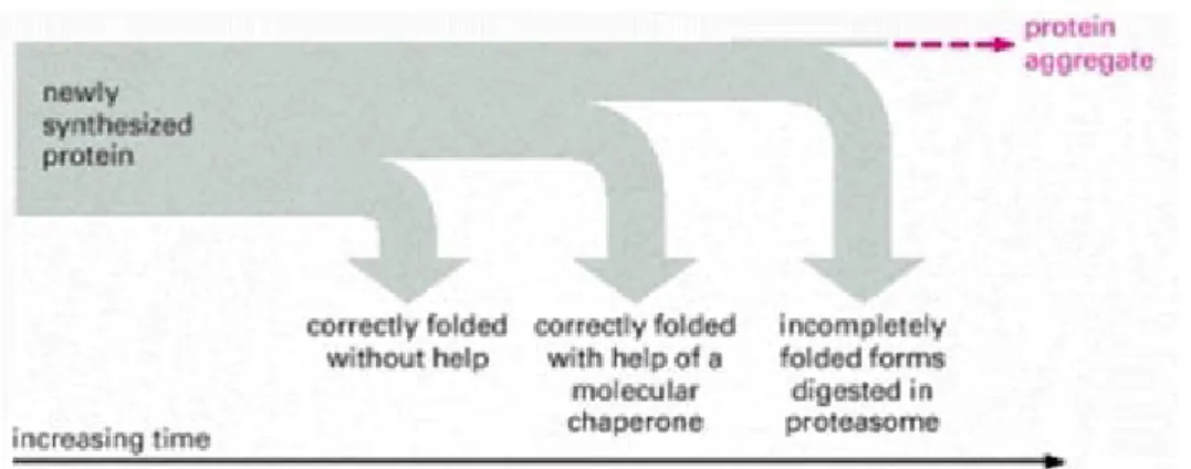
that is achieved by their correct folding. The protein folding initiates when nascent chain is
still attached to ribosome. These nascent chains, or the newly synthesized proteins, might
fold correctly into their three-dimensional structure on their own and maintain these native
states throughout their lifetime. However, mutations or errors in transcription, RNA
splicing and translation may lead to slowly folding protein or to a protein that will never
achieve the correct folding. Incompletely or abnormal folded proteins expose their
hydrophobic patches. Thus, cells have evolved mechanisms that recognize and remove the
hydrophobic patches helping proteins to refold. Molecular chaperones, such as heat-shock
proteins (HSP) recognized the exposed hydrophobic patches acting as a first defense to
help on protein folding. The incompletely folded proteins, accumulates in endoplasmic
reticulum (ER) when attempts to refold a protein fails, it is also marked for destruction by
ER-association protein degradation (ERAD), a process that involves the protein
retrotranslocation into cytosol and proteasome-mediated proteolysis (Barral et al., 2004).
The molecular chaperones and the proteasome-mediated proteolysis processes function
concurrently to prevent massive protein misfolding accumulation (Figure 1) (Alberts et al.,
2002).
Figure 1. The cellular mechanisms of protein quality control after protein synthesis
(from Alberts et al., 2002).
4
The misfolded proteins may be nonfunctional or suboptimally funtional, or they may
be degraded by cellular machinery or the misfolding ones may expose epitopes which lead
to dysfunctional interactions with other proteins causing protein aggregation in the aqueous
cellular environment. Accumulation of aggregates can disrupt cellular trafficking and
degradation process leads to the formation of toxic protein precipitates, inactivation of
functional proteins and ultimately cell death (Gao and Hu, 2008).
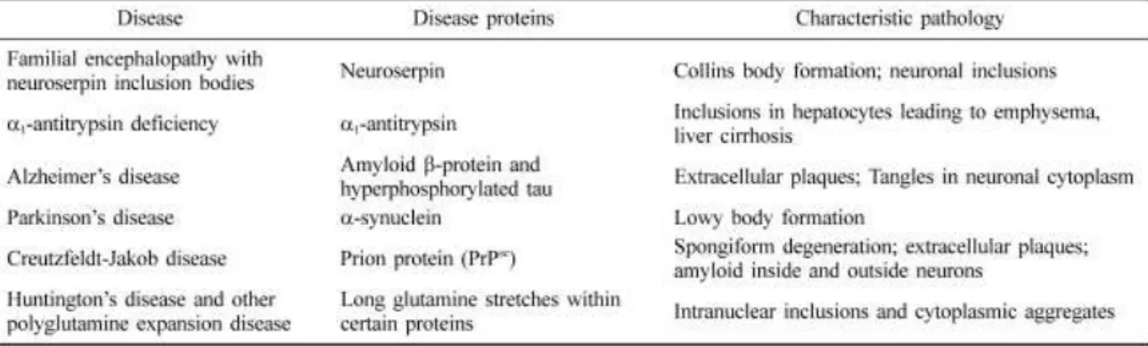
Protein misfolding leads to the malfunctioning of the living systems. There are a
number of serious diseases which have a common aspect that appear to involve
inappropriate folding of a particular protein. Neurodegenerative diseases (Table I) are
directly or indirectly caused by aberrant protein folding and aggregation (Lee and Yu,
2005). The hallmark of these disorders is the presence of cytosolic, nuclear and
extracellular protein aggregates, which are toxic for the cell leading to the alterations of
cellular activities and promoting the activation of cell death signaling pathways.
Furthermore, it is also thought that these disorders are the result of the acquisition of
dominant toxic functions by aberrantly folded proteins (Barral et al., 2004) as a gain of
function of the protein related disease. The accumulation of toxic misfolded proteins, in
these disorders, may overload the cellular chaperone capacity, thus giving rise to disease
phenotypes. Moreover, these disease phenotypes increase in severity with age, when
changes in the intracellular environment reduce the efficiency of the quality control
machinery (Barral et al., 2004).
Table I. Examples of human conformational diseases caused by protein deposits (Lee
5
I. Introduction
Among the different neurodegenerative diseases caused by aberrant protein folding,
the polyglutamine-expanded disorders, Huntington‟s and Machado-Joseph diseases will be
focused in the next section.
2. Machado-Joseph’s disease
Machado-Joseph‟s disease was first reported in North American families of
Portuguese-Azorean ancestry. This is a slowly progressive neurodegenerative disease and
the patients present a wide range of clinical manifestations that include cerebellar ataxia,
pyramidal signs, dystonia, progressive external ophtalmoplegia and peripheral neuropathy
and will became confined to a wheelchair and later bedridden (Kawai et al., 2004). The
neuropathology consists in the neuronal loss of gray matter on multiple areas, such as the
pontine noroadrenegic system and the dopaminergic and cholinergic midbrain, in white
matter degeneration was restricted to the cerebellum, spinal cord and brainstem (reviewed in D‟Abreu et al., 2010). It is a hereditary autossomal dominant disease, meaning that the inheritance of one copy of the ATXN3 gene, from an affect parent, will lead to the
development of the disease. Takiyama and colleagues, using highly polymorphic
microsatellite DNA polymorphism, assigned the MJD1 (ATXN3) gene to the long arm of
chromosome 14 (14q24.3-q32.1) by genetic linkage to microsatellite loci D14S55 and
D14S48 (Takiyama et al., 1993).
Figure 2. CAG tract in both normal and disease allele. The CAG repeats encodes for a
glutamine tract in ataxin-3 protein, show here as Q, which is the single-letter amino acid
6
A CAG repeat expansion found in the ATXN3 gene that encodes an expanded polyQ
tract in the disease protein (Figure 2), known as ataxin-3 or MJDp, includes MJD into a
class of related genetic disorders, the so-called expanded polyglutamine (polyQ) diseases
(Kawaguchi et al, 1994).
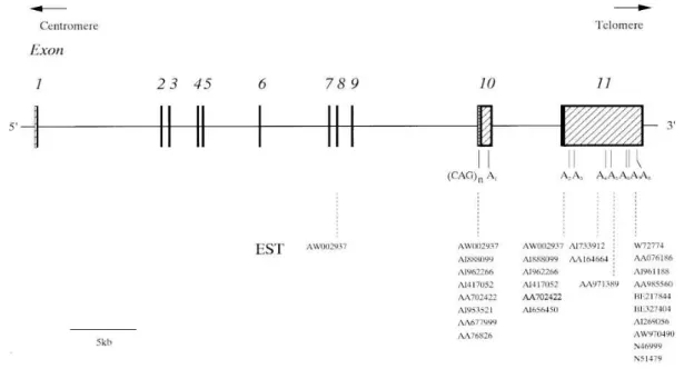
The ATXN3 gene, responsible for MJD, is approximately 48,2 kb in size and is
composed of 11 exons (Figure 3). The CAG repeats are found in exon 10, as represented in
figure 3 (Ichikawa et al., 2001).
There are five described variants of ATXN3 gene (Ichikawa et al., 2001). The first
one, MJD1a,use exon 10 to provid the 3‟-terminal sequence (Kawagushi et al., 1994). The
second group was previously described by Goto (Goto et al., 1997), MJD2-1, is similar to
MJD1a and carries a single nucleotide substitution in the stop codon.
Figure 3. Genomic structure of ATXN3/MJD gene. Exons are numbered 1 to 11 and are
represented as boxes. Filled boxes represent the coding region and hatched boxes represent the 5‟- and 3‟-untranslated regions (UTRs). A1 to A8 are polyadenylated consensus sequences (from Ichikawa et al., 2001), shift in the open reading frame. And the last group
is represented by a collection of clones that cannot be included in other groups, which
7
I. Introduction
The third group was described by Goto (Goto et al., 1997), includes the MJD1-1 and
MJD5-1, which skip 3‟ regions of exon 10 and employed exon 11 as 3‟terminal sequence.
In this group two distinct poliadenylation sites are created in the gene. There is a four
group were exon 2 is absent, presumably by alternative splicing, but that do not cause a
shift in the reading frame. The last group is represented by a collection of clones that
cannot be included in other groups, which from the chimera cDNA (Ichikawa et al., 2001).
Ichikawa and colleagues observed that ATXN3gene is ubiquitously transcribed in all adult
human tissues, as human organs, brain and non-nervous tissues (Ichikawa et al., 2001).
More recently, Conceição Bettencourt and colleagues proposed that ATXN3 gene presents
at least 56 possible alternative splicing variants generated by four types of splicing events
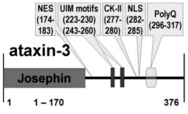
(Bettencourt et al., 2009). Despite the different isoforms, the ATXN3 gene encodes for a
42kDa protein, the ataxin-3. This protein presents a conserved globular deubiquitinating
N-terminal Josephin domain (JD), followed by two or three Ubiquitin Interacting Motifs
(UIMs), depending of the protein isoform and in C-terminal presents the CAG repeat tract
(Figure 4) and the longest slice isoform of ataxin-3 has 376 amino acids (Albrecht et al.,
2004).
Figure 4. Protein architecture of human ataxin-3 (from Albrecht et al., 2004).
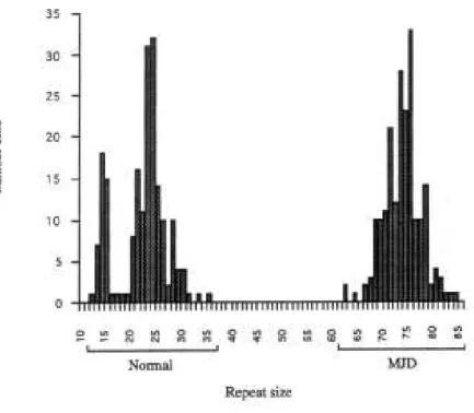
The individuals who are not affected present alleles containing from 12 to 37 repeats,
whereas the affected individuals have at least one expanded allele, with sizes varying
8
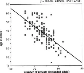
These affected individuals have an inverse correlation between age of disease onset
and the CAG repeat length, meaning that these individuals have a tendency for age at onset
to decrease as the CAG repeat length increases (Figure 5). During transmission from parent
to offspring the CAG repeat length presents some instability (Figure 6) (Maciel et al.,
1995). The poly-glutamine expansions (polyQ) are thought to cause a conformational
chance in the polypeptide sequence promoting misfolding and aggregation of the disease
protein. In MJD, it was described that ataxin-3 forms ubiquitinated intracellular aggregates
or nuclear inclusions in selected populations of disease neurons (Paulson et al., 1997).
Although the pathogenic role of aggregations is controversial, protein misfolding, of
disease protein, is generally thought to be crucial to pathogenesis raising the possibility
that the cellular machinery that normally recognizes and handles misfolded proteins may
play a role MJD.
Several studies were performed to dissect the machinery involving ataxin-3 protein
on MJD.
Figure 5. Distribution of CAG repeat sizes in unaffected control individuals and in
9
I. Introduction
Figure 6. Correlation between CAG repeat lengths in the MJD chromosomes and age
of onset. To this analysis were used 156 affected individuals with age at onset of disease
(from Maciel et al., 1995).
Burnett and colleagues, using an ubiquitin protease assay with 125I-Lysozyme, demonstrated that ataxin-3 has ubiquitin protease activity, removing ubiquitin from
growing chains and that cystein-14, from Josephin domain, is required for its activity
(Burnett et al., 2003). Using similar in vivo studies, Berke and colleagues not only
confirmed that ataxin-3 has a ubiquitin protease activity and that binds polyubiquitin
chains but also that normal and expanded ataxin-3 are degraded by the proteasome,
defining that ataxin-3 has a function in the ubiquitin proteasome pathway (UPP) and that it
is also regulated by this process (Berke et al., 2005). In fact, it was described that
ubiquitinated proteins and the proteasome complex redistributes into polyglutamine
aggregates formed by the ataxin-3 (Chai et al., 1999). These studies raise the possibility
that expansion on CAG tract modulates ataxin-3 protease activity and/or the range of its
substrates, being possible that expanded ataxin-3 acquires new properties that alter the
UPP. Several proteins can bind ataxin-3, therefore is possible that ataxin-3 may act at the
10
Furthermore, some studies have shown that ataxin-3 interact with Rad23, that is a
yeast homolog of human HHR23A and with Valosin-Containing Protein (VCP), that is a
mammalian homolog of yeast cell cycle division protein Cdc48p (Doss-Pepe et al., 2003
and Zhong and Pittman, 2006). These two proteins, Rad23 and VCP are involved on UPP. Rad23 play a role as a “shuttle factor” that translocate proteins to proteasome for degradation binding polyubiquitin chains through its ubiquitin-associated (UBA) domain.
VCP is a member of AAA family of ATPases associated proteins with diverse cellular
activities, including control of cell cycle division, vesicle-mediated transport and the UPP.
In UPP, it is involved on ubiquitin-mediated proteolysis and has a role on endoplasmatic
reticulum (ER)-associated degradation (ERAD) promoting the retranslocation of substrates
from ER to the cytosol to be degraded (Zhong and Pittman, 2006). It was suggested that
ataxin-3 compete with Ufd1 (protein from heterodímero responsible for extraction of
proteins from ER) for binding to VCP impairing the binding of ubiquitilated ERAD
substrates and decreasing their extraction from ER, promoting new cycles of folding and
sorting the substrates into other quality control (QC) pathway. Biochemical data showed
that mutated ataxin-3 binds VCP more efficiently than wild-type ataxin3, promoting a
greater effect on ERAD inhibition, which may contribute to disease pathogenesis (Zhong
and Pittman, 2006). The ataxin-3 properties link their normal function to protein
surveillance pathways and suggest that alteration in normal ataxin-3, caused by the CAG
expansion, could compromise protein homeostasis in the cell.
Pathological ataxin-3 is also involved on transcription process. Using transgenic
mice expressing polyglutamine-expanded ataxin-3-Q79, Chou and colleagues, performed a
microarray analysis and real-time PCR assays, from cerebellum, to test the involvement of
expanded ataxin-3 on transcription. The microarrays indicated that genes involved in
glutamatergic transmission, transcription factors, intracellular Ca++ signaling, heat shock proteins, GABA receptors and MAP kinase pathways are downregulated. While genes
involved in pro-apoptotic response, cyclin and cyclin-dependent kinase 5, G protein
subunits, proteasome subunits and RNA polymerase subunits are upregulated (Chou et al.,
2008). It was previous described that ataxin-3 has a role in transcriptional repression,
binding to CBP, p300 and PCAF coactivators, regulating transcription. N-terminal of
11
I. Introduction
coactivators/HATs, acting on a new mechanism of transcriptional repression (Li et al.,
2002). Moreover, it is known that in early stages of disease the expanded ataxin-3 is
associated with the mitochondrial-mediated cell death, enhancing the release of
cytochrome c and decreasing the Bcl-2 expression, which promotes defects on
transcription (Tsai et al., 2004).
It is widely thought that age-related changes may be involved on MJD pathogenesis,
as others neurodegenerative diseases. It is known that over decades neurons that express
polyQ-containing ataxin-3 might be functional until an imbalance of cellular homeostasis
that was promoted by ageing. The age-related phenomena such as activation of calpain and
caspases and the decreased on chaperone activity, have been proposed to be involved on
the initial steps of the disease (Barral et al., 2005 and Haacke et al., 2006). In fact, ataxin-3
suffers proteolytic cleavage, by calpain and caspases, forming a shortest fragment
containing the polyQ tract (Haacke et al., 2006). These fragments have the ability to recruit
polyQ-containing proteins to aggregates, which include non-pathological ataxin-3. The
incorporation of non-pathological ataxin-3 into aggregates and the corresponding reduction
of soluble intracellular ataxin-3 levels could in turn contribute to MJD pathogenesis
(Berke, et al., 2004 and Haacke, et al., 2006).
Taken together these data proposed multiple mechanisms for the implication of
ataxin-3 in transcriptional dysregulation and alteration of genes expression patterns. The
first one involves the interaction of mutated ataxin-3 with polyglutamine-rich nuclear
factors or co-factor, like p300 and CBP. The second involves the transcriptional repression
function of ataxin-3 recruiting histone deacetylase 3 and inhibiting histone
acetyltransferase activity of co-activators. The third mechanism involves the effect of
mutated ataxin-3 on UPP that affect the normal turnover of transcription factors.
Regardless of the existing knowledge on ataxin-3 interactions, there are still many
questions to be addressed. Having this in mind, we herein present an approach for the
development of a new yeast model for MJD that will ultimately allow us to increase our
12
3. Huntington’s disease
Huntington‟s disease (HD) is a late-onset autosomal dominant disorder, characterized by neuropsychiatric symptoms, progressive loss of motor coordination and
cognitive decline. In neuropathology of this disease occurs the selective dysfunction and
death of specific neuronal subpopulations in the central nervous system, being
gamma-aminobutyric acid (GABBA)-releasing spiny-projecting neurons, from striatum, the most
affected cells. During disease progresses, there is general neuronal loss in different brain
regions (reviewed in Borrell-Pagès, et al., 2006).
The causative gene of Huntington‟s disease was first assigned by Gussella and colleagues in 1983, on chromosome 4p16.3, using linkage analysis of polymorphic DNA
markers of humans (Gussela et al., 1983). After 10 years, Huntington Disease Research
Group, using an exon amplification approach, identified a large gene, the IT15 gene, that
contains in the open reading frame a polymorphic (CAG)n trinucleotide repeat tract,
varying from 11 to 34 CAG in normal population (The Huntington‟s Disease Collaborative
Research Group, 1993).
HD is caused by a mutation in the IT15 gene (HTT gene), characterized by an
expansion of a variable stretch of CAG triplet in the first exon, promoting a
polyglutamine-enconding tract in the N-terminal region of the huntingtin protein. Huntingtin is a 350-kDa
protein characterized for the presence of different domains, including the polymorphic
glutamine/proline rich domain in its amino-terminus (figure 7). This protein was observed
in and outside of the nervous system. It was associated with the embryonic development,
being essential for this process. In cells, it was associated with different compartments, as
Golgi, endoplasmatic reticulum and mitochondria (reviewed in Landles and Bates, 2004
and Cattaneo et al., 2005). Specifically in neurites and synapses, this protein is associated
with vesicular structures such as clathrin-coated vesicles, endosomal compartments and
microtubules (reviewed in Cattaneo et al., 2005).
As described for Machado-Joseph‟s disease, in HD the age of onset is inversely
correlated with the CAG repeat length (Figure 8) and that affected individuals have more
13
I. Introduction
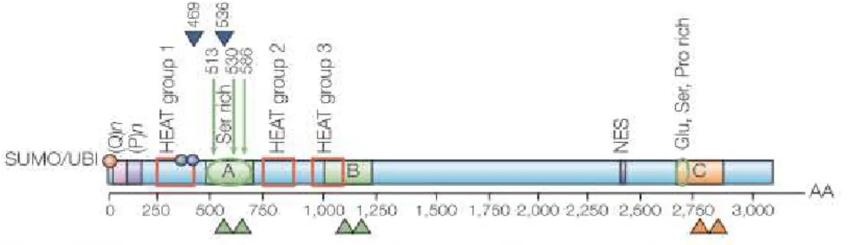
Figure 7. Schematic diagram of huntingtin amino acid sequence. (Q)n represents the
polyglutamine tract; (P)n represents the polyproline sequence. The green arrows represent
the caspase cleavage sites and the blue arrowheads represent the calpain cleavage sites.
The green and orange arrowheads point to the approximate amino acid region for protease
cleavage. The red squares represent the three main clusters of HEAT repeats. B identifies
the region cleaved preferentially in the cerebral cortex, C the region mainly cleaved in
striatum and A the region cleaved in both regions of brain. The red cycle indicates the
post-translational modification, ubiquitilation (UBI) and/or sumoylation (SUMO) and the
blue circles indicate the phosphorylation at serine 421 and serine 434. NES is the
nuclearexport signal. The glutamic acid (Glu)-, serine (Ser)- and proline (P)- rich regions
are indicated. (from Cattaneo et al., 2005).
Several studies were performed attempting to determine the role of normal and
pathogenic huntingtin in cells. It was reported that the normal and mutated protein had the
ability to bind several proteins having a flexible and multifunctional structure. These
abilities lead to specific conformations and different activities depending on its cellular
localization (reviewed in Cattaneo et al., 2005). It was suggested that the
neurodegeneration observed in this pathology is related to the gain of function of mutated
huntingtin and the loss of function of normal huntingtin (reviewed in Landles and Bates,
14
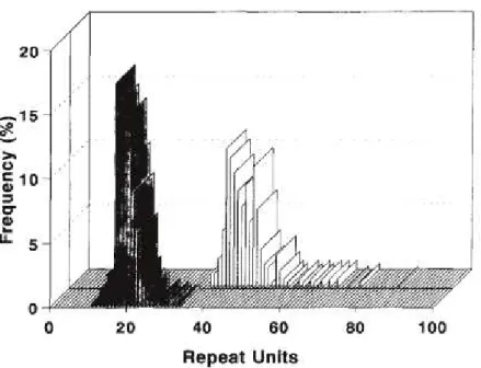
Figure 8. Comparison of CAG repeat units on control and HD chromosomes. Shaded
bars, control chromosomes; open bars, HD chromosomes (from Duyao et al., 1993).
Figure 9. Relationship between repeat unit length and age of onset (from Duyao et al.,
15
I. Introduction
As described for others polyQ disorders, such as Machado-Joseph‟s disease,
huntingtin protein also suffers proteolytic cleavage, resulting a fragment containing the
first exon of HTT gene. These fragments were described as being more toxic than the
full-length protein. Moreover, huntingtin flanking sequences and its protein interactions also
determines its toxicity (Duennwald et al., 2006a and Duennwald et al., 2006b). Huntingtin
was found in the nucleus of specific brain regions. Nevertheless, this protein lacks the
classic importin beta-dependent polypeptide sequence, the nuclear localization sequence
(NLS), to enter to nucleus. Were then hypothesized that polyglutamine tract has NLS
activity or huntingtin has high affinity to NLS-containing protein, such as transcription
factors, forming complex that allow its import into the nucleus (reviewed in Truant 2003
and Truant et al. 2006). The full-length protein has a nuclear export sequence (NES)
(Figure 7) that is not present in the huntingtin fragment leading to a highest toxicity of the
fragment compared to full-length huntingtin. The fragment toxicity was related to the
capacity of the protein to bind several proteins and its nucleocytoplasmic transport (Figure
10) (reviewed in Truant 2003).
Figure 10. Potential biological roles of huntingtin protein in nucleocytoplasmic
transport. Huntingtin interacts with diverse proteins involved on transcription, trafficking
16
In attempt to dissect the role of huntingtin fragment in the disease, Rockabrand and
colleagues found that the first 17 amino acids of exon 1 are involved in the huntingtin
fragment association with cytosolic organelles, such as mitochondria and ER (Rockabrand
et al., 2007). Furthermore these amino acids when mutated enhance the propensity of
fragment to aggregate (Atwal et al., 2007 and Rockabrand et al., 2007) and also may be
involved in the disruption of [Ca2+] homeostasis (Rockabrand et al., 2007). Towards the understanding of the effects of the first amino acid sequences of huntingtin, Atwal and
colleagues, observe that this region, that are well conserved in all vertebrate species, may
be involved in huntingtin sub-cellular localization, having a membrane targeting domain
that can mediate the association of huntingtin with the ER. When the association of
huntingtin/ER is disrupted, by point mutations, was observed the capacity of huntingtin to
enter in the nucleus. With these observations the authors suggest that polyglutamine
expansion may inhibit proper ER-targeting or nuclear shuttling of huntingtin leading to
increased protein levels in the nucleus (Atwal et al., 2007).
Using a yeast model, Solans and colleagues demonstrated that the expression of a
mutant fragment of huntingtin (first exon) affects the mitochondrial homeostasis soon after
large SDS-insoluble aggregate start to form (Solans et al., 2006). They described that this
fragment impairs the OXPHOS complex II+III activities, leading to deficits in respiratory
capacity and leading to an increased production of free radicals. They also observed that
the distribution and morphology of the mitochondrial network are progressively disturbed
and may be the result of the disruption of actin cytoskeleton (Solans et al., 2006). These
data support the idea that mutant hintingtin toxicity was associated with the apoptotic
process (Leavitt et al., 2001) and that wild-type huntingtin can reduce the apoptotic
toxicity of mutant huntingtin, suggesting that wild-type protein may normally have an
anti-apoptotic function (Leavitt et al., 2001). Moreover, Zhai and colleagues, using an in vitro
transcription assay with different huntingtin N-terminal fragments, demonstrate that
several components from transcription machinery are target by mutant huntingtin. Also, it
was demonstrated that soluble form os mutant huntingtin directly dysregulate transcription
interfering with specific components, such as TFIID and TFIIF (Zhai et al., 2005). The
mutant huntingtin is also associated with inhibition in the production of BNF
17
I. Introduction
Wild-type huntingtin bind repressor element-1 silencing transcription factor/neuron
restrictive silencer factor (REST/NRSF), sequester this transcription repressor in cytosol
promoting the expression of BDNF (brain-derived neurotrophic factor), important for the
survival of striatal neurons. In HD this mechanism is affected, the mutant huntingtin had
reduced capacity to bind REST/NSRF lead to their entrance to nucleus and repress the
expression of BDNF (reviewed in Truant et al., 2006).
As in others polyglutamine diseases, in HD a role on UPP has been demonstrated.
Using cellular and trangenic mouse models, it was demonstrate that the proteasome is
involved in degradation of polyglutamine-expanded truncated N-terminal huntingtin and
that the rate of degradation was inversely dependent on glutamine-repeat length (Jana et
al., 2001). Moreover, it was shown that altered proteasome activity is associated to the
disruption of mitochondrial membrane potential, leading to the release of cytochrome c
into cytosol and activation of caspase3- and caspase 9-, culminate in to apoptotic cell death
process (Jana et al., 2001). The impairment of proteasome activity has also been reported
to induce the expression of several cytoplasmatic heat-shock proteins and ER-resident
chaperones (Cowan et al., 2003 and Jana et al., 2001).
The data here reported lead us to raise new questions on the mechanisms underlying
the disease. Having this in mind, we herein present new insights on the role of autophagy
in HD disease.
4. Models for study neurodegenerative disorders
A growing number of completed sequencing genomes have revealed that the sequence
information per se is highly uninformative. Nevertheless, the determination of the
pathogenesis of diseases is not only based in the basic units of information, the genes, but
involves more complex systems (van Ham et al., 2009). To understand the systems
involving the disease pathogenesis, different models can be used. To develop a model for
studying the different neurodegenerative diseases must be taken into account the proposal
18
to dissect the neurodegenerative diseases pathogenesis (Table II). The different models
used were described in the next sections.
Table II. Conservation of genetics of human disease in models used to protein
aggregation research (from van Ham et al., 2009).
4.1. Mammal models
Mouse and cell cultures were the most commonly used models, to complement
clinical study of these disorders, due to their complexity and simplicity, respectively.
In mouse case, it is possible to use transgenic or knock-in mouse models to further
provide new valuable insights into the mechanisms associated with the development of the
diseases, particularly the early changes and into preclinical testing of therapeutic strategies
(reviewed in Rubinstein, 2002). These models also permit an in-depth analysis of the
different stages of the disease process and are helpful to identify the factors related to the
somatic repeat instability (reviewed in Bates and Davies, 1997). In long term both models
19
I. Introduction
Although these models will be invaluable in developing or understanding of the
molecular pathways by which a polyQ tract can cause cell death (reviewed in Bates and
Davies, 1997).
4.2. Invertebrate models
The invertebrate models encompass the Drosophila melanogaster and Caenorhabditis
elegans organisms (Brignull et al., 2006), being one of the classical tools used for unbiased
genetic screens in several neurodegenerative disease studies (Table III).
Drosophila melanogaster has been used as a model for investigating complex
biological processes, that are common with humans, such as regulation of gene expression,
membrane trafficking, cell signaling and cell death (Sang and Jackson, 2005 and Guo,
2010). This model has a compact genome (1/30th of human genome), limited genetic redundancy, and a short generation time (10 days). The sequencing genome has revealed
that 77% of human disease genes are conserved in the fly (Guo, 2010) and has several
cellular pathways shared with humans (van Ham et al., 2009). Furthermore an availability
of a number of genetic manipulations that are impossible or impractical to carry out on
mammals is possible to perform in this model.
It was used the fly eye, as a model, given that the adult eye phenotype are easy to
detect. Moreover, the eye is tolerant to genetic disruption of basic biological processes and
under laboratory conditions it is necessary to fly survival (Sang and Jackson, 2005). Using
the fly eye is possible study the cell control, cell proliferation and differentiation, neuronal
connectivity (Link, 2001) and the processes involving cell death and age-associated
neurodegeneration (Sang and Jackson, 2005).
The potential of the screens performed using Drosophila model has stimulate the
development of other invertebrate models as C. elegans. C. elegans model has sufficient
complexity and simplicity to allow investigation of cellular and behavioral phenotypes
which facilitates the high‐throughput testing of hypotheses (Brignull et al., 2006). The
20
modifiers genes or the screens of libraries at a speed that might be difficult if not
impossible to accomplish in mammalian models (Link, 2001)
Although, these models show late-onset and progressive pathology, not been possible
directly address the age-associate nature of the neurodegenerative disease (Link, 2001).
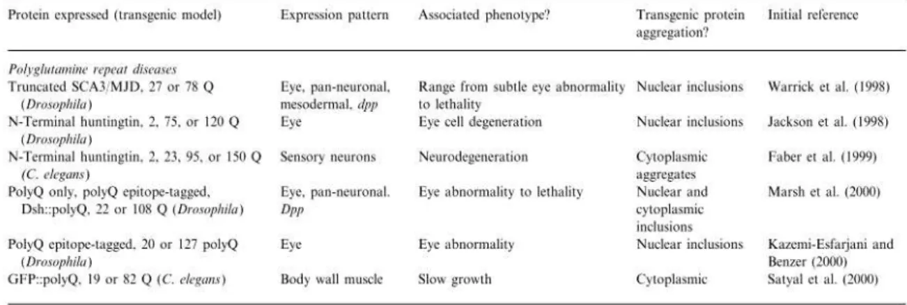
Table III. Description of several studies using invertebrate models to dissect the role
of polyQ disease related-proteins (adapted from Link, 2001).
4.3. Yeast models (Nervous Yeasts)
Several studies were performed using yeast as a model to study neurodegenerative
diseases (Duennwald, et al., 2006a; Duennwald, et al., 2006b and Outeiro and Linquist,
2003). Saccharomyces cerevisiae was the first eukaryote organism to be fully sequenced in
1996 (Goffeau et al., 1996). After 14 years about 80% of the proteins predicted to be
encoded in the yeast genome have been characterized functionally. Furthermore, the yeast
research community built valuable biological tools to work with this model (Table IV) and
the data obtained from different genomic approaches are well organized and actualized in
21
I. Introduction
Table IV. Some examples of the yeast collection available for genetic screens (from
Tenreiro and Outeiro, 2010).
Yeast models have been instrumental for our current understanding of conserved
cellular mechanisms such as cell division, the DNA replication, the metabolism, the
protein folding and the intracellular transport. Moreover, this model express numerous
genes with human orthologs that can be rapidly manipulated using established genetic
techniques (reviewed in Chen et al 2005).
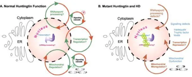
The yeast model is widely used to understand the molecular mechanisms involved in
different neurodegenerative diseases, because it is known that the basic mechanisms and
pathways involved in these processes are highly conserved between yeasts and humans,
such as mitochondrial dysfunction, transcriptional dysregulation, trafficking defects and
proteasomal impairment (Figure 11) (Winderickx et al., 2008 and reviewed in Tenreiro and
Outeiro, 2010). Moreover, there others advantages in using this model as the reduced
complexity of yeasts compared to neurons and the all the cDNA libraries available to study
the mechanism behind the disease. Nevertheless, such as others models yeast cells have
some limitations. Yeasts compared to neurons are not embedded in tissues neither
functionally linked to other cells, and lack the neuron-specific morphological structures,
such as dendrites, axons and synapses. Specially compared to neurons, yeasts have a less
diverse collection of molecular players, nevertheless the basic molecular process were
conserved between neurons and yeasts (reviewed in Braun et al., 2009). The high
22
in yeasts cells using the genetic and postgenomic tools, such as collections of strains and
plasmids, enable to identify novel modifiers of aggregation and toxicity, which are central
hallmarks of these diseases (reviewed in Tenreiro and Outeiro, 2010). Furthermore the
yeast models are also being implicated in drug discovery efforts.
In yeasts there are two different approaches to study the human neurodegenerative
diseases. If the gene implicated in the disease has a yeast homolog, it is possible to study
its function directly. If, on the other hand, the gene underlying the disease is absent in yeast
genome, it can be modeled by the heterologous expression of the human gene in yeast
cells. Nevertheless, the results obtained in yeast models need to be confirmed in other in
vitro and in vivo models, such as neuronal cells, worms or flies to validate the putative
targets for therapeutic intervention (Figure 12) (reviewed in Tenreiro and Outeiro, 2010).
23
I. Introduction
Figure 12. Schematic representation of yeast Saccharomyces cerevisiae being a eukaryotic model in combination with other cell and animal models (from Tenreiro
and Outeiro, 2010).
4.3.1 Yeast and Aging process
Aging is an important factor to develop neurodegenerative diseases and yeasts are a
widely used model for study the cellular aging. Using this model is possible to study the
mechanism associated to aged diseases through two mechanisms, the replicative life span
(RLS) and the chronological life span (CLS or chronological aging) (Figure 13). The RLS
was defined as the number of dougther cells produced by a mother cell before senescence
and the CLS, which was recently developed in yeast, is defined as the length of time a
population maintains viable in a nondividing state (stationary phase), mimicking the
situation of postmitotic cells, such neurons (Figure 13). These two models of aging provide
a unique opportunity to compare the aging processes of both proliferating and
nonproliferating cells in (reviewed in Braun et al., 2009; Chen et al., 2005 and Kaeberlein
24
Figure 13. Schematic representation of yeast replicative and chronological life span
(from Kaeberlein et al., 2007).
5. Autophagy, proteasome and aging on polyglutamine diseases
5.1. Autophagic process
Autophagic process was described more than 50 years ago, but only in the past
decade the molecular basis were elucidated, achieved through genetic approaches in yeast
mutants defective in autophagy (reviewed in Kundu and Thompson, 2008).
Autophagy is a cellular process in which cellular components are sequestered in
double membrane vesicles and delivered to lysosome for degradation and recycling of
bioenergrtic components (reviewed in Kundu and Thompson, 2008). This process is
extremely well regulated and it is involved in turnover of long-lived proteins and in the
elimination of supernumerary or damage organelles, such as mitochondria and ER
(reviewed in Maiuri et al., 2007). Moreover, autophagy is a process that represents a
25
I. Introduction
generates metabolic substrates to provide bioenergetics components to cells and thereby
allows the adaptative protein synthesis (reviewed in Maiuri et al., 2007 and Mizushima et
al., 2008).
The basic steps and the molecular machinery involved in autophagy are conserved
between yeasts and humans (reviewed in Kundu and Thompson, 2008). There are seven
well described steps in the autophagic process (reviewed in Kundu and Thompson, 2008).
The initiation process includes induction or selection/packaging of cargo, depending of the
activated pathway, if the stimuli promote the activation of selective or nonselective
pathways. Subsequent steps include nucleation, vesicle expansion, completion, fusion,
degradation and export of metabolic components (Figure 14-A) (reviewed in Kundu and
Thompson, 2008).
Autophagy is induced when cells enters in starvation, where the target of rapamycin
(TOR) kinase is inactive. Nevertheless, there are others pathways that lead to a autophagy
induction. The basal levels of autophagy are important for maintain normal cellular
homeostasis. The TOR regulates the switch between autophagy (Figure 14-B) and the
cytoplasm-to-vacuole targeting (CTV) pathway, changing the interaction between
serine-threonine kinase ATG1 with other components of the induction complex, including ATG13,
ATG17 and ATG11 (reviewed in Høyer-Hansen and Jäättelã, 2007 and Kundu and
Thompson, 2008).
When autophagic process is activated, specific autophagic proteins are recruited
leading to the formation of the double membrane autophagosomes, contain cargo proteins
or organelles, such as mitochondria, ER and peroxisomes, which will dock and fuses with
lysosomal (mammals)/vacuolar membrane (yeasts) and starts the degradation process
(reviewed in Kundu and Thompson, 2008). Finally, the bioenergetic components resulting
from degradation are exported from lysosome/vacuole (reviewed in Kundu and Thompson,
2008). Yeast genome presents more than 30 autophagy-related (ATG) genes involved in
autophagic process were identified in yeast cells. Some of them participate in all types of
autophagy while others only participate on one or few particular types of autophagy
(Figure 14-B) (reviewed in Kraft et al., 2009).
Some studies suggest that the apoptotic and the autophagic processes inhibit each
26
the autophagy. The autophagic process has a dual ellefct on cells, can act as cytoprotective
process or a cellular death process (reviewed in Maiuri et al., 2007).
Figure 14. A) Schematic representation of autophagic process. The signal to initiate the
autophagy comes from nonselective pathway, relative to changes in nutrient availability, or
selective pathway, relative to cargo proteins or organelles (from Kundu and Thompson,
2008). B) Schematic representation of autophagic regulation (from Høyer-Hansen and
27
I. Introduction
The involvement of autophagy in cellular suicide may be explained by the direct
self-destruction of massive autophagy or, alternatively, by hardwiring of the autophagic process
to pro-apoptotic signal (reviewed in Maiuri et al., 2007).
There are two major degradation pathways in cells, the ubiquitin proteasome pathway
(UPP) and autophagy.
The ubiquiton proteasome pathway (UPP) is responsible for the degradation of
short-lived proteins. In this pathway proteins are ubiquitilated and then shuttled to 26S
proteasome. The 26S proteasome is a multimeric structure composed of a 20S core
subunit, with four rings, capped by two 19S regulatory subunits. The proteolytic
degradation of ubiquitilated proteins takes place within the 20S core subunit following
removal of the ubiquitin monomers by deubiquitination enzymes (DUB) and folding of the
substrate protein (reviewed in Lehman, 2009).
Ding and colleagues, using transfected cells, observed a relation between autophagy
and UPP (Ding et al., 2007). They demonstrated that if the UPP is inhibited then there is an
activation of autophagy. When an impairment of the UPP occurs there is an accumulation
of polyubiquitinated proteins that causes the accumulation of misfolded proteins in the ER,
leading to ER stress. In this situation seems that autophagy is activated to compensate the
UPP impairment and ameliorates de ER stress (Ding et al., 2007). With these studies it was
demonstrate that diverse pathways may be involved on relation between autophagy and
UPP.
5.2. Autophagy, aging and neurodegenerative diseases
Growing evidences point a role for autophagy in diverse human diseases, such as in
neurodegeneration, cancer, infection and immunity, heart disease, myopaties and liver
diseases (Mizhushima et al., 2008). There are several antibiotics that block or enhance the
autophagic process, as shown in figure 15 that contributed to the understanding of the
effects of autophagy in neurodegenerative and other diseases (reviewed in Maiuri et al.,
28
Among these antibiotics, one of the best described is rapamycin. This substance is an
enhancer of autophagy (figura 15) by inhibiting the mTOR pathway leading to the
induction of autophagy. Several studies were performed using this enhancer in order to
understand its effect on neurodegeneration. More recently, Pan and colleagues, using PC12
cell line with proteasomal dysfunction, induced by lactacystin, and C57BL/6 mice, demonstrated that these cells presents neuronal death with formation of α-synuclein of ubiquitin-positive cytoplasmatic inclusion and inducing autophagy (Pan et al., 2008).
When cells were treated with rapamycin, the lactacystin-induced apoptosis was attenuated
and cells were rescued from lactacystin induced loss of dopaminergic neurons (Pan et al.,
2008).
Figure 15. Schematic representations of autophagy and its inhibitors (from Maiuri et
29
I. Introduction
In mouse model of Machado-Joseph disease it was shown that the induction of
autophagy leads to a reduction on mutant ataxin-3 levels (Menzies et al., 2010).
Furthermore, in Caenorhabditis elegans model, using a siRNA to knock down
autophagy-related genes, it was shown an increase of aggregate-containing cells and that this
aggregates were focus on nucleus (Khan et al., 2008).
These, as well as others studies, indicate that the induction of autophagy has a
neuroprotective role, when the ubiquitin-proteasome was impaired (Pan et al., 2008).
Taken together these studies reveal a close kink between autophagy and
neurodegeneration. Moreover, these processes are related with aging process.
There is an accumulation of damage proteins and damage organelles with age,
caused by different factors, such genetic factors, environmental influences and certain
diseases (Figure 16) (reviewed in Vellai, 2009), leading to an imbalance between the rate
of protein damage and protein turnover. The accumulation of damage organelles or
structures lead to the reduction of cellular efficiency of biological processes that are
required to maintain homeostasis and survival, which could contribute to aging process
(reviewed in Cuervo et al., 2005 and Rajawat and Bossis, 2010). Nevertheless, the precise
mechanism of aging is not yet completely understood.
Figure 16. Environmental clues and evolutionary conserved pathways that regulate
30
Lysosomes have a pivotal role on aging. Age-related alterations of lysosomal system
leads to the accumulation of lipofuscin (pigmented product) as a result of incomplete
digestion of engulf components (reviewed in Cuervo, 2008 and Rajawat and Bossis, 2010).
The rate of lipofuscin formation is inversely related to age, and lysosomes with lipofuscin
have a reduced ability to fuse with autophagic structures. Mitotic cells can eliminate
lipofuscin by diluting the pigment in each mitotic cycle. However pos-mitotic cells, like
neurons, do not have this ability (reviewed in Cuervo, 2008 and Rajawat and Bossis,
2010). Lipofuscin, thus, seems to be an important contributor to cellular degeneration, and
not only a hallmark of aging (reviewed in Cuervo et al., 2005). Furthermore
macroautophagy dysregulation in old organisms could also be a consequence of its
persistent activation as demonstrated in rodent liver (reviewed in Cuervo, 2008).
An important limitation of the studies linking autophagy and aging is that autophagy
blockage is induced early in life (invertebrates and mammalian models), whereas the
age-dependent decrease in this pathway does not begin in most organisms until middle age
(reviewed in Cuervo, 2008). Further studies, using yeasts as a model, should be done take
this limitation in account.
6. Objectives
The general goal of the studies described in this thesis is to improve the knowledge
on polyglutamine expansion diseases, using a yeast model.
The first goal of this study was the development of yeast model for
Machado-Joseph disease (MJD) with variants 1 and 2 genes under the control of Tet On system.
The second goal was the characterization of the yeast model for Huntington‟s
disease (HD). For the characterization of Huntington‟s model we want to address the effect
of autophagy on the pathogenesis. For this purpose, we used chloroquine to inhibit the
autophagic process in cells expressing normal and mutated huntingtin protein at
35
II. Materials and Methods
1. Construction Machado-Joseph disease models
1.1 Construction of ATXN3 variants on pUG35 plasmid
1.1.1 DNA manipulations and Cloning
The ATXN3 variants, 1-1, 1-1E, 2-1 and 2-1E were obtained from plasmidic DNA,
pCMV and pEGFP-C, respectively, kindly provided by Dra. Patrícia Maciel
(ECS/ICVS-UM) via PCR amplification using the primers 1 and 2 or 3 (Results, Table I) that included,
respectively, BamHI and HindIII restriction site for cloning into the pUG35 plasmid, which
will allow the genes fusion with GFP. To amplify the fragments, 200 ng of DNA were
added to 49.5 μl of reaction mixture defined by reaction buffer 1x with 1.5 mM MgCl2, 0.2
mM dNTP, 1 mM of each primer and 2.5 units High Fidelity PCR enzyme (Fermentas), the PCR amplification was performed on My Cycler™ (Bio-Rad) following the condition described in Table II (Results). The fragments were extracted from a 0.8% agarose gel
prepared on TAE buffer (40 mM Tris-Base, 2 mM EDTA, Glacial Acetic Acid to pH 0.8)
and purified gel using Silica Bead DNA Gel Extraction Kit (Fermentas). The amplified
fragments and plasmid was digested using the referred restriction enzymes, BamHI and a
HindIII (Fast Digest enzyme, Fermentas) during 3hours. Followed by new products
purification, performed with Silica Bead DNA Gel Extraction Kit (Fermentas), afterwards
the products were run in a 0.8% agarose gel and extracted from the gel. The purified
products were then quantified on NanoDrop® ND-1000 Spectrophotometer (Alfagene). To
perform the ligation between gene fragments and linear plasmids was used 2 units of T4
DNA Ligase (Fermentas), during 1 hour at 26ºC. The products resulting from ligation
reaction were transformed in Escherichia coli, using the Inoue Method. Briefly, ~25ng of
ligation product DNA were added to 90μl of competent E.coli cells, which were then
submitted to 30 minutes incubation on ice, followed by the heat shock during 45 seconds at
42ºC and incubation on ice for 10 minutes. Afterwards, to the mix were added 800 μl of
SOC (Super Optimal broth with catabolite repression) medium and incubated during1
36
0.5% Yeast extract, 0.05% NaCl, 0.0186% KCl, at pH 6.7) the cells are centrifuged for 1
minute at 13,000 rpm, ressuspended in 100 μl of SOC medium and plated in LB
amplicillin medium (Sambrook and Russell, 1989).
1.1.2 Analysis of transformants
The plasmid isolation was performed on overnight cultures by Fast Alkaline Lysis.
Therefore 3 ml of LB ampicillin liquid medium with the bacterial cells were centrifuged
and resuspended in 250 μl of resuspension buffer, followed by the addition of 250 μl of
Solution 2 (1% SDS and 200 mM NaOH), after 5 minutes it was added 300 μl of Solution
3 (3 mM Potassium acetate). The Mix was centrifuged and it was added 600 μl of
Isoporanol (100%) to perform the DNA precipitation. To carry out the DNA wash, 200 μl
of 70% Ethanol were added to the pellet. After ethanol evaporation, the DNA was
resuspended in 50 μl of TE buffer (1 mM EDTA, 10 mM Tris-HCl, pH 8.0) with 10 μg/ml
RNase A. The plasmidic DNA of transformants was analyzed for the introduction of the
variant genes by restriction digestion analysis. The pUG35::ATXN3 variant genes were
digested with BamHI and KpnI, or only KpnI, and with EcoRI and EcoRV during 3 hours.
The resultant digested products were run in 0.8% agarose gel.
1.2 Construction of ATXN3-GFP variants on pCM252 plasmid
1.2.1 DNA manipulations and Cloning
The ATXN3 variants, 1-1, 1-1E, 2-1 and 2-1E fused with GFP were obtained from
pUG35 plasmidic DNA, described in the precious section, via PCR amplification using the
primers 1 and 4 (Results, Table I) that included, respectively, a BamHI and a ApaI
restriction site for cloning into the pCM252 plasmid. To amplify the fragments, 200 ng of
37
II. Materials and Methods
MgCl2, 0.2 mM dNTP, 1 mM of each primer and 2.5 units High Fidelity PCR enzyme
(Fermentas), the PCR amplification was performed on My Cycler™ (Bio-Rad), following
the condition described in Table III (Results). The resultant fragments were extracted from
a 0.8% agarose gel prepared on TAE buffer (40 mM Tris-Base, 2 mM EDTA, Glacial
Acetic Acid to pH 0.8) and purified using Silica Bead DNA Gel Extraction Kit
(Fermentas). The amplified fragments and plasmid were digested using the referred
restriction enzymes, BamHI and ApaI (Fast Digest enzyme, Fermentas) during 3hours. The
ligation and transformation conditions were the same described above in the section 1.1.1.
1.2.2 Analysis of transformants
The plasmid extractions were performed as described in the section 1.1.2. Plasmidic
DNA of transformants was analyzed for the introduction of the variant genes fused with
GFP by restriction digestion analysis. The pUG35::ATXN3-GFP variant genes were
digested with BamHI or ApaI during 3 hours. The resultant digested products were run in
0.8% agarose gel.
2. Studies on yeast model of Huntington’s disease
2.1 Strains, media and treatments
In this study it was used the Saccharomyces cerevisiae wild-type strain W303-1a
(MATa; ura3-52; trp1Δ 2; leu2-3,112; his3-11; ade2-1) harboring pCM252 plasmids with
exon 1 fragment of IT15 gene (gene associated to Huntington‟s disease) with 25 CAG and
103 CAG, this fragments were C-terminally tagged with GFP gene (kindly provided by
Dr. Fulvio Reggiori). All yeast cultures were inoculated in selective YNB medium,
containing 0.67 % (w/v) Yeast Nitrogen Base (Difco Laboratories), 2% (w/v) glucose as