PhD Thesis
Analysis of the effect of Ischemia and Reperfusion on Neutrophil Proteome in Rats
Muhammad Tahir
Supervisor: Prof. Dr. Wagner Fontes
Brasilia, June – 2014 University of Brasília
Department of Cell Biology
Postgraduate in Molecular Biology
II
Muhammad Tahir
PhD Thesis
Analysis of the effect of Ischemia and Reperfusion on Neutrophil Proteome in Rats
Supervisor: Prof. Dr. Wagner Fontes
Brasilia, June – 2014 University of Brasília
Department of Cell Biology
Postgraduate in Molecular Biology
Laboratory of Biochemistry and Protein Chemistry
Thesis presented as partial requirement for the degree of Doctor of Philosophy in Molecular Biology, to the Department of Cell Biology at the Institute of Biological Sciences, University of Brasilia.
This work was performed in the laboratory of Biochemistry and Protein Chemistry (LBQP) University of Brasilia in collaboration with Laboratory of Surgical Physiopathology (LIM-62), Faculty of Medicine University of São Paulo and Protein Research Group (PRG) Southern Denmark University, Odense, Denmark.
IV
ACKNOWLEDGMENT
All praises for Almighty Allah the most compassionate, the most beneficent and ever merciful, who gives me the power to do, the sight to observe and mind to think and judge. Peace and blessings of Almighty Allah be upon His Prophet Hazrat Muhammad
(P.B.U.H) who exhorted his followers to seek knowledge from cradle to grave.
This thesis is based on results from Laboratório de Bioquímica e Química de Proteínas (LBQP), University of Brasília, Brazil, Laboratory of Surgical Physiopathology (LIM-62), of the Faculty of Medicine (LIM-(LIM-62), University of São Paulo, Brazil, Protein Research Group of Southern Denmark University (SDU), Odense.
I want to express my deepest gratitude for my supervisor, Prof. Dr. Wagner Fontes, for his kind supervision, useful suggestions, consistent encouragement, friendly behavior and dynamic supervision which enabled me to perform my research work in ensuring my academic, professional and moral well being.
I would like to extend my vehement sense of gratitude and sincere feelings of reverence and regards to Prof. Dr. Peter Roepstorff to receive me in his research group under his supervision, for his co-operation in providing me all possible facilities during my research at Protein Research Group, (SDU). Many thanks also go to my honorable teachers, especially Prof. Dr. Belchor Fontes and Prof. Dr. Edna Montero for their support and providing me all possible facilities during research at LIM 62.
I am grateful to TWAS-CNPq for the financial assistance in the form of PhD Fellowship.
Also many thanks to my wife Samina Arshid, a PhD student at FMUSP, for her kind cooperation and help during my research.
Furthermore, I am deeply obliged and thankful to my lab fellows at LBQP and LIM62 especially to Alison da Silva Alexandre, Adelson Silva, Rayner Queiroz, Muhammad Faheem, Michaella Pereira, Carlos Garcia, Everton da Rosa, Diana Gomez, Elaine Nascimento, Anne Dian, Nuno Domingues, Ana Maria Cattani, Marcos, Mario, Luci, and others for their co-operation and sympathetic attitude during my study.
Also, during my stay at SDU, Odense, I am very much thankful to Simone Sidoli and Giuseppe Palmisano for their help in Mass Spectrometric Analysis, Veit Schwämmle for statistical analysis of data, Lene Jakobsen for my initial training at SDU, Odense, Maria Ibáñez-Vea for nice discussions and guidelines for the sample preparation, Prof. Dr. Martin Larsen, Karin Hjernø, Helle Marquard Mortensen, Marco Milioli, Philipp Holzinger, Marcela Braga, Thiago Braga, Richard Hemmi Valente and other members of PR-Group.
Finally, I wish to express my deepest gratitude to my family for continuous support and love during these years. I wish special thanks to my dear parents especially to my father
Taj Merin for his love, encouragement and support in every part of my life and my son Ayan Tahir for unconditional love, patience and encouragement.
May Almighty Allah shower His blessings and prosperity on all those who assisted me in any way during my work.
VI ABSTRACT
Intestinal Ischemia and reperfusion injury are widely used models, which results into tissue dysfunction and organ failure especially after trauma and surgery. Whereas neutrophils play an important role in the mechanism of injuries caused by ischemia and reperfusion. However, the effect of intestinal ischemia and reperfusion on stimulation and activation of neutrophils is still unclear. Proteomic analysis has been ratified as an appropriate tool to studying complex systems, such as stimulation and activation of neutrophils, so the objective of this work was to evaluate the effect of ischemia and reperfusion on neutrophil proteome in Wistar rats. For proteomic neutrophils from three groups; control, laparotomy, and intestinal ischemia/reperfusion were separated. Protein was extracted according to FASP protocol, trypsin digested, iTRAQ-labeled. Phosphopeptides were enriched by TiO2-SIMAC-HILIC procedure. Non-phosphorylated and mono-phosphorylated peptides were HILIC fractionated prior to LC-MS/MS analysis. LC-MS/MS and bioinformatics procedures were applied to peptides for their quantitation, phosphosite assignment and protein identification. A total of 3816 proteins containing 2231 non-phosphorylated unique proteins and 892 unique phosphorylated proteins whereas 693 phosphorylated proteins were identified in both total and phosphoproteome. In total 1585 phosphorylated proteins were identified, representing 41.5% of protein phosphorylation of the total neutrophil proteome identified in this study. We aimed to bring further light to this process by analyzing hematological parameters after ischemia and preconditioning. Gene ontologies for all the regulated proteins represented changes confer by the experimental groups on neutrophil proteome. We could identify the relevant predicted enzyme activities and pathways involved in neutrophils activation after intestinal ischemia and reperfusion. Most of the regulated predicted enzymes of the rat neutrophil proteome are transferases, hydrolases and oxidoreductases. Important pathways from KEGG analysis include ribosomal, regulation of actin cytoskeleton, Fc gamma R-mediated phagocytosis, chemokine signaling, metabolic and oxidative phosphorylation pathways. We also aimed to bring further light to this process by analyzing hematological parameters after ischemia and preconditioning. Our findings provide platform for the first time to understand the effects
of intestinal ischemia and reperfusion on neutrophil proteome and mechanism involve in this process.
Keywords: Ischemia Reperfusion, Neutrophils, Proteomics, Systemic Inflammatory Response, Phosphoproteomics.
VIII RESUMO
Lesão por isquemia e reperfusão é um modelos amplamente usados que resulta em disfunção tecidual e falha do órgão especialmente após trauma e cirurgia, enquanto que neutrófilos desempenham papel importante no mecanismo de lesão causada por isquemia e reperfusão. No entanto, o efeito da isquemia e reperfusão na estimulação e ativação de neutrófilos ainda não é claro. A análise proteômica tem sido ratificada como uma ferramenta adequada no estudo de sistemas complexos, tais como a estimulação e ativação de neutrófilos, portanto o objetivo desse trabalho foi avaliar os efeitos da isquemia e reperfusão no proteoma de neutrófilos e nos parâmetros hematológicos em ratos Wistar. Três grupos de neutrófilos foram separados: controle, laparotomia e isquemia/reperfusão intestinal. Proteínas foram extraídas de acordo com o protocolo FASP, digeridas com tripsina e marcadas com iTRAQ. Fosfopeptídios foram enriquecidos pelo procedimento TiO2-SIMAC-HILIC. Peptídios fosforilados e não fosforilados foram fracionados por HILIC antes da análise por LC-MS/MS. Procedimentos de LC-MS/MS e bioinformática foram aplicados para quantificação dos peptídeos, designação do sitio de fosforilação e identificação proteica. Foi identificado um total de 3816 proteínas contendo 2231 proteínas exclusivamente não fosforiladas, 892 exclusivamente fosforiladas e 693 presentes tanto no proteoma total quanto no fosfoproteoma. No total 1585 proteínas fosforiladas foram identificadas, representando 41,5% da fosforilação de proteínas no proteoma total de neutrófilos identificado nesse estudo. A classificação das proteínas reguladas por ontologia genética mostrou mudanças no proteoma de neutrófilos conferidas pelos grupos experimentais. Nós pudemos identificar atividades enzimáticas e vias de sinalização relevantes envolvidas na ativação de neutrófilos após isquemia intestinal e reperfusão. A maior parte das enzimas reguladas no proteoma de neutrófilos de rato são transferases, hidrolases e oxidoredutases. Vias identificadas na base de dados KEGG e contendo diversas proteínas reguladas incluem a ribossomal, regulação de actina do citoesqueleto, fagocitose mediada por Fc-gama-R, sinalização por quimocina, vias de fosforilação oxidativa e metabólica. Nossos resultados fornecem base para compreender pela primeira vez os efeitos de isquemia e reperfusão no proteoma de neutrófilos e os mecanismos envolvidos no processo.
Palavras-chave: Isquemia/Reperfusão, Neutrófilos, Proteômica, Resposta Inflamatória Sistêmica, Fosfoproteômica.
X CONTENTS INTRODUCTION ... 1 1.1 Polymorphonuclear Neutrophils (PMNs) ... 2 1.1.1 Rolling ... 3 1.1.2 Tight adhesion ... 3 1.1.3 Tansendothelial Migration/Diapedesis ... 3 1.1.4 Chemotaxis ... 4
1.2 Neutrophils Granules and Degranulation ... 4
1.3 Ischemia ... 6
1.3.1 Cellular effects of Ischemia ... 6
1.4 Ischemic Reperfusion injury (IRI) ... 8
1.5 Intestinal Ischemic Reperfusion injury and PMNs ... 9
1.6 Mediators and receptors of IRI ... 11
1.7 Mass Spectrometry Based Proteomics ... 13
1.7.1.1 Ion source ... 14
1.7.1.2 Mass analyzer ... 15
1.7.1.3 Peptide fragmentation ... 17
1.8 Quantitative Proteomics ... 18
1.8.1 Isobaric tags for relative and absolute quantitation (iTRAQ) ... 19
1.9 Phosphoproteomics ... 21
1.9.1 PTM-Specific Enrichment ... 23
1.10 Proteomics in Neutrophils ... 26
1.10.1 Phosphorylation of neutrophil Proteins ... 27
1.11 Hematological analysis ... 28
OBJECTIVE ... 30
2 Global Objective ... 31
2.1 Specific Objectives ... 31
MATERIAL & METHODS ... 32
3.1 Experimental Subjects: ... 33
3.1.1 Experimental groups ... 33
3.3 Surgical procedures and sample collection: ... 34
3.4 Hematological analyses ... 34
3.5 Ficoll Gradient Protocol for Neutrophils separation from Rat Blood: ... 35
3.7 Filter-aided sample preparation (FASP) ... 35
3.8 Amino Acid Composition Analysis ... 37
3.9 iTRAQTM Labelling ... 37
3.10.1 First TiO2 Purification ... 38
3.10.12 Enzymatic Deglycosylation ... 38
3.10.3 SIMAC Purification of the Multi-Phosphorylated Peptides ... 39
3.10.4 Second TiO2 Purification of the Mono-Phosphorylated Peptides ... 39
3.11 Sample Desalting ... 40
3.12 HILIC fractionation of Mono-phosphorylated and non-phosphorylated peptides ... 40
3.13 Nano-Liquid Chromatography Tandem Mass Spectrometry (nano-LC-MS) ... 40
3.14 Database Searching and Bioinformatics ... 43
RESULTS & DISCUSSION ... 46
Hematological Analysis ... 47
Results & Discussion ... 47
4 Results and Discussion of Hematological Analysis ... 48
Proteomics Analysis ... 54
Results & Discussion ... 54
5.1 Identification of proteins by mass spectrometry ... 55
5.2 Statistical analysis of data with R package ... 57
5.3 Principal Component Analysis (PCA): ... 59
5.4 Phosphoproteome of rat neutrophil ... 60
5.5 GO Slim analysis of total and regulated proteins of rat neutrophils ... 62
5.6 Predicted Enzyme activity for the total rat neutrophil proteome ... 67
5.7 KEGG Pathway Analysis of Rat Neutrophil Proteome ... 85
5.8 Kinases and Phosphatases ... 102
5.8.2 Phosphorylated Phosphatases in Neutrophil ... 113
6 Summary of pathway results ... 117
7 Conclusion and Future perspectives ... 119
8 Portuguese Version………...……….………...121
9 Introdução………...122
10 Resultados………...151
11 Abstracts & Articles published during PhD………...273
XII
List of Figures
Figure 1 Neutrophil recruitment to sites of inflammation ... 2
Figure 2 Neutrophil granules and their characteristic proteins. ... 5
Figure 3 A Cellular effect of Ischemia. ... 7
Figure 4 Scheme representing the steps performed in a mass spectrometer ... 13
Figure 5 Ionization techniques for MS ... 15
Figure 6 Mass spectrometers used in proteome research. ... 16
Figure 7 Schematic view of the LTQ Orbitrap Velos ... 17
Figure 8 The Roepstorff-Fohlman-Biemann peptide fragment ion nomenclature ... 18
Figure 9 Structure of the iTRAQTM reagents. ... 20
Figure 10 A general scheme and example data for a 4-plex iTRAQ experiment. ... 21
Figure 11 Overall SIMAC workflow ... 25
Figure 12 Experimental groups and their time of ischemia and reperfusion. ... 33
Figure 14 The raw files searched against Mascot database by using Proteome Discoverer. ... 45
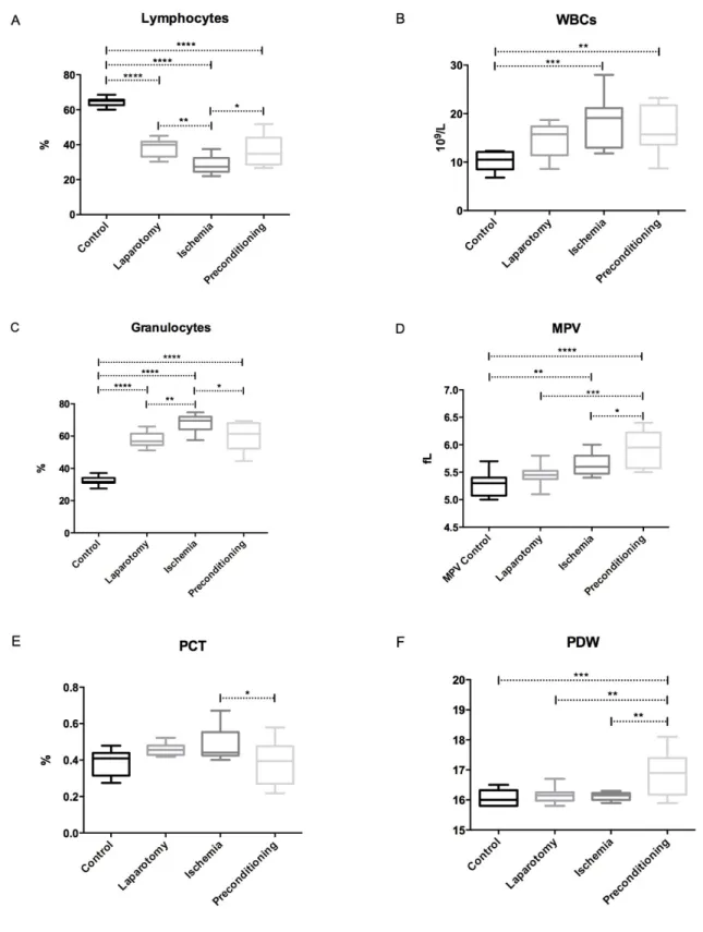
Figure 15 Distribution of the hematimetric parameters in the four experimental groups. ... 49
Figure 16 supplementary. Distribution of hematimetric parameters in the four experimental groups. ... 52
Figure 17 Common and exclusive regulated proteins in laparotomy (Lap) and intestinal ischemia reperfusion (IR) groups. ... 55
Figure 18 Number of Regulated and non-regulated proteins in LAP and IR groups. ... 56
Figure 19 A 4-way Venn diagram illustrating unique and overlapping up and down regulated proteins in neutrophil after laparotomy and intestinal ischemia/reperfusion. ... 57
Figure 20 Expression profile of regulated proteins and phosphopeptides after laparotomy and Ischemia reperfusion in neutrophils. Control (114), Laparotomy (115), and Ischemia reperfusion (117). ... 58
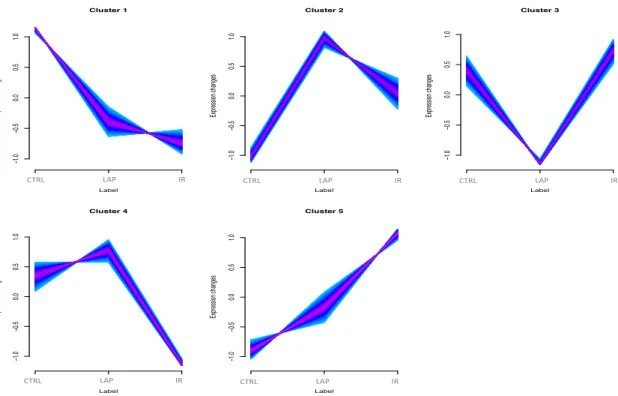
Figure 21 Regulated Proteins distribution among the five clusters in neutrophils. ... 58
Figure 22 Principal component analysis (PCA). ... 59
Figure 23 Phosphoproteome of rat neutrophil. ... 61
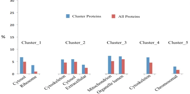
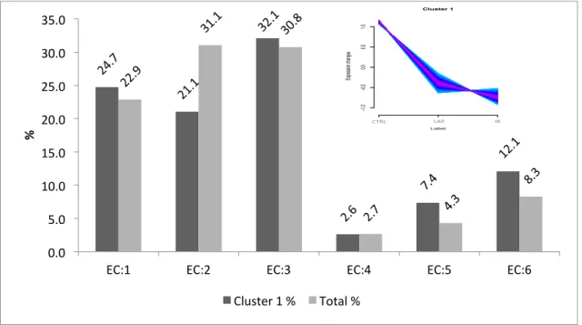
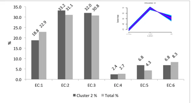
Figure 24 Bar chat of GO slim terms of cellular component of total and all regulated neutrophil proteins of (A) Cluster 1, (B) Cluster 2, (C) Cluster 3, (D) Cluster 4 (E) Cluster 5. ... 62
Figure 25 GO slim terms of more abundant cellular component of total and all regulated neutrophil proteins of (A) Cluster 1, (B) Cluster 2, (C) Cluster 3, (D) Cluster 4 (E) Cluster 5. .... 63
Figure 26 GO Slim terms of Biological Process of total and all regulated neutrophil proteins of (A) cluster 1, (B) Cluster 2, (C) Cluster 3, (D) Cluster 4 (E) Cluster 5. ... 65
Figure 27GO Slim terms of more abundant Biological Process of total and all regulated neutrophil proteins of all clusters. ... 65
Figure 28 GO Slim terms of Molecular functions of total and all regulated neutrophil proteins of (A) cluster 1, (B) Cluster 2, (C) Cluster 3, (D) Cluster 4 (E) Cluster 5. ... 66
Figure 29 GO Slim terms of more abundant proteins for Molecular functions for all clusters. ... 67
Figure 30 Enzyme prediction of total rat neutrophil proteome. ... 68
Figure 31 Enzyme prediction of cluster 1 proteins. ... 69
Figure 32 Enzyme prediction of cluster 2 proteins.. ... 74
Figure 33 Enzyme prediction of cluster 3 proteins. ... 74
Figure 34 Enzyme function prediction of cluster 4 proteins. ... 75
Figure 37 Computer graph visualization of the enrichnet of the dataset from cluster 1 overlap (green) is shown for the regulation of the ribosomal pathway. ... 89
Figure 38 Interaction network of proteins from cluster 1 identified in ribosomal KEGG pathway. ... 89
Figure 40 Computer graph visualization from enrichnet.org of uploaded dataset for clusters 2, 4 and 5 overlap (green) is shown for regulation of actin cytoskeleton pathway. ... 91
Figure 41 Interaction network of proteins from cluster 2, 4 and 5 identified in regulation of actin cytoskeleton KEGG pathway. ... 92
Figure 42 The enriched Fc gamma R-mediated phagocytosis pathway for the differentially
expressed proteins (DEPs) from cluster 2, 4 and 5. ... 93
Figure 43 Computer graph visualization from enrichnet of uploaded dataset from cluster 2, 4 and 5 overlap (green) is shown for Fc gamma R-mediated phagocytosis pathway. ... 95
Figure 44 STRING network analysis of proteins from Fc gamma R-mediated phagocytosis pathway from cluster 2, 4 and 5. ... 95
Figure 46 Computer graph visualization from enrichnet of uploaded dataset from cluster 2, 4 and 5 and overlapping genes in green for chemokine signaling pathway. ... 97
Figure 47 STRING network for proteins from Fc gamma R-mediated phagocytosis pathway from cluster 2, 4 and 5. ... 98
Figure 48 Computer graph visualization from enrichnet of uploaded dataset from cluster 3. ... 99
Figure 49 STRING network analysis of proteins from metabolic pathways from cluster 3. ... 99
Figure 50 The enriched Oxidative Phosphorylation pathway for differentially abundant proteins (DAPs) from cluster 3. The purple highlighted show the overlapping proteins. ... 100
Figure 51 Computer graph visualization from enrichnet of uploaded dataset from cluster 3 and over lapping proteins (green) for oxidative phosphoryalation pathway. ... 101
Figure 52 STRING network analysis for proteins from Oxidative Phosphorylation pathway of cluster 3. ... 102
Figure 53 A shows protein phosphorylation and de-phosphorylation by protein kinase and phosphatase respectively. Fig. B. shows distribution of regulated kinases and phosphatases with and without regulation of phosphorylation in domains identified in total rat neutrophils. ... 103
XIV List of Tables
Table 1 Haematological analyses, expressed as mean ± standard deviation, median and range (min. - max.) of (Wistar Rat) control, laparotomy, intestinal ischemia/reperfusion and ischemic
preconditioning. ... 50
Table 2 Number and percentage of all regulated proteins in LAP and IR group. ... 55
Table 3 Predicted isomerase function of cluster 1. ... 72
Table 4 Predicted ligase function of cluster 1 ... 73
Table 5 Predicted transferase function of cluster 4. ... 78
Table 6 Predicted oxidoreductase function of cluster 5. ... 83
Table 7 Predicted KEGG pathways for all clusters. ... 86
Table 8 Phosphorylated Kinases with regulated expression after laparotomy and ischemia. ... 105
Table 9 Phosphorylated Phosphatases with regulated expression after laparotomy and ischemia. ... 115
List of Abbreviations
1DE One-dimensional gel electrophoresis 2DE Two-dimensional gel electrophoresis
6PGDH 6-phosphogluconate dehydrogenase
AA Arachidonic acid
AdoRA2B Adenosine A2B-receptors
ALI Acute Lung Injury
AMI Acute mesenteric ischemia
AR Aldose reductase
ARTs Adp-ribosyl transferase
ATP Adenosine triphosphate
BAL Bronchoalveolar lavage
BLT-1 Leukotriene B (4) receptors.
BPI Permeability-increasing protein BTK Bruton tyrosine kinase
C3a Complement 3a
CaMKs Calmodulin-dependent protein kinase
CG Cathepsin
CI Chemical ionization
CID Collision-induced dissociation
CINC-1 Cytokine-induced neutrophil chemoattractant-1
XVI
CXCR CXC chemokine receptors
CyPs Cyclophilins
DEGs Differentially expressed genes
DGKζ Diacylglycerol kinase ζ
DIGE Differential gel electrophoresis
ECD Electron capture dissociation
ECs Endothelial cells
ERK1/2 Extracellular signal-regulated protein kinase-1/2
ESI Electrospray ionization
ESI-MS/MS Electrospray tandem mass spectrometry
ETD Electron transfer dissociation
FAB Fast atom bombardment
FAK Focal adhesion kinase
FGAM Phosphoribosyl formyl glycin amidine
Fhs Formate tetrahydrofolate ligase
fMLP Formyl-Methionyl-Leucyl-Phenylalanine
G-CSF Granulocyte colony-stimulating factor
GAPDH Glyceraldehyde-3-phosphate dehydrogenase
GROs Growth-related oncogenes
Gsr Glutathione reductase
H2O2 Hydrogen peroxide
HK3 Hexokinase-3
HNP-1 Human neutrophil peptides
HPLC High-performance liquid chromatography
ICAMs Intercellular Adhesion Molecule
ICAT Isotope coded affinity tag
ILs Interleukins
IMAC Immobilized metal affinity chromatography
IPC Ischemic Preconditioning
IRAK-4 Interleukin-1 receptor-associated kinase-4
iTRAQ Isobaric tags for relative and absolute quantitation
KATP channel ATP-sensitive potassium channel
KEGG Kyoto Encyclopedia of Gens and Genomes
LDH Lactate dehydrogenase
LPS Lipopolysaccharide
LSI-MS Liquid secondary ion mass spectrometry
LTB4 Leukotriene B4
LTQ Linear ion trap
MALDI-TOF Matrix-assisted laser desorption/ionization tandem mass spectrometry
MAPK Mitogen-activated protein kinase
MAPKAPK 2 Map kinase-activated protein kinase 2
MIF Macrophage migration inhibitory factor
XVIII
MOAC Metal oxide affinity chromatography
MOF Multiple organ failure
MPO Myeloperoxidase
MRP-14 Myeloid-related protein-14
MTP Mitochondrial transition pore
NADPH oxidase Nicotinamide adenine dinucleotide phosphate-oxidase
NAP2 Neutrophil activating peptide-2
NE Neutrophil elastase
NF-κB Nuclear factor-κB
NQO1 Nad(p)h quinone oxidoreductase 1
PAF Platelet-activating factor
PAK2 P21-activated kinase
PAR1 Proteinase-activated receptor 1
PD Plasma desorption
PDI Protein disulfide-isomerase
PECAM-1 Platelet endothelial cell adhesion molecule
PGE Prostaglandin-e synthase
PGG2 Prostaglandin g2
PGH2 Prostaglandin h2
PIP5K Phosphatidylinositol 4 phosphate 5 kinase
PMNs Polymorphonuclear Neutrophils
PR3 Proteinase 3
PSGL-1 P-selectin glycoprotein ligand-1
PTM Post translational modifications
RA Rheumatoid arthritis
Raf-1 Serine/threonine-specific protein kinases
RALDH Retinal dehydrogenase
RANTES Regulated on Activation Normal T-cell Expressed and Secreted
ROS Reactive oxygen specie
RPLC Reversed Phase liquid chromatoraphy
SDC Sodium deoxycholate
SDS Sodium dodecyl sulfate
SDSPAGE Polyacrylamide gel electrophoresis
SILAC Stable isotope labeling of Amino acids in cell culture
SIMAC Sequential elution from IMAC
SIRS Systemic inflammatory response syndrome
SMAO Superior mesenteric artery occlusion
SOD Superoxide dismutase
Sph Serine residue
TEAB Triethylammonium bicarbonate
TEM Trans endothelial migration
TFA Trifluoroacetic acid
XX
TLR Toll like receptors
TMT Tandem Mass Tags
TNF-α Tumor necrosis factor α
Tph Threonine
TrxR NADPH-dependent thioredoxin reductases
VEGF Vascular endothelial growth factor
XDH Xanthine dehydrogenase
INTRODUCTION
2 1.1 Polymorphonuclear Neutrophils (PMNs)
Neutrophils are part of peripheral blood and play important role in microbe clearance and participate in systemic inflammatory response after activation [1]. As soon as pathogen invasion occurs neutrophil become activated and infiltrate to site of injury. To control infection neutrophil take part in phagocytosis and produce free radicals. These cells can be primes by hypoxia, microbial products, and cytokines, and on complete activation results into superoxide production, endothelial adherence, and membrane receptor expression [2]. Neutrophil recruitment is multistep process and requires three classes of adhesion receptors, including selectins, integrins and adhesion receptors of the immunoglobulin superfamily [3]. These steps are as shown the figure 1.
Figure 1 Neutrophil recruitment to sites of inflammation
A step-wise process involving leukocytes rolling, activation, and adherence to endothelium, transmigration across endothelium and pierce basement membrane migrate towards chemoattractants emanating from source of injury. Adapted from [4].
1.1.1 Rolling
Selectins are glycoprotein surface adhesion molecules, as L-selectin (leukocytes), E-selectin (endothelial cells (EC)), and P-selectin (platelets and EC) which not only take part in rolling but also has impact on adherance [5]. The ligands for neutrophil L-selectin are multiple sialylated carbohydrate determinants, which are linked to mucin-like molecules [5]. Activation of toll like receptor (TLR-2) and complement system, ROS and thrombin production and a high intracellular calcium concentration causes maximum increase in expression of endothelial P-selectin from Weibel–Palade within 10-20 min of reperfusion. Interaction of P-selectin with P-selectin glycoprotein ligand-1 (PSGL-1) expressed by neutrophils results into weak and reversible interaction between neutrophil and endothelium i-e, adherence followed by rolling [6, 7]. In an ischemia/reperfusion model experiment blocking of L-selectin and/or P-selectins has decreased both neutrophil rolling and adherence [8].
1.1.2 Tight adhesion
Integrins are heterodimeric proteins, having α-subunits and β-subunits that are expressed on cell surface. β2 integrins expresses on leukocytes and consist of three distinct α-subunits such as CD11a, CD11b, and CD11c, which bind to a common β-subunit such as CD18. Relative expression of α-subunit differs according to the stimulus causing leukocyte adherence and transmigration [9]. Endothelial cells molecules like intercellular adhesion molecule (ICAM-1) act as the ligand for both CD11a/CD18 and CD11b/CD18, but ICAM-2 binds only with CD11a resulting into strong adhesion of neutrophils to endothelium [10]. Increase in adhesion and migration of PMNs to post capillary venules has been noticed during study of tissue underwent IRI [11]. ROS, platelet-activating factor (PAF), IL-1, TNF-α and leukotriene B4 (LTB4) released by endothelium and immune cells after reperfusion causes increase in expression of neutrophil β2 integrins from intracellular granules resulting into tight adhesion [12, 13].
1.1.3 Tansendothelial Migration/Diapedesis
Platelet endothelial cell adhesion molecule (PECAM-1) expresses on the lateral borders of EC as well as on neutrophil and is involved in transfer of neutrophils towards the interstitium called as diapedesis. Blocking PECAM-1, lead to inhibition of diapedesis but stronger
4
adhesion between neutrophil and endothelium [14]. In another study, up-regulation of adhesion molecules have been observed following IRI which can promote diapedesis of neutrophil by further contributing to muscle dysfunction [15]. CD11/CD18–ICAM-1 interaction and ROS also facilitate diapedesis by decreasing the expression of cadherin and inducing phosphorylation of vascular endothelial-cadherin and catenin which lead to loosening of intercellular junctions resulting into neutrophil transmigration [16, 17].
1.1.4 Chemotaxis
Transmigrating cells moves towards increasing gradient of chemoattractants in a process known as chemotaxis [18]. Chemoattractants for different leukocyte population include N-formylated peptides produced by bacteria, such as Formyl-Methionyl-Leucyl-Phenylalanine (fMLP), polypeptides (e.g. C5a), and lipids (e.g. leukotriene-B4) [19]. Cytokines, or chemokines, are a novel family of include IL-8 [20], neutrophil activating peptide-2; growth-related oncogene (GRO)-α, GRO-β and GRO-δ; and macrophage inflammatory protein MIP-2α and MIP-2β. These chemokines belongs to CXC chemokines/α chemokines and are similar in structure. Another family of chemokines is the CC chemokines/β chemokines, that includes RANTES/CCL5 (regulated on activation, normal T cell expressed and secreted), Monocyte chemoattractant protein-1, -2 and -3 (MCP-1/2/3); and Macrophage inflammatory proteins, MIP-1α and MIP-1β [21].
Along with massive ROS production, proteases released from neutrophilic granules and metabolites of arachidonic acid (AA) such as PAF and LTB4 are also involve in neutrophil related tissue injury. PAF and LTB4 are strong chemokines that stimulate neutrophil degranulation [22].
1.2 Neutrophils Granules and Degranulation
There are four fundamental types of granules in neutrophils (Fig-2) as below. 1.2.1 Azurophilic granules
Azurophilic granules or primary granules are produced first during neutrophil maturation, and contain myeloperoxidase (MPO) which is very important enzyme of oxidative burst [23]. Defensins, lysozyme, permeability-increasing protein (BPI), and a number of serine
proteases: neutrophil elastase (NE), proteinase 3 (PR3), and cathepsin G (CG) are also stored in primary granules [24].
1.2.2 Specific (or secondary) granules
Specific granules contain the glycoprotein lactoferrin and different antimicrobial compounds including neutrophil gelatinase-associated lipocalin (NGAL), human cathelicidin antimicrobial protein (hCAP-18), and lysozyme [23, 24].
1.2.3 Gelatinase (tertiary) granules
The gelatinase granules contain few antimicrobials, and stores metalloproteases, such as gelatinase and leukolysin [25].
1.2.4 Secretory vesicles:
Secretory vesicles consist of albumin and stores membrane-bound molecules required during neutrophil migration [26].
6
Neutrophil granules carry a rich variety of antimicrobials and signaling molecules. Adapted from [27].
1.2.5 Degranulation
Neutrophil activation, causes alteration in molecular composition of membrane of granules resulting into mobilization of granules, fusion with the plasma membrane or the phagosome and secretion of the contents to outside of neutrophil. The order of mobilization is first secretory, tertiary, primary and then secondary granules while intera-cellular calcium level play role in this type of mobilization [28].
Mobilization of secretory vesicles, is necessary for continued activation of the neutrophil because its membrane contains the β2 integrins, complement and fMLP receptors, as well as the FcγRIII receptor [25]. This results into firm adhesion of neutrophil with endothelium further mobilizing gelatinase granules, which releases metalloproteases [29]. After extravasation, oxidative burst starts with mobilization of the azurophilic and specific granules, which contain flavocytochrome b558, a component of the NADPH oxidase machinery. As a result assembly of the NADPH oxidase complex and production of ROS takes place inside the phagolysosome and outside the cell creating antimicrobial environment [30].
1.3 Ischemia
Ischemia is insufficient blood supply to an organ that leads to cellular dysfunction and necrosis. It occurs mostly in case of trauma, shock, surgery, or organ transplantation [31].
1.3.1 Cellular effects of Ischemia
Prolonged ischemia results in a variety of cellular metabolic and ultra-structural changes (fig-3A). During ischemia, anaerobic metabolism produces a decrease in cell pH by accumulating hydrogen ions, and then Na+/H+ exchanger excretes excess hydrogen ions, resulting into large influx of sodium ions [32]. Cellular ATP depletes which inactivates ATPases, reduces active Ca2+ efflux, and limits the re-uptake of calcium by the endoplasmic reticulum, thereby produce calcium overload in the cell. As a result mitochondrial permeability transition (MPT) pore open that further disrupt ATP production by interrupting mitochondrial membrane potential. Magnitude of blood flow
and duration of ischemia effect the degree of tissue injury [33] (fig-3A). Hypoxia-inducible factor 1 (HIF-1) induces increase in transcription of vascular endothelial growth factor (VEGF), which also plays important role in angiogenesis [34, 35].
Figure 3 A Cellular effect of Ischemia.
Modified from [32]. (1) Excretion of H+ due to pH lowering, (2) deactivation due to loss of ATP, and (3) reduction of Na+/Ca2+ exchange due to lowered extracellular pH and intracellular accumulation of Na+.
8
Figure 3-B: Cellular effect of Reperfusion.
Modified from [32]. (1) Robust excretion of H+ due to prompt recovery of extracellular pH, (2) “reverse mode” excretion of accumulated Na+ and Ca2+ influx in turn, and (3) re-excretion of Ca2+ followed by recovery of ATP synthesis.
1.4 Ischemic Reperfusion injury (IRI)
Reperfusion results into infiltration of activated neutrophil to site of injury which participate along with acute inflammatory response [36]. The mechanism underlying reperfusion injury is complex, multi-factorial (Fig-3B) and involve
1. Generation of reactive oxygen species (ROS) upon re-introduction of molecular oxygen,
2. Calcium overload,
3. Opening of the MPT pore, 4. Endothelial dysfunction, 5. Hypoxanthine accumulation,
6. Expression of certain pro-inflammatory gene products such as leukocyte adhesion molecules, cytokines,
7. Repression of protective gene products like constitutive nitric oxide synthase, thrombomodulin and bioactive agents i-e prostacyclin, nitric oxide [37].
Vasoconstriction after reperfusion injury occurs as a result of reduction in bioavailability of endothelial mediators that results into more production of adhesion molecules and cytokines. These adhesion molecules and cytokines supports recruitment of inflammatory cells which release more inflammatory mediator leading to more endothelial dysfunction and tissue damage [38, 39].
1.5 Intestinal Ischemic Reperfusion injury and PMNs
Intestine is most sensitive organ to IRI and is caused by many clinical conditions like acute mesenteric ischemia, intestinal obstruction, incarcerated hernia, small intestine transplantation, neonatal necrotizing enterocolitis, trauma, and shock which can result into severe clinical syndrome, even death [40, 41]. For example acute mesenteric ischemia (AMI) has overall mortality of 60% to 80%, and the reported incidence are increasing with time because the major reason is the continued difficulty in recognizing the condition [42, 43].
IRI of intestines alters absorption of intestines resulting into inadequate absorption of nutrients causing infarction of the bowel, short-bowel syndrome and even death [44]. Bacterial translocation through epithelial mucosa to extra-intestinal sites can occur after IRI which subsequently can produce sepsis, shock, or multiple organ failure (MOF). Bacterial translocation has been noticed in 44% of the pediatric patients after small bowel transplantation [45, 46]. Similarly endotoxin lipopolysaccharide (LPS), (part of outer membrane of gram-negative bacteria) binds to TLR4 and at the end amplify the production of cytokines [47-49]. Reactive oxygen species such as hydrogen peroxide, superoxide, and cytokines leads to the development of systemic inflammatory response syndrome (SIRS), which can progress to Multiple organ failure (MOF) [50]. Pulmonary infiltration of neutrophils is another well studies process, which contributes to the development of acute respiratory distress syndrome (ARDS) and Acute Lung Injury (ALI) [51].
10
The intestine consists of labile cells that are sensitive and easily injured by ischemia and reperfusion [52]. Mucosa of the intestine becomes the site for the production of various acute-phase proteins [53], gut hormones [54], cytokines [55], reactive oxygen species [11], nitric oxide [56], AA derivatives [57], and cell adhesion molecules [58]. Molecular and cellular inflammatory responses like transcription of nuclear factor-κB (NF-κB) [59], induction of granulocyte colony-stimulating factor (G-CSF) and interleukin (IL)-6, and recruitment of neutrophils to the intestinal muscularis takes place after IRI [55].
Following studies prove that PMNs are involved in the pathophysiology of IRI. Intestinal reperfusion injury is primarily due to leukocyte and EC interactions in the mucosa of the transplanted intestine [60]. Depletion of PMNs from blood before reperfusion has shown decrease IRI in the human small bowel [61]. Intra-vital microscopic studies of tissues following IRI showed an acute inflammatory response due to increase protein efflux and PMNs adhesion in post capillary venules [11]. Riaz et al. showed that following IRI of the mouse intestine, both P- and E-selectins were over expressed. Blocking of P-selectin reduced PMN rolling and adhesion, so attenuating the injury [62]. PMNs causes damage by different ways like secretion of proteolytic enzymes from cytoplasmic granules [63], free radicals production by respiratory burst [64], and damage to microcirculation and extension of ischemia [65]. A study confirmed that PMNs are the initial source of free radicals in a rat model of IRI of the intestine [66]. This intestinal reperfusion injury causes not only local acute inflammatory response, but also noteworthy pulmonary injury and systemic inflammatory changes [67]. Pharmacological strategies that reduced neutrophil infiltration also reduce IRI [67, 68]. Although numerous modalities and substances have been tested to reduce ischemia/reperfusion-induced mortality, none has been entirely successful. Furthermore, the molecular mechanisms and networks underlying IRI are still poorly known. Neutrophil is an important player in IRI and how the neutrophil takes part in whole story is still not known. For a better understanding of the molecular mechanisms taking place during IRI, further understanding of the neutrophil biology is necessary. That would benefit from the knowledge of the neutrophil proteome.
1.6 Mediators and receptors of IRI
1.6.1 Leukotriene (LT) B4
Leukotriene (LT) B4 is one of the inflammatory mediators known to activate neutrophils and induce their recruitment in intestinal tissues following severe ischemia and reperfusion injury of the superior mesenteric artery. But pharmacological study using LTB4 receptor antagonist, showed no effect on neutrophil hence BLT receptor plays a minor role in the local, remote and systemic injuries after severe IRI in rats [69]. LTB4 is produced as a result of arachidonic acid (AA) metabolism. It is produced by a reaction catalyze by 5-lipoxygenase and leukotriene A4 hydrolase [70] binds to the G protein-coupled receptor BLT-1 and promotes chemotaxis in neutrophils [71, 72].
1.6.2 TNF-α
TNF-α is released during IRI and mediates inflammatory cascade. Decrease in neutrophil recruitment and tissue injury has been observed after inhibition of TNF-α [73]. TNF-α also induced apoptosis in neutrophils [74]. In a study TNF-α and IL-1β production has been found associated with inflammatory response mediated in part by Toll-like receptor (TLR) signaling in IR model. This pulmonary and intestinal inflammation was dependent on TLR/MyD88 signaling suggesting involvement of p38 kinase and NF-kB [75].
1.6.3 Thrombin
Thrombin induces leukocyte rolling and adhesion after IR and antithrombin markedly decreased the lung injury characterized by decrease extravasation, leukocyte sequestration, and MPO activity [76]. It induce PAR1 pathway via G protein-coupled receptors resulting into release of rat cytokine-induced neutrophil chemoattractant-1 (CINC-1) (chemokine) during IR [77].
1.6.4 Potassium channels (KATP)
In epithelial cells of small intestine, K+ channels provide the driving force for electrogenic transport processes across the plasma membrane, and they are involved in cell volume regulation. Fine-tuning of salt and water transport and of K+ homeostasis
12
occurs in colonic epithelia cells, where K+ channels are involved in secretory and reabsorptive processes [78].
Potassium channels (KATP) blockers, has been shown to suppress neutrophil migration and chemotaxis during acute inflammatory responses. Local, remote and systemic injury was prevented after treatment with glibenclamide (KATP blocker) suggesting important role of KATP channels in neutrophil associated injury in intestine rat model [79].
1.6.5 Complement system
Complement system is a well-known mediator involved in ischemia/reperfusion (I/R) injury and exerts its effects in a number of ways. For example, anaphylatoxins (C3a and C5a) and membrane attack complement complex (C5b-9) induce increased expression of ICAM-1, endothelial E-selectin, P-selectin, IL-8, MCP-1, and ROS. As a result neutrophil attraction and aggregation, chemotaxis, cytotoxic activity, and the release of RO metabolites and proteases occur [80, 81]. In addition, C5a is a potent chemoattractant for other immune cells and leads to the up-regulation of vascular adhesion molecules [82]. C5a receptor (C5aR) knockout in mice before SMA occlusion leads to protection of intestinal injury and diminishes intestine-derived pulmonary neutrophil sequestration [83].
1.6.6 CXC receptors 1 and 2 (CXCR1/2)
CXCR1 and CXCR2 belong to G protein-coupled receptors (GPCR), act as receptors for C5a, LTB4, PAF, and fMLP [84]. Blockage of CXCR1 and CXCR2 significantly reduced neutrophil infiltration of the jejunal lamina and lung parenchyma, and vascular leakage into the airways (BAL protein) but had no effects on expression of myeloperoxidase, IL-1, IL-6, GRO, MIP-2 and MMP-9 [85, 86].
1.6.7 Reactive oxygen species (ROS)
ROS such as superoxide anion (O2− •), hydrogen peroxide (H2O2) and hydroxyl radical (OH−) can cause oxidation of proteins, DNA, phospholipids and other biological structures. During reperfusion, PAF, TNF-α, IL-6, IL-1β, GMCSF, C5a and the ROS themselves stimulate endothelial and neutrophil ROS production [87, 88]. The main
sources of ROS in neutrophils are NADPH-oxidase, xanthine oxidase (XO), mitochondria and the Arachidonic acid metabolism after reperfusion [89, 90]. ROS directly lead to structural damage, enhance MTP opening, activate immune and ECs and induce apoptosis [91].
1.7 Mass Spectrometry Based Proteomics 1.7.1 Introduction to mass spectrometry
The mass spectrometer is an instrument that measures the mass-to-charge ratios (m/z) of ions in order to find molecular mass. A typical mass spectrometer consists of an ion source, a mass analyzer and a detector. The ion source creates gas phase ions from different analytes. The mass analyzer resolves the ions according to their mass to charge ratio (m/z). The detector sequentially detects the ions. Fig.4 is a scheme graph of the mass spectrometer.
Figure 4 Scheme representing the steps performed in a mass spectrometer
(blue boxes) and some steps related to sample preparation and data analysis (orange boxes). Most boxes represent mass spectrometers’ components. The boxes named prefractionation and sample represent steps performed to prepare the sample for mass
14
spectrometry. To the side of the boxes are represented some of the most common types of mass spectrometer components.
Adapted from [92].
1.7.1.1 Ion source
There are many ionization methods including Chemical ionization (CI) and plasma desorption (PD), which were introduced in 1966 and 1974 [93]. Later, fast atom bombardment (FAB) [94], liquid secondary ion mass spectrometry (LSI-MS) [95], electrospray ionization (ESI) (Fig.5-A) [96] and matrix-assisted laser desorption ionization (MALDI) (Fig.5-B) [97] were developed, being the last two the predominant methods currently applied to proteomics. In MALDI analysis, the analyte is first co-crystalized with a matrix. Afterwards, laser radiation is applied to this mixture (matrix-analyte) leads to the sublimation of matrix and analyte, and the matrix donates a charge to the analyte [98]. The generated ions from MALDI are mostly singly charged and accelerated by an electrostatic field towards the analyzer. The main disadvantage of MALDI is the lower reproducibility (different from shot-to-shot) due to sample preparation and its non-homogenous deposit on the probe. However, MALDI allows the ionization of analytes with very high molecular mass; up to 300,000 Da. ESI ionizes the analytes in solution. Application of a strong electric field results in the evaporation of the sample solution into gas phase of highly charged electrospray (ES) droplets. While the charged droplets evaporate, the analyte is ionized and transferred to the gas phase and MS lenses with opposite charge attract these ions. ESI has the advantage to produce multiply charged ions from large biomolecules. In addition, coupling pre-fractionation of molecules using high-performance liquid chromatography (HPLC) with the ESI-MS has made this hyphenated technique proficient in analyzing different sizes of molecules with various polarities in a complex sample mixture [99].
Figure 5 Ionization techniques for MS
A- Ion formation in electrospray ionization technique and B- Matrix-Assisted Laser Desorption Ionization process, Adapted from [92].
1.7.1.2 Mass analyzer
A mass analyzer separates ions according to their m/z values. There are different physical principles used for the separation of ions such as electrically driven traditional analyzers (i.e., magnetic sectors) that employs magnetic field. Currently, widely used mass analyzers are quadrupole (Q), quadrupole ion trap (QIT), time-of-flight (ToF), and Fourier transform ion cyclotrone resonance (FT-ICR) analyzers (Fig. 6).
16
Figure 6 Mass spectrometers used in proteome research.
Adapted from [100]. (a) Time-of-flight (TOF) instruments, (b) TOF-TOF instrument, (c) Quadrupole mass spectrometers, (d) Quadrupole TOF instrument, (e) The (three-dimensional) ion trap and (f) FT-MS instrument.
The characteristics of a mass analyzer are determined by resolution (the ion separation efficiency, through their m/z ratio), mass accuracy (confidence in the m/z values), mass range, MS/MS acquisition and precision (the ability to reproduce a mass measurement of a given compound) including dynamic range (abundance ratio from the most abundant to the least abundant detected ion within a single scan guaranteeing specified mass accuracy). The most recent acquisition of the FT/MS family is the Orbitrap analyzer, which is a modification of the QIT, where the Orbitrap works with static electrostatic fields while the QIT uses a dynamic electric field typically oscillating at ~1 MHz. Orbitrap mass spectrometers fundamentally differ from the most FT-ICR mass spectrometers because of their built in excitation-by-injection mechanism [101]. Orbitraps have a high mass accuracy (1–2 ppm), a high resolving power (up to 240,000 at m/z 400), a high dynamic range (around 5000) and high sensitivity [102-104].
LTQ-Orbitrap Velos. It includes an electrospray ion source (ESI), and two LTQ ion traps. It can eject the ions in the Orbitrap mass analyzer or in the collision cell (Fig. 8).
Figure 7 Schematic view of the LTQ Orbitrap Velos Adapted from [105].
1.7.1.3 Peptide fragmentation
To obtain information about the amino acid sequence of a peptide from a proteolytically digested protein using mass spectrometry, the precursor peptide ion needs to be fragmented into a “ladder” of consecutively smaller fragment ions, each having lost one or more amino acids. The most common peptide fragmentation technique in MS, collision-induced dissociation (CID), leads to N- and C-terminal fragment ions resulting from a cleavage of the C-N bond in the peptide backbone, producing y- and b-ions, respectively (Fig. 8). The other important types of fragmentation methods in peptide analysis are electron capture dissociation (ECD)/electron transfer dissociation (ETD), which produce N- and C-terminal fragments of a backbone bond between the nitrogen and alpha carbon atom, thereby generating c- and z-ions, respectively [106, 107]. ETD fragmentation can identify CID-labile posttranslational modifications (PTMs). Ideally, for peptides with PTMs, it can provide both the sequence information and the localization of the modification sites. Another alternative type of fragmentation method is the
beam-18
type CID or high-energy collision dissociation (HCD). The fragmentation pattern of HCD is featured with higher activation energy and shorter activation time compared to the traditional ion trap CID. HCD also generates b- and y-type fragment ions. While the higher energy for HCD leads to a predominance of y-ions; b-ions can be further fragmented to a-ions or smaller species. Without the low mass cut-off restriction and with high mass accuracy MS2 spectra, HCD has been successfully applied for de novo peptide sequencing, providing more informative ion series. As for PTMs studies, certain diagnostic ions specific for HCD could be recognized for PTMs identification. Efficient fragmentation with detection over a wide m/z range makes HCD a powerful tool for sequencing and quantitation of iTRAQ labeled peptides. In particular, the combination of high quality accurate mass MS/MS spectra with the high abundance and resolution of the iTRAQ reporter ions is well suited for the simultaneous peptide identification and quantitation in complex protein digests [108, 109].
Figure 8 The Roepstorff-Fohlman-Biemann peptide fragment ion nomenclature [107, 110].
A. Cleavage of a single Cα-C, C-N, or N-C αbond results in a, b, and c-ions, respectively, when the charge is retained on the N-terminal side, while x,y, or z-ions are produced, respectively, if the charge is retained on the C-terminal fragment. B. Double backbone cleavage resulting in an immonium ion only containing a single side chain.
1.8 Quantitative Proteomics
identification of functional modules and pathways or monitoring of disease biomarkers. Quantitative results can be obtained using stable isotope labels or label-free methods [104, 111]. Isotope labels techniques have higher reproducibility whereas label-free techniques require highly reproducible LC-MS/MS platforms [112]. Several labeling methods based on heavy isotopes (2H, 13C, 15N, and 18O) have been developed such as stable isotope labeling by amino acids in cell culture (SILAC) [113], Isotope coded affinity tag (ICAT) [114] and Isobaric tags for relative and absolute quantitation (iTRAQ) [115].
In contrast to ICAT and SILAC, where two or three samples are compared, iTRAQ allows simultaneous labeling and quantitation of four or eight samples [115, 116]. By combining multiple samples in one run, the instrument time for analyses can be reduced, and variations between different LC/MS runs do not affect the results. Comparative studies for different isotope labels including differential gel electrophoresis (DIGE), ICAT, and iTRAQ showed that iTRAQ is more sensitive than ICAT [117]. High-throughput quantitative proteomics experiments produce large datasets [115, 116, 118].
1.8.1 Isobaric tags for relative and absolute quantitation (iTRAQ)
Relative and absolute quantitation can be performed by iTRAQ by using internal standards (for absolute quantitation) [115, 116].
1.8.2 iTRAQTM reagent chemistry
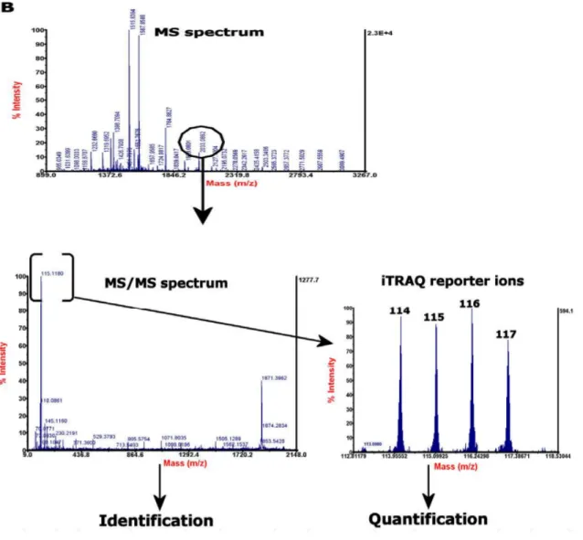
The iTRAQTM tags are isobaric labels that can label most of the peptides and proteins in as ample as it reacts with primary amines of amino termini including the N-terminus and the ε-amino group of the lysine side-chain. Each label contains a unique reporter group, a peptide reactive group, and a neutral balance group, these together maintaining a total mass of145Da (Fig. 9). During peptide fragmentation in MS/MS, iTRAQTM reporter groups separate from isobaric tags and produce distinguishable ions with m/z 114, 115, 116 and 117. In this way, the relative intensities of the reporter ions give the relative abundances of each peptide in the samples. This MS/MS fragmentation of iTRAQTM -tagged peptides also produces strong y- and b-ion signals for more confident identification [119].
20
Figure 9 Structure of the iTRAQTM reagents. Adapted from [120].
1.8.3 iTRAQTM work-flow
Samples are first reduced, alkylated and then trypsin digested. The set of peptides obtained after digestion are then labeled with one of the four iTRAQTM tags. Then the labeled peptides are pooled, separated by liquid chromatography (LC), and fractions obtained at the end are analyzed through MS (Fig-10).
Figure 10 A general scheme and example data for a 4-plex iTRAQ experiment. Adapted from [120].
iTRAQTM based quantification can be performed on LTQ-Orbitrap and include Pulsed Q Dissociation (PQD) [121] and higher energy C-trap dissociation (HCD) [122]. Sensitive and accurate iTRAQTM quantification of a total proteome can be done now using HCD fragmentation method in combination with the CID fragmentation technique [123].
1.9 Phosphoproteomics
Protein phosphorylation, an essential post-translational modification, affects most cellular activities including signal transduction, gene expression, cell cycle progression and other biological functions [124]. Enzymes dedicated to protein phosphorylation are among the
22
largest class of PTM (post transaltional modification) enzymes. This superfamily of protein kinases has been termed the kinome, with over 500 members in the human kinome. Protein kinases catalyze the transfer of a phosphoryl group from a high-energy organic compound such as adenosine triphosphate (ATP) to the side chain of serine, threonine, tyrosine or histidine amino acid residues in mammalian proteins. Kinases have been predicted to encompass 1.7% of the human genes [125, 126]. It has furthermore been estimated that the relative abundances of phosphoserine, -threonine, and -tyrosine in the human proteome are 90%, 10%, and 0.05%, respectively [127]. By hydrolyzing the covalent phosphoester bond, thereby releasing orthophosphate, protein phosphatases catalyze the enzymatic removal of these phosphate groups from proteins, returning them to their non-phosphorylated state [128]. Phosphorylation affects protein structure by changing the charge and hydrophobicity of the amino acid side chain. Negatively or positively charged amino acids in close proximity to the phosphorylated residue will be repelled or attracted, respectively, and the phosphoryl moiety will furthermore introduce a very hydrophilic and polar region in the affected protein. Thereby, conformational changes can be induced, affecting the properties of the protein [129].
Phosphopeptide analysis is a challenge for mass spectrometry for several main reasons [130]:
1. High activity of phosphatase requires sensitive careful sample preparation to avoid losing the modified proteins;
2. The phosphorylation is a labile PTM, which can be lost during the MS/MS fragmentation process;
3. Incomplete peptide fragmentation complicates the correct localization of the phosphorylation site;
4. The low stoichiometry of phosphopeptides implies the use of enrichment strategies, which have their limitations (see next section);
5. Some of multi-phosphorylated peptides display less ion intensities compared to the less phosphorylated peptides by conventional RPLC-MS/MS analysis [131, 132];
6. The phosphoproteome is dynamic and complex. Currently, it is predicted that more than 100,000 phosphorylated sites are present in mammalian proteins, distributed in a wide dynamic range [133]. However, large-scale studies currently reach no more than 20,000 sites using phosphopeptide enrichment and LC-MS. Different sample fractionation and phosphopeptides enrichment strategies can address the problem of low stoichiometry of the phosphopeptides.
1.9.1 PTM-Specific Enrichment
There are several specific protein and peptide enrichment methods including PTM-directed affinity chromatography or immune precipitation with PTM specific antibodies. Phosphoproteins can be purified by PTM-specific affinity resins [134] or anti-phospho amino acid antibodies [135]. Subsequent digestion of protein and LC-MS/MS analysis of peptides gives phosphorylation sites in the recovered proteins. However, it is more useful to enrich PTM peptides before MS analysis that can improve sensitivity and specificity. Immobilized metal affinity chromatography (IMAC) [136], or TiO2 columns [137] are used to recover enriched PTM peptides before MS/MS analysis. Strong cation exchange and anion exchange chromatography methods are also helpful for reducing peptide complexity including phosphopeptides. Combinations of these methods have revealed thousands of phosphorylated sites in proteins from different species. IMAC showed stronger sensitivity for multiply phosphorylated peptides, so, buffer exchange by reversed phase chromatography before IMAC has been used with a high risk of losing phosphorylated peptides [138, 139].
1.9.1.1 Titanium dioxide affinity chromatography-TiO2:
TiO2 is a resin that can create an ionic interaction with the phosphate groups of biomolecules and therefore, it is mixed (or used in microcolumns) together with the sample to trap peptides that are phosphorylated. The subsequent washing procedure can eliminate all the non-binding peptides and elution step can create a mixture of peptides containing only the fraction of interest. The approach is comparable to others but it is cheaper, faster, and reproducible and it is also MS compatible due to the buffers used for the elution. Regarding the application of TiO2 for phosphopeptide enrichment, first the ratio between the amount of titanium and the concentration of peptides needs to be
24
optimized in order to achieve the higher efficiency of enrichment [140]. In this enrichment strategy, the pH value of the loading buffer needs to be acidic to allow the acidic residues in the peptides become neutral while, the phospho groups remain negatively charged. Several studies have been done to achieve the highest selectivity of enrichment by comparing different loading buffers, all of which enable the acidic conditions [141].
1.9.1.2 Sequential elution from IMAC (SIMAC)
SIMAC (Fig. 12) is a phosphopeptide-enriching method combining both MOAC (Metal Oxide Affinity Chromatography) and IMAC [142]. It separates both multiply and singly phosphorylated peptides and first acidic conditions are used to elute monophosphorylated peptides from IMAC material, whereas subsequent basic elution recovers the multiply phosphorylated peptides that are normally not readily detected. Singly phosphorylated peptides as well as flow through peptides then pass on TiO2-MOAC. Finally the two distinct phosphopeptide pools can now be analyzed separately using tandem mass spectrometry parameters that are optimized for each type of sample [143]. Second TiO2enrichment help to separate non-phosphorylated peptides in the first eluate and having phosphorylated peptides in the flow-through [143].
SIMAC results into a larger amount of phosphopeptides identification than MOAC itself and broadened the phosphopeptide spectrum [142]. The sequential elution is another advantage, since having a greater amount of less complex phosphopeptide fractions increases the chances for ionization and identification of phosphopeptides by MS [144]. Overall SIMAC schematic workflow is given below in fig.12.
Figure 11 Overall SIMAC workflow Adapted from [142].
26 1.10 Proteomics in Neutrophils
There is need to identify protein expression of cells as not all the genes are translated into proteins which is also true for neutrophils, where correlation between mRNA and protein expression is inconsistent too. There are limited proteomic studies of the neutrophil particularly explaining effect of the inflammatory response on the neutrophil proteome [77].
The First global analysis of rat neutrophil proteins analysis done by two-dimensional gel electrophoresis (2DE) and MS approaches identified 52 proteins [145]. Later 250 proteins were identified through a combination of 1DE followed by ESI-MS/MS from bovine neutrophils. Proteins identified belong to cell metabolism, cell motility, immune response, protein synthesis, cell signaling, and membrane trafficking [146].
Proteomic analysis of gelatinase, specific, and azurophil granules by 2DE and MALDI-TOF identified 87 proteins including one membrane-spanning protein. Although the detection ability of 2DE was limited yet it identified differential expression of actin associated with all granules [147]. Cytoskeletal, structural, and membrane fusion proteins (247 Proteins) were identified from human neutrophil azurophil granules lipid rafts by 10% SDSPAGE and LC-MS/MS [148]. A similar study identified total of 23 proteins from plasma membrane lipid rafts using gradient gel electrophoresis and MALDI-MS or MALDI-MS/MS. Nine of the proteins belonging to the cytoskeleton were common to a previous study of human neutrophil azurophil granules lipid rafts [149]. Fessler et al. [150] identified 1200 proteins from neutrophils after exposure to lipopolysaccharide (LPS) for 4 h, which resulted in to 50% increase in expression of 100 proteins, and 50% decrease in expression of another 100 proteins. 2DE followed by MALDI-TOF-MS identified substrates for MMP2 and MMP9 from the BAL fluid of mice. These substrates include Ym1, S100A8, and S100A9, which showed chemotactic activity [151]. Proteomic analysis of rat intestinal mucosa after ischemic preconditioning in IRI model identified 10 proteins using 2DE in combination with MALDI-TOF-MS and these proteins were involved in anti-oxidation, apoptosis inhibition and energy metabolism. This study also revealed up-regulation of aldehyde dehydrogenase and aldose reductase in IPC group [152]. A similar study used 2-DE combined with MALDI-MS to analyze
the proteome of the intestinal mucosa subjected to I/R injury in the absence or presence of IPC pretreatment in rats. Total 16 proteins were differentially expressed belonged to cellular energy metabolism, anti-oxidation and anti-apoptosis of which aldose reductase which removes ROS, was significantly down regulated in IR and up regulated in IPC [153].
Our group has been working on proteomics approach for the identification and characterization of a set of proteins in neutrophils. In order to obtain a broader view of the protein profile in human neutrophils activated by PAF, fMLP and PMA, 2DE and MS of human neutrophils was performed with 22 protein identifications [154]. Comparison of neutrophils proteome from normal individuals and multiple trauma and revealed 114 spots, with 27 up regulated and 87 down regulated in trauma conditions [155].
1.10.1 Phosphorylation of neutrophil Proteins
Eight substrates of MAPKAPK2 (a downstream kinase in the p38 MAPK pathway) were identified by combination of in vitro phosphorylation of 32P-orthophosphate-loaded neutrophil lysates, one-dimensional SDS-PAGE, and peptide identification by MADLI-TOF/MS. One of these substrates, 14-3-3ζ was found to be phosphorylated at Ser-58, and this phosphorylation regulated 14-3-3ζ dimerization and binding to Raf-1 [156]. Similar analysis identified the p16-Arc subunit of the Arp2/3 complex as a MAPKAPK2 substrate. The functional consequences of its phosphorylation are not yet known, but may explain the participation of the p38 MAPK pathway in actin-dependent cellular functions [157]. Another similar approach to identify the calcium-binding protein myeloid-related protein-14 (MRP-14) as a target for p38 MAPK phosphorylation in neutrophils stimulated with fMLP, suggesting a role of MRP-14 in stimulated exocytosis [158]. The RhoGTPase regulator, LyGDI, was identified by 2DE, immunoblot and MS/MS and was found to be tyrosine phosphorylated during fibronectin-accelerated TNF-α-mediated neutrophil apoptosis. Phosphorylation was followed by increased caspase-3-mediated LyGDI cleavage, and this cleavage was a signaling event in TNF-α-mediated apoptosis [74]. Pacquelet et al. [159] used LC-MS/MS analysis of neutrophil lysates separated by SDS-PAGE to find out that interleukin-1 receptor-associated kinase-4 (IRAK-4) is downstream of TLR-4 and serine and threonine residues were phosphorylated on p47phox