https://doi.org/10.1590/0004-282X20180012
ARTICLE
OxLDL plasma levels in patients with
Alzheimer’s disease
Níveis plasmáticos de LDL-ox em pacientes com doença de Alzheimer
Marina Felipe Grossi1, Maria das Graças Carvalho1, Josianne Nicácio Silveira1, Gisele Santos Gonçalves1,2, Karina Braga Gomes1, Maria Aparecida Bicalho3,4, Ieda de Fátima Oliveira Silva1
Several studies have correlated dyslipidemia and Alzheimer’s disease (AD), mainly by considering the increase of both total cholesterol (TC) and low density lipoprotein choles-terol (LDL-C) and reduced high density lipoprotein cholescholes-terol (HDL-C)1,2. In addition, oxidative stress in the central nervous
system can cause oxidation of LDL-C (oxLDL) and very low-density lipoprotein (oxVLDL), which determines cytotoxicity3.
Studies have suggested that oxidative stress may be involved in the pathogenesis of AD4,5. According to Pirillo et al.6, oxLDL
plays an important role in the initiation and progression of ath-erosclerotic plaques contributing to endothelial cell activation and dysfunction, foam cell formation, and migration and
pro-liferation of smooth muscle cells. Therefore, oxLDL induces endothelial dysfunction and shows pro-inflammatory and
1Universidade Federal de Minas Gerais, Faculdade de Farmácia, Departamento de Análises Clínicas e Toxicológicas, Belo Horizonte MG, Brasil;
2 Centro Universitário Newton Paiva, Belo Horizonte MG, Brasil;
3Universidade Federal de Minas Gerais, Hospital das Clínicas, Instituto Jenny de Andrade Faria, Ambulatório de Idosos, Belo Horizonte MG, Brasil;
4Universidade Federal de Minas Gerais, Faculdade de Medicina, Departamento de Clínica Médica, Belo Horizonte MG, Brasil.
Correspondence: Iêda de Fátima Oliveira Silva; Faculdade de Farmácia da UFMG; Avenida Antônio Carlos, 6627; 31270-901 Belo Horizonte MG, Brasil; E-mail: [email protected]
Conflict of interest: There is no conflict of interest to declare.
Support: FAPEMIG (Fundação de Amparo à Pesquisa de Minas Gerais) and CNPq (Conselho Nacional do Desenvolvimento da Pesquisa).
Received 13 July 2017; Received in final form 12 December 2017; Accepted 17 December 2017. ABSTRACT
Objective: The objective of this study was to characterize the conventional lipid profile, oxLDL levels and ApoE polymorphism in patients with Alzheimer’s disease (AD) and in elderly individuals without cognitive impairment. Methods: Eighty elderly individuals were selected and the levels of oxLDL were determined using the ELISA kit, and ApoE gene polymorphism was investigated using polymerase chain reaction-restriction fragment length polymorphism. Results: Significantly reduced levels of oxLDL were observed in patients with AD compared to the control group. A higher frequency of the ApoE ε4 allele was observed in patients with AD compared to controls. No difference was observed for total cholesterol, HDL-C, and LDL-C levels between the two groups, while triglyceride levels were higher in controls compared with patients with AD. Conclusion: The data analyzed together did not reveal significant differences in lipid profiles, including oxLDL levels. However, the importance of lipid changes in the genesis of the disease cannot be ruled out. Nevertheless, the ApoE
ε4 allele was significantly more frequent in patients with Alzheimer’s dementia in agreement with previous findings in the literature, but this genetic component did not change the levels of oxLDL.
Keywords: Alzheimer’s disease; Cholesterol, LDL.
RESUMO
Objetivo: O objetivo deste estudo foi caracterizar o perfil lipídico convencional, os níveis de LDL-ox e o polimorfismo da ApoE em pacientes com doença de Alzheimer (DA) e em indivíduos idosos sem comprometimento cognitivo. Métodos: Foram selecionados oitenta indivíduos idosos. Os níveis de LDL-ox foram determinados usando o kit ELISA e a investigação do polimorfismo do gene da ApoE por PCR-RFLP. Resultados: Níveis significativamente reduzidos de LDL-ox foram observados em pacientes com DA comparado ao grupo controle. Uma maior frequência do alelo ε4 da ApoE foi observada nos pacientes com DA em relação aos controles. Nenhuma diferença foi observada para os níveis de colesterol total, HDL-C e LDL-C entre os dois grupos, enquanto níveis de triglicérides foram mais altos em controles comparados aos pacientes com DA. Conclusão: Os dados analisados em conjunto não revelaram diferenças significativas no perfil lipídico, incluindo os níveis de LDL-ox. No entanto, não se pode excluir a importância de alterações lipídicas na gênese da doença. Não obstante, o alelo ε4 da ApoE foi signicativamente mais frequente nos pacientes com demência de Alzheimer em concordância com achados prévios da literatura, mas esse componente genético não interferiu nos níveis de LDL-ox.
pro-atherogenic effects7. Apolipoprotein E (ApoE), a plasma
protein that transports cholesterol, is involved in synapse repair, especially in response to tissue injury, and plays an important role in the maintenance of neuronal structure and cholinergic function8. Furthermore, the identification of the
variant ε4 of the gene ApoE as the most common genetic risk factor for late onset AD9 suggests that cholesterol may have a
direct role in the pathogenesis of the disease.
Based on the important role of dyslipidemia in athero-sclerosis and its possible association with AD10, a
comple-mentary study of the lipid profile addressing oxLDL levels,
which is not part of the universe of conventionally-evaluated
variables, was justified.
As AD is a multifactorial degenerative disease, and con-sidering the evidence of the importance of dyslipidemias in neurodegenerative processes, the present study aimed to characterize a small set of patients with Alzheimer’s
demen-tia with respect to conventional lipid profile variables, plasma
levels of oxLDL and ApoE polymorphism genotyping, com-pared with a group of elderly people without the disease.
METHODS
Population study
An observational case-control study was conducted.
The groups comprised 80 elderly outpatients recruited between
2010 and 2011 at the Jenny de Andrade Faria Institute of Elderly, of the Clinical Hospital, aged between 60 and 90 years including: a) patients who showed no cognitive or functional impairment (control group; n = 40) who attended the same institution and
b) patients with a diagnosis of AD (AD group; n = 40). The clin -ical characteristics of the patients studied were obtained from
medical records. The characteristics of the groups according to gender, age and smoking showed that there was no difference between the control and AD groups (Table 1). Also no difference
was found between the groups in relation to co-morbidities such as hypertension, diabetes mellitus and hypoglycemia (Table 2).
The use of statins and antidepressants showed significant differences between the groups. The use of a statin was more
frequent in the control group than in AD group, while this lat-ter group, in turn, used more antidepressants than the con-trol group (Table 3).
All AD patients were on cholinesterase inhibitors agents such as rivastigmine, galantamine and donepezil. Other drugs, accord-ing to their need, such as statins, antihypertensives, antidepres-sants, and hypoglycemic agents, among others, were also used.
Selection criteria
Individuals who participated in the study were selected
by convenience. They met the criteria for clinical diagnosis
performed by a multidisciplinary team including a geriatri-cian and neuropsychologists.
Participants aged younger than 60 or older than 90 years were excluded, as well as those with a severely altered health status that could compromise cognition, neuropsychological assess-ments, and complementary examinations. Participants with clinical and neuroimaging evidence of a vascular component and delirium were also excluded, as well individuals with major mobility, visual, or auditory disabilities, because such conditions would not enable us to fully apply the tests. Patients with demen-tia were also excluded if they had demendemen-tia secondary to other causes, non-Alzheimer dementia and moderate or advanced AD.
The diagnosis of sporadic “probable” AD was estab -lished according to the National Institute of Neurological and Communicative Disorders and Stroke and by the Alzheimer’s
Table 1. Description and distribution of individuals involved in the study.
Variable C AD p-value
Gender (%) n = 40 n = 40
Female 31 (50.8%) 30 (49.2%)
0.7931
Male 09 (47.4%) 10 (52.6%)
Age (years)
Median (Q3–Q1) 76.50 (7.0) 78.00 (7.0) 0.7652
Smoking (%) n = 37 n = 31
0.2511
Yes 17 (63%) 10 (37%)
No 20 (48.8%) 21 (51.2%)
n: sample number; C: control group; AD: Alzheimer’s dementia group; 1- Asymptotic Power of Pearson’s Chi-square test; 2- Mann-Whitney test.
Table 2.Relationship comorbidities observed in the studied participants.
Diseases C AD p-value
Arterial hypertension (n = 40) (n = 40)
Yes 29 (45.3%) 35 (54.7%)
0.094
No 11 (68.8%) 05 (31.2%)
Diabetes mellitus
Yes 08 (44.4%) 10 (55.6%)
0.592
No 32 (51.6%) 30 (48.4%)
Hypoglycemia
Yes 08 (42.1%) 11 (57.9%)
0.431
No 32 (52.5%) 29 (47.5%)
n: sample number; C: control group; AD: Alzheimer’s dementia group; Asymptotic Power of Pearson’s Chi-square test.
Table 3. Use of statins and antidepressants by the study participants.
Drugs C AD p-value
Statin (n = 40) (n = 40)
Yes 18 (66.7%) 09 (33.3%)
0.033*
No 22 (41.5%) 31 (58.5%)
Antidepressant
Yes 07 (21.9%) 25 (78.1%)
< 0.0001*
No 33 (68.8%) 15 (31.3%)
Disease and Related Disorders Association11 criteria, based on
geriatric and neuropsychological assessments. The control group
consisted of patients with other clinical problems who attended
the same reference center. The elderly control group showed no
neurological and neuropsychiatric diseases and no cognitive or functional decline. All participants (AD and control groups) were submitted to the same study protocol and evaluated by a geriatri-cian and neuropsychologist with experience in applying cognitive tests. Only those participants with agreement between the clinical
and neuropsychological diagnosis were included in the study. The study coordinator verified the agreement between the profession
-als involved in the research. The following tests were applied for the
assessment of cognition, mood and functionality12-17: Mini-mental
State Examination, Geriatric Depression Scale (15 item version),
Pfeffer Functional Activities Questionnaire, Neuropsychiatric Inventory Questionnaire, Clinical Dementia Rating scale, Mattis
Dementia Rating Scale, digit span test, Corsi cubes, Token test, Rey Auditory Verbal Learning Test, frontal assessment battery, and the London Tower test. Due to the low educational level of the population in the study, the neuropsychological assessment battery included cognitive tests suitable for this aspect of the sam-ple. All the neuropsychological/functional tools were adapted to
and/or validated for Brazilian Portuguese. The cut-off points were
considered according to their education level. After clinical evalu-ation, participants were submitted to laboratory tests to identify clinical conditions, and to exclude other causes for cognitive or
functional impairment. The AD group was submitted to struc -tural neuroimaging (computed tomography and nuclear mag-netic resonance) to exclude another structural etiology and to
contribute to the confirmation of the AD diagnosis.
Laboratory evaluation
Blood samples of all participants were collected in the morning after 12-14 hours of fasting, to obtain serum and plasma. Samples were processed within two hours
post-col-lection. The EDTA-plasma samples were frozen at -80°C until
the time of testing.
The level of oxLDL was determined using the OX-LDL-C
Kit/MDA adduct ELISA kit from Immundiagnostik, Germany. Determining absorption of the samples was performed with an ELISA reader at 450 nm, using a Versamax Microplate Reader-Molecular Device®. The TC, HDL-C and triglycerides
(TG) were measured in serum using BIOCLIN® reagents. ApoE
gene polymorphism was investigated using polymerase chain reaction-restriction fragment length polymorphism with primers and the methodology described by Tsukamoto et al.18
Statistical analysis
Data were analyzed using the Statistical Software Package for Social Sciences version 13.0, with p < 0.05 considered
sig-nificant for all analyses. Data were analyzed for distribu
-tion by the Shapiro-Wilks test. The results were presented as
mean and standard deviation when there was normal distri-bution and as median and interquartile range, in the case of
non-normal distribution. The following tests for comparison
between the groups were used:
a) qualitative variables, such as statin and antidepres-sant use, and the presence or absence of the ε4 allele, were analyzed by the Asymptotic Power of Pearson’s Chi-square test; b) continuous variables were ana-lyzed using the Student’s t-test when the data showed normal distribution and the Mann-Whitney test for non-normal distribution.
Ethical considerations
The study was approved by the Research Ethics
Committee of the Federal University of Minas Gerais (ETIC 0118.0.203.000-10) and the Department of Research and Extension in Education of the Hospital das Clínicas of the Federal University of Minas Gerais. Informed consent was obtained from all participants or their caregivers before being included in the study sample.
RESULTS
Eighty individuals were included between 2010 and 2011; 40 in the AD group and 40 in the control group.
Lipid plasma levels, oxLDL profile and polymorphism of the ApoE gene
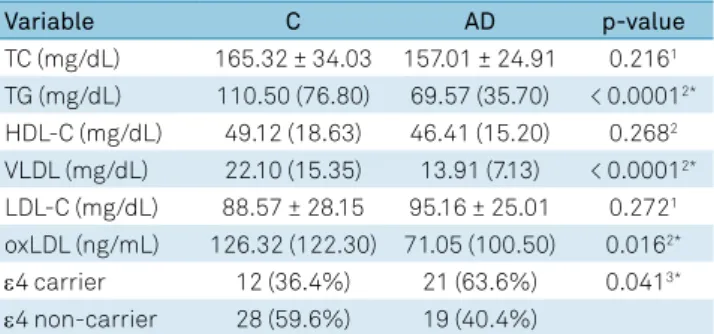
Plasma levels of TG, TC and fractions, as well as oxLDL are shown in Table 4. It was observed that TG, VLDL and
oxLDL had significantly higher values in the control group
when compared to the AD group, while the TC, HDL-C and
LDL-C levels were not different between the groups. When
comparing oxLDL levels in the control and AD groups
sep-arately according to comorbidities, no significant difference
was observed between the groups (data not shown). A higher
Table 4. Lipid plasma levels, oxLDL profiles and ApoE ε4 carriers in control and AD groups. (n = 40).
Variable C AD p-value
TC (mg/dL) 165.32 ± 34.03 157.01 ± 24.91 0.2161 TG (mg/dL) 110.50 (76.80) 69.57 (35.70) < 0.00012* HDL-C (mg/dL) 49.12 (18.63) 46.41 (15.20) 0.2682 VLDL (mg/dL) 22.10 (15.35) 13.91 (7.13) < 0.00012* LDL-C (mg/dL) 88.57 ± 28.15 95.16 ± 25.01 0.2721 oxLDL (ng/mL) 126.32 (122.30) 71.05 (100.50) 0.0162*
ε4 carrier 12 (36.4%) 21 (63.6%) 0.0413*
ε4 non-carrier 28 (59.6%) 19 (40.4%)
frequency of the ApoE ε4 allele carrier was observed in the AD group when compared to the control group (Table 4).
DISCUSSION
In our study the patients were sequentially selected according to their order of admission in the Reference Center for the Elderly, with a clear predominance of women, which is explained by the feminization process of aging. Regarding gender, age, comorbidities and some of the drugs used by
participants of the present study, there was no difference
between the control and AD groups, which reduces bias interpretation of the main results of this report, for the
con-ventional and oxLDL lipid profiles (Table 1, 2 and 4).
The main objective of the present study was to evaluate the lipid profile and oxLDL levels in AD patients compared to
controls. It was observed that values were within the
recom-mended cut-off points in both groups. However, the AD group
showed lower levels of TG and VLDL compared to the con-trol group. On other hand, the use of polypharmacy may have provoked the analytical and biological interference of some of these drugs on the levels of some of the biomarkers evaluated. In this context, it is noteworthy that the use of certain drugs by patients diagnosed with AD may interfere with the level of certain biomarkers, and the oxLDL may be an example of the interference of drugs on these levels. According Sinem et al.19,
short-term therapy (7.5±1.5 months) with acetylcholinester-ase inhibitors in AD patients resulted in a reduction of oxLDL
compared to the baseline. These authors also reported that
the use of antipsychotics combined with acetylcholinesterase inhibitor drugs may lead to reduced oxLDL levels in patients with AD, compared with the group taking only cholinester-ase inhibitors19. On other hand, oxLDL levels may interfere
with the efficacy of acetylcholinesterase inhibitors, since they
can increase the acetylcholinesterase activity, resulting in the increased production of reactive oxygen species20. According
to Yamchuen et al.20, mildly- and fully-oxidized LDL were
cyto-toxic in dose- and time-dependent patterns in SH-SY5Y neu-roblastoma cell culture, which reduces central cholinergic transmission. According to their report, oxLDL (10-200ng/ mL) is capable of increasing the activity of
acetylcholinester-ase after four and 24 hours of treatment. This increacetylcholinester-ased activ -ity has been implicated in the progression of AD. It should be noted that acetylcholinesterase inhibitors have been widely used to improve cholinergic transmission at the brain level21,
thus enhancing cognitive status. Therefore, there are indica -tions that drugs such as acetylcholinesterase inhibitors may interfere with oxLDL levels. It is important to note that all AD patients were using this medication. However, further
stud-ies are necessary to confirm these previous findings and their
clinical importance as, in cases where plasma levels of oxLDL are high, conventional treatment with acetylcholinesterase
inhibitor agents may not achieve the desired effect.
With regard to therapy, treatment should be initiated through the use of a cholinesterase inhibitor when an individ-ual meets the diagnostic criteria for AD. On the other hand, due to the depressive symptoms in many of these elderly patients, antidepressants were prescribed. Some evidence has suggested that statins, drugs widely used in the treatment of cardiovascular disorders to lower cholesterol levels, present a therapeutic potential in AD22. Elderly controls who use statins
in a significantly different way to AD patients may be benefit -ing in some way from the use of this drug, especially if they began their use in middle age. Based on the literature2, it has
been suggested that the use of statins may be delaying and/
or softening the neurodegenerative process. Thus, the optimi -zation of therapy for AD might be obtained by incorporating pharmacogenomic and pharmacogenetic protocols23.
Atherosclerosis has also been associated not only with an increased incidence of AD, but also with vascular dementia24.
Therefore, risk factors for atherosclerosis, such as increased oxLDL,
may also predispose the patient to AD and vascular dementia. Usually, AD patients are under the guidance of their caregivers and also use lipid-lowering drugs for control of TC and
athero-genic fractions, which may explain, in part, the lack of difference
in TC, HDL-C and LDL-C levels between the groups in this study. Consistent with the main tone of this study, it is also impor-tant to emphasize that TG values were higher in the elderly without AD compared with the values obtained for those with AD. In this respect, it should be mentioned that the living
hab-its of AD patients are quite restrictive. The intake of high-cal
-orie foods and others raising the lipid profile components are
reduced, which has often contributed to weight loss in these patients. In addition, it should be also considered, that a ten-dency towards higher levels of LDL-C in the AD group
com-pared to the control group, although not significant, may be a reflection of the increased use of statins in the latter.
In the present study, a higher frequency of the ε4 allele of the ApoE gene in patients with AD was also observed, compared to the frequency in the control group, which rein-forces the importance of genetic factors in the development
of AD. The mechanisms linking AD to the ε4 allele are not
yet fully understood. The data in the literature suggest a
correlation between infarcts of small brain vessels and pro-tease activity degrading the β-amyloid peptide in carriers of the ε4 allele25. Koffie et al.26, in a more detailed analysis,
revealed that patients with AD carrying the ApoE ε4 allele
have a significantly higher β-amyloid oligomeric load leading to exacerbated synapse losses compared to ApoE ε3 patients. However, it should be noted that the levels of oxLDL were
not different between those with and without the ε4 allele (p = 0.363). In this respect, this genetic factor does not seem to interfere with the levels of oxLDL according to our data.
An important limitation of this study is the lack of
infor-mation on the levels of the different parameters of the lipid profile when the patients investigated in the present
comorbidities were diagnosed in the AD patients at the
time of blood collection. The lack of data related to drugs
used by patients in middle-age may also be an important limitation of the present investigation. Another limitation of this study is the lack of a group of patients with AD who were not using acetylcholinesterase inhibitor drugs, which prevented a comparative analysis of the levels of oxLDL
between patients who did, or did not, use the drug. Thus,
our data suggests that the evaluation of oxLDL does not add any additional information to the prognosis and/or monitoring of AD. Moreover, the statin use interfered in the
lipid profile interpretation in both groups.
Thus, further studies are needed to elucidate the associa -tion between oxLDL and AD and, especially, the interac-tion
between the lipid profile and drugs prescribed in conven -tional treatment protocols.
In conclusion, the data analyzed did not reveal significant differences in the lipid profile, including the levels of oxLDL.
However, the importance of lipid changes in the genesis of the disease cannot be excluded. Nevertheless, the ApoE ε4 allele
was found significantly more frequently in patients with AD, which is in agreement with previous findings in the literature,
but this genetic component did not change the levels of oxLDL.
ACKNOWLEDGMENTS
The authors thank the medical team and neuropsychol -ogists of the Elderly Clinic of Instituto Jenny de Andrade Faria de Atenção à Saúde do Idoso, Hospital das Clínicas da Universidade Federal (UFMG), Brazil, as well as to the patients and elderly who generously contributed to this study.
References
1. Launer LJ, White LR, Petrovitch H, Ross GW, Curb JD. Cholesterol and neuropathologic markers of AD: a population-based autopsy study. Neurology. 2001 Oct;57(8):1447-52. https://doi.org/10.1212/WNL.57.8.1447
2. Wanamaker BL, Swiger KJ, Blumenthal RS, Martin SS. Cholesterol, statins, and dementia: what the cardiologist should know. Clin Cardiol. 2015 Apr;38(4):243-50. https://doi.org/10.1002/clc.22361
3. Escargueil-Blanc I, Salvayre R, Nègre-Salvayre A. Necrosis and apoptosis induced by oxidized low density lipoproteins occur through two calcium-dependent pathways in lymphoblastoid cells. FASEB J. 1994 Oct;8(13):1075-80.
4. Aldred S, Bennett S, Mecocci P. Increased low-density lipoprotein oxidation, but not total plasma protein oxidation, in Alzheimer’s disease. Clin Biochem. 2010 Feb;43(3):267-71. https://doi.org/10.1016/j.clinbiochem.2009.08.021
5. Huang WJ, Zhang X, Chen WW. Role of oxidative stress in Alzheimer’s disease. Biomed Rep. 2016 May;4(5):519-22. https://doi.org/10.3892/br.2016.630
6. Pirillo A, Norata GD, Catapano AL. LOX-1, OxLDL, and atherosclerosis. Mediators Inflamm. 2013;2013:ID152786. https://doi.org/10.1155/2013/152786
7. Tsai NW, Lee LH, Huang CR, Chang WN, Chang YT, Su YJ et al. Statin therapy reduces oxidized low density lipoprotein level, a risk factor for stroke outcome. Crit Care. 2014 Jan;18:R16. https://doi.org/10.1186/cc13695
8. Munoz DG, Feldman H. Causes of Alzheimer’s disease. CMAJ. 2000 Jan;162(1):65-72.
9. Laws SM, Hone E, Gandy S, Martins RN. Expanding the association between the APOE gene and the risk of Alzheimer’s disease: possible roles for APOE promoter polymorphisms and alterations in APOE transcription. J Neurochem. 2003 Mar;84(6):1215-36. https://doi.org/10.1046/j.1471-4159.2003.01615.x
10. Sato N, Morishita R. Roles of vascular and metabolic components in cognitive dysfunction of Alzheimer disease: short- and long-term modification by non-genetic risk factors. Front Aging Neurosci. 2013 Nov;5:64. https://doi.org/10.3389/fnagi.2013.00064
11. Blacker D, Albert MS, Bassett SS, Go RC, Harrell LE, Folstein MF; The National Institute of Mental Health Genetics Initiative. Reliability and validity of NINCDS-ADRDA criteria for Alzheimer’s disease. Arch Neurol. 1994 Dec;51(12):1198-204. https://doi.org/10.1001/archneur.1994.00540240042014
12. Folstein MF, Folstein SE, McHugh PR. “Mini-mental state”: a practical method for grading the cognitive state of patients for the clinician. J Psychiatr Res. 1975 Nov;12(3):189-98. https://doi.org/10.1016/0022-3956(75)90026-6
13. Almeida OP, Almeida SA. [Reliability of the Brazilian version of the Geriatric Depression Scale (GDS) short form]. Arq Neuropsiquiatr. 1999 Jun;57 2B:421-6. Portuguese. https://doi.org/10.1590/S0004-282X1999000300013
14. Pfeffer RI, Kurosaki TT, Harrah CH Jr, Chance JM, Filos S. Measurement of functional activities in older adults in the community. J Gerontol. 1982 May;37(3):323-9. https://doi.org/10.1093/geronj/37.3.323
15. Camozzato AL, Godinho C, Kochhann R, Massochini G, Chaves ML. Validity of the Brazilian version of the Neuropsychiatric Inventory Questionnaire (NPI-Q). Arq Neuropsiquiatr. 2015 Jan;73(1):41-5. https://doi.org/10.1590/0004-282X20140177
16. Montaño MB, Ramos LR. Validity of the Portuguese version of clinical dementia rating. Rev Saude Publica. 2005 Dec;39(6):912-7. https://doi.org/10.1590/S0034-89102005000600007
17. Mattis S. Dementia rating scale: professional manual. Florida: Psychological Assessment Resources; 1988.
18. Tsukamoto K, Watanabe T, Matsushima T, Kinoshita M, Kato H, Hashimoto Y et al. Determination by PCR-RFLP of apo E genotype in a Japanese population. J Lab Clin Med. 1993 Apr;121(4):598-602.
19. Sinem F, Dildar K, Gökhan E, Melda B, Orhan Y, Filiz M. The serum protein and lipid oxidation marker levels in Alzheimer’s disease and effects of cholinesterase inhibitors and antipsychotic drugs therapy. Curr Alzheimer Res. 2010 Aug;7(5):463-9. https://doi.org/10.2174/156720510791383822
20. Yamchuen P, Aimjongjun S, Limpeanchob N. Oxidized low density lipoprotein increases acetylcholinesterase activity correlating with reactive oxygen species production. Neurochem Int. 2014 Dec;78:1-6. https://doi.org/10.1016/j.neuint.2014.07.007
21. Tayeb HO, Yang HD, Price BH, Tarazi FI. Pharmacotherapies for Alzheimer’s disease: beyond cholinesterase
inhibitors. Pharmacol Ther. 2012 Apr;134(1):8-25. https://doi.org/10.1016/j.pharmthera.2011.12.002
23. Cacabelos R. Pharmacogenomics in Alzheimer’s disease. Methods Mol Biol. 2008;448:213-357. https://doi.org/10.1007/978-1-59745-205-2_10
24. Hofman A, Ott A, Breteler MM, Bots ML, Slooter AJ, Harskamp F et al. Atherosclerosis, apolipoprotein E, and prevalence of dementia and Alzheimer’s disease in the Rotterdam Study. Lancet. 1997 Jan;349(9046):151-4. https://doi.org/10.1016/S0140-6736(96)09328-2
25. Zhu H, Bhadelia RA, Liu Z, Vu L, Li H, Scott T et al. The association between small vessel infarcts and the activities of amyloid-β peptide degrading proteases in apolipoprotein E4 allele carriers. Angiology. 2013 Nov;64(8):614-20. https://doi.org/10.1177/0003319712462125