www.scielo.br/aabc
Haematophagous arthropod saliva and host defense system:
a tale of tear and blood
BRUNO B. ANDRADE1,2, CLARISSA R. TEIXEIRA1,2, ALDINA BARRAL1,2,3 and MANOEL BARRAL-NETTO1,2,3
1Centro de Pesquisas Gonçalo Moniz (FIOCRUZ-BA) Rua Waldemar Falcão, 121, 40295-001 Salvador, BA, Brasil 2Faculdade de Medicina, Universidade Federal da Bahia/UFBA Av. Reitor Miguel Calmon s/n, Vale do Canela, 40110-100 Salvador, BA, Brasil
3Instituto de Investigação em Imunologia (iii) – Instituto do Milênio
Manuscript received on June 20, 2005; accepted for publication on June 23, 2005;
contributed byALDINABARRAL*ANDMANOELBARRAL-NETTO*
ABSTRACT
The saliva from blood-feeding arthropod vectors is enriched with molecules that display diverse functions that mediate a successful blood meal. They function not only as weapons against host’s haemostatic, inflammatory and immune responses but also as important tools to pathogen establishment. Parasites, virus and bacteria taking advantage of vectors’ armament have adapted to facilitate their entry in the host. Today, many salivary molecules have been identified and characterized as new targets to the development of future vaccines. Here we focus on current information on vector’s saliva and the molecules responsible to modify host’s hemostasis and immune response, also regarding their role in disease transmission.
Key words:saliva, bites, hemostasis, host, vector, infection.
INTRODUCTION
Blood-feeding arthropods can require vertebrate host blood for nutrition, egg development, and sur-vival. The medical and public health importance of these ectoparasites is evident because of the alarm-ing emergence of new vector-borne infectious agents and the resurgence of previously known ones. The morbidity and mortality of infectious diseases trans-mitted by blood-feeding arthropods were more ex-pressive than all other causes in the last centuries (Gubler 1998).
*Member, Academia Brasileira de Ciências Correspondence to: Manoel Barral-Netto, MD E-mail: [email protected]
Haematophagous vectors of disease are not regarded simply as tools for the delivery of their pathogens. Advances in biomedical research fo-cused on the role of blood-feeding arthropods saliva in the transmission of some infectious diseases have shown the presence of a co-evolutionary relation-ship between these vectors and the pathogen they transmit. Rather, vector’s saliva seems to be a po-tent pharmacologically active fluid that directly af-fects the haemostatic, inflammatory and immune re-sponses of vertebrate host (Ribeiro 1995a).
hem-orrhagic pools upon which it feeds. Such insects as triatomine bugs feed directly from inside venules and arterioles, after been guided by an initial hem-orrhagic pool (Lavoipierre 1965, Ribeiro 1987b). During this process insect’s saliva is injected into the host’s skin at the site of the bite. This saliva contains a great variety of haemostatic, inflamma-tory and immunomodulainflamma-tory molecules such as pro-teins, prostaglandins, nucleotides, and nucleosides that locally modify the physiology of the host, mak-ing an adequate microenvironment for parasitism. Pathogens transmitted by these vectors interact with both saliva components and host mediators taking advantage of the altered host physiology to become established (Belkaid et al. 2000, Jones et al. 1992, Titus and Ribeiro 1988).
Understanding mammalian response to in-sect’s saliva is of utmost importance in several ways. Besides being related to allergy (Reunala et al. 1994, Shan et al. 1995) insect’s saliva is known to facili-tate parasite survival (Belkaid et al. 1998, Kamhawi 2000, Samuelson et al. 1991). Arthropod saliva is also related with specific antibody production by humans and other vertebrates against its compo-nents (Brummer-Korvenkontio et al. 1994, Feingold and Benjamini 1961, Wikel 1996). Conversely, host immunity to vector saliva may decrease infectivity of the transmitted pathogens (Belkaid et al. 1998, Bell et al. 1979). These responses can be used as epidemiological markers of vectors exposure (Bar-ral et al. 2000, Schwartz et al. 1990, 1991) and also support the possibility to prevent and treat allergic responses and to develop anti-arthropod vaccines.
Accordingly, the purpose of this review is to expose the salivary molecules that have been identi-fied and characterized in various blood-feeding arthropods and its activities related to host’s de-fense, including hemostasis and immune response. Indeed, we also focus on the role of saliva in parasite transmission and recent data suggesting that salivary peptides are an alternative target for the control of pathogen transmission through the development of effective vaccines.
ARTHROPOD SALIVA AND HOST HEMOSTASIS: THE BLOOD QUEST
Attempting to probe and feed, blood-sucking arthro-pods must circumvent the host haemostatic system. Host hemostasis is highly sophisticated and effi-cient process that includes several redundant path-ways geared towards overcoming blood loss; among which are blood-coagulation cascade, vasocons-triction, and platelet aggregation (Ribeiro 1987b, 1995a). These components act together leading to the arrest of blood flow at the site of vessel lesion. To overcome these obstacles, blood-feeding arthro-pods have evolved within its salivary secretions an array of potent pharmacological components, such as anticoagulants, anti-platelet and vasodilators (Champagne 1994, Ribeiro 1995a, Stark and James 1996b). As a rule, blood-suckers’ saliva contains at least one anticlotting, one antiplatelet, and one vasodilatory substance (Ribeiro and Francischetti 2003). In many cases, more than one molecule ex-ists in each category and in some, a molecule alone is responsible for more than one anti-haemostatic effect. For example, compounds such as adenosine and nitric oxide that are once antiplatelet and va-sodilatory are found in saliva. Salivary molecules responsible for these effects on host hemostasis have been characterized and some proteins were isolated, indicating the possibility to neutralize these mecha-nisms.
PLATELETAGGREGATION
The first host’s mechanism to avoid blood loss during tissue injury seems to be platelet aggrega-tion. Platelets can be activated by diverse stimulus including collagen exposure, thrombin interaction, thromboxane A2and ADP. After activated, platelets
es-says (Valenzuela et al. 1999). The salivary gland homogenate of the tickRhodnius prolixus(Fig. 1d) presents a 19kDa protein named Rhodnius proli-xus aggregation inhibitor 1 (RPAI-1) that inhibits collagen-induced platelet aggregation by binding to ADP (Francischetti et al. 2000), the same effect ob-served by a molecule with similar sequence and structure (pallidipin) isolated from saliva of Tria-toma pallidipennis (Fig. 1d) (Noeske-Jungblut et al. 1994). The deerflyChrysopsspp. saliva (Fig. 1a, b and d) can prevent platelet aggregation induced by ADP, thrombin and collagen, and also inhibits fibrinogen, binding to the glycoprotein IIb/IIIa re-ceptor on platelets (Grevelink et al. 1993). ADP has a key function in hemostasis through induction of platelet aggregation and derives from activated platelets and injured cells (Vargaftig et al. 1981). Thus, it is not surprisingly that the most common molecule involved in inhibition of platelet aggre-gation encountered on the majority of blood feed-ing arthropods seems to be the salivary apyrase en-zyme, that hydrolyses ATP and ADP to AMP and orthophosphate, preventing the effect of ADP on hemostasis. However, at least two different fami-lies of this enzyme exist and both known famifami-lies require Ca2+
and/or Mg2+
for their action. Aedes aegypti(Champagne et al. 1995b), Anopheles (Arca et al. 1999) and Culex mosquitoes (Fig. 1e) (Nasci-mento et al. 2000) present in their saliva apyrases from the same family of 5’-nucleotidases. A novel apyrase enzyme sequence was found recently in the salivary glands of the haematophagous bed bug
Cimex lectularius(Valenzuela et al. 1998) and ho-mologous sequences were found in the sand flies
Lutzomia longipalpis(Charlab et al. 1999) and Phle-botomus papatasi (Valenzuela et al. 2001), indi-cating that this family of enzymes is widespread among arthropod species (Fig. 1e). This novel apyrase functions exclusively with Ca2+
. It is im-portant to show that, in the sand flies salivary com-ponents analyzed, a salivary 5’- nucleotidase was also found in L. longipalpisbut not inP. papatasi
(Fig. 1e) (Charlab et al. 1999). Finally, the salivary
apyrase fromTriatoma infestans(Fig. 1e) also be-longs to the 5’-nucleotidase family (Faudry et al. 2004) and are peculiarly dependent of Mn2+
and Co2+
(Ribeiro et al. 1998).
Platelet function can also be antagonized by substances that increase platelet cyclic adenosine monophosphate (cAMP) or cyclic guanosine mono-phosphate (cGMP). Previous work had demon-strated that prostaglandin E2(PGE2)and
prostacy-clin obtained from tick’s saliva can increase platelet cyclic nucleotides (Higgs et al. 1976). Nitric Oxide (NO) released within saliva of the bugs Rhodnius prolixus andCimex lectularius (Fig. 1g) activates the cytosolic guanylate cyclase enzyme, causing an anti-clotting effect (Ribeiro et al. 1993, Vogt 1974).
BLOOD-COAGULATIONCASCADE
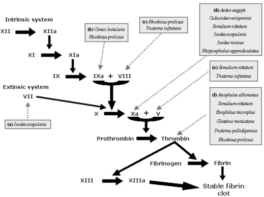
Fig. 1 –Vector’s saliva acting on platelet activation and aggregation: (1)Blood feeding vectors induce vessel laceration and tissue injury resulting in collagen exposure when probing for a blood meal. (2)Thus, platelets aggregate, promoting clotting, and release of vasoconstrictor mediators promoting hemostasis. Blood feeders can inhibit platelet aggregation by preventing fibrinogen, thrombin (Anopheles albimanusand
Chrysops spp.) or cAMP/cGMP stimulation (Boophilus microplus).(3)Platelet activation and degranulation also occur after thromboxane A2that results in vasoconstrictor response and(4)the NO present within bug’s
saliva can prevent haemostatic effect (Cimex lectulariusandRhodnius prolixus).(5)They can also bind to ADP (Rhodnius prolixus, Triatoma pallidipennisandChrysops spp.) or(6)Prevent the action of ADP through salivary apyrase to prevent platelet aggregation (Aedes aegypti, Anopheles gambiae, Culex quinquefasciatus, Lutzomyia longipalpis,Phlebotomus papatasi, Triatoma infestansandCimex lectularius).
family of serine protease inhibitors (Stark and James 1998). Salivary gland extract of Culicoides vari-ipennis(the primary North America vector of blue-tongue viruses) (Fig. 2d) contains a factor Xa in-hibitor similarly to all the subfamily of culicine mosquitoes (Perez de Leon et al. 1997). It has been proposed that despite variation in the degree of inhi-bition, all anophelines have thrombin directed anti-coagulants and culicine mosquitoes have factor Xa directed anticoagulants. Differences in the site of action of the anticoagulants must likely reflect the
long period of independent adaptation of the two subfamilies to the challenges presented by ver-tebrate hemostasis (Stark and James 1996a).
A potent and specific low molecular mass (3,530 Da) anticoagulant peptide purified from salivary gland of Glossina morsiatans morsiatans
(Fig. 2f) is a thrombin inhibitor (Cappello et al. 1996, 1998). This peptide is a stoichiometric in-hibitor of thrombin and also a potent inin-hibitor of thrombin-induced platelet aggregation.
biochem-ical approaches was used to discover activities in the salivary glands of the haematophagous sand fly
Lutzomyia longipalpis (Charlab et al. 1999). Se-quences of nine full-length complementary DNA (cDNA) clones were obtained and five were pos-sibly associated with blood meal acquisition, each having cDNA similarity to: (a) the bed bugCimex lectulariusapyrase, (b) a 5’-nucleotidase/phospho-diesterase, (c) a hyaluronidase, (d) a protein contain-ing a carbohydrate-recognition domain (CRD), and (e) a unique RGD-containing peptide. This work was the first to identify a hyaluronidase activity in a haematophagous insect salivary gland. The CRD-protein and the RGD containing peptide seem to be involved in anticlotting activities.
Triatomine bugs also evolved potent anti-coagulants, as factors V and VIII inhibitors from
Triatoma infestans (Fig. 2c and e) (Pereira et al. 1996) and triabin, a salivary 142-reside protein of Triatoma pallidipennis(Fig. 2f) that selectively interacts with thrombin, exclusively via its fibrino-gen recognition exosite (Fuentes-Prior et al. 1997). Prolixin S (nitrophorin 2), from salivary gland ex-tracts ofRhodnius prolixus(Fig. 2c) inhibits coag-ulation factor VIII-mediator activation of factor X and accounts for all the anti-clotting activity ob-served in its saliva (Ribeiro et al. 1995). Saliva of the hard tick and Lyme disease vector,Ixodes scapularis
(Fig. 2d), was genetically sequenced in a cDNA li-brary. In this process, a clone with sequence homol-ogy to tissue factor pathway inhibitor was identified and this cDNA codes for a mature protein, herein called ixolaris, with 140 amino acids. Observations of ixolaris function evidenced the blockage of fac-tor Xa generation by endothelial cells expressing tissue factor. This work also demonstrated that ixo-laris uses factor X and Factor VIIa as scaffolds for the inhibition of factor VIIa/Tissue factor complex (Fig. 2a) (Francischetti et al. 2002).
VASOCONSTRICTION
Arachdonic acid is released by activated platelets when blood vessels are lacerated by arthropods’
mouthparts and is converted by other platelet en-zymes into thromboxane A2, a powerful
platelet-aggregating, platelet-dagranulating, and vasocons-tricting substance (Ribeiro 1987b). Activated pla-telets also release serotonin, which together with thromboxane A2is responsible for the early
vaso-constrictor response in local inflammation caused by tissue injury (Weigelt et al. 1979). Saliva from blood feeder insects presents vasodilatory sub-stances or molecules that antagonize vasoconstric-tors produced on the site of tissue injury caused by inoculation of mouthparts during probing. These molecules act directly or indirectly on smooth-muscle cells activating intracellular enzymatic path-ways that lead to cAMP or cGMP formation. Sia-lokinin, a tachykinin decapeptide from Aedes ae-gypti, is a vasodilator through activating nitric oxide production by endothelial cells via cGMP induction (Champagne and Ribeiro 1994).
Fig. 2 –Blood-coagulation cascade (intrinsic and extrinsic system) activated in response to tissue injury is also blocked by salivary molecules.The blood-coagulation cascade is activated after blood vessels injury resulting in the production of active thrombin, which cleaves fibrinogen to fibrin that polymerizes forming a stable clot blocking blood loss. Salivary anticoagulants from blood feeding arthropods inhibit specific targets of the coagulation cascade. They target components such as factor IXa (Cimex lectulariusand Rhod-nius prolixus); VIII (Rhodnius prolixusandTriatoma infestans); Xa (Aedes aegypti, Culicoides variipennis, Simulium vittatum, Ixodes scapularis, Ixodes ricinusandRhipicephalus appendiculatus); V (Simulium vitta-tumandTriatoma infestans), VII (Ixodes scapularis) and thrombin (Anopheles albimanus, Simulium vittatum, Boophilus microplus, Glossina morsiatans, Triatoma pallidipennisandRhodnius prolixus) resulting in inhi-bition or delayed blood-thrombin (Anopheles albimanus,Simulium vittatum, Boophilus microplus, Glossina morsiatans, Triatoma pallidipennisandRhodnius prolixus) and coagulation response.
than the last separation of the continental plates, stresses the diversity of compounds found in the salivary glands of blood-feeder arthropods (Ribeiro et al. 1999). Finally, the black fly Simulium vitta-tum salivary gland has a 15 kDa vasodilator that acts on ATP-dependent K-channels and has no struc-tural similarity to other known proteins (Cupp et al. 1994, 1998).
Another example of salivary vasodilator is prostaglandin E2 (PGE2) and prostaglandin F2
(PGF2) demonstrated from salivary gland
homo-genate of different tick species (Dickinson et al.
1976, Ribeiro et al. 1985). PGE2 and prostacyclin
dilate host’s blood vessels, thus antagonizing the vasoconstrictor component of hemostasis throm-boxane A2. The triatomine bugRhodnius prolixus
Cimexnitrophorin is a member of the inositol phos-phatase family (Valenzuela et al. 1995). Because
Cimex lectulariusandRhodnius prolixusbelong to different hemipteran families (Cimucidae and Re-duvidae, respectively) and evolved independently to blood-feeding, Cimex lectulariusandRhodnius prolixusnitrophorins may represent a case of con-vergent evolution (Valenzuela et al. 1995). In the case ofRhodnius prolixus, four NO-carrying pro-teins were isolated and named N1-N4 nitrophorins (Champagne et al. 1995a). Interestingly, the main nitrophorin from this triatomine has a very high affinity to histamine, a common autacoid found by blood-feeding insects on the skin of allergic hosts. Histamine binds to nitrophorin and further displaces NO at the site of injury. Thus, this nitrophorin also works as an anti-histaminic substance (Ribeiro 1995a).
Anopheline mosquitoes do not produce vaso-dilatory substances, but rather secrete a peroxidase enzyme that has significant NADPH oxidase activ-ity. The NADPH oxidation produces H2O2, which
is used by the enzyme to destroy serotonin and cat-echolamines, thus inactivating host’s physiologic vasoconstrictor substances that may interfere with insect feeding (Ribeiro 1995a, Ribeiro and Valen-zuela 1999).
Indeed, haematophagy evolved independently in several orders of insects and ticks. For this reason, a variety of salivary anti-haemostatic compounds are found in these diverse groups of arthropods. The combined effects of apyrases, prostaglandins, an-tithrombotics, anti-clotting and many classes of va-sodilators effectively counteract host hemostasis and increase the chance of blood-suckers survivor.
SALIVA AND HOST IMMUNE SYSTEM: BREAKING DOWN THE ENEMY
IMMUNOMODULATORYPROPERTIES OFBLOOD
-FEEDERARTHROPODSSALIVACOMPONENTS
After repeated exposure to salivary antigens, host immune system may elaborate cellular
(delayed-type hypersensitivity, DTH) and/or humoral reac-tions that will alter the local site of probing that may result on rejection of the ectoparasite (Wikel 1982). This host’s resistance is related to a Th1 immune response, with significant production of in-terferon (IFN)-γ, interleukin (IL)-2 and IL-12. To face this problem, blood-feeding arthropods have evolved salivary immunomodulatory factors which prevent host from becoming sensitized to the vaso-modulatory substances of saliva that facilitate blood meal (Gillespie et al. 2000) or even retard delete-rious host responses. Such factors induce a Th2 deviation of host’s immune response, which favors insect survivor. Many types of immunomodulatory molecules have been isolated from different blood-feeding arthropod species. Most of these mediators act directly or indirectly on immune effectors cells, like macrophages, T cells, B cells, Natural Killer (NK) cells and granulocytes.
INNATE IMMUNE RESPONSE
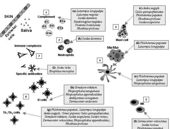
Innate immune system consists of all the immune defenses that lack immunologic memory. Innate responses frequently involve complement, acute-phase proteins besides granulocytes, mast cells, dendritic cells, macrophages, and NK cells. Com-plement components, prostaglandins, leukotrienes and other inflammatory inductors all contribute to the recruitment of inflammatory cells to the site of ectoparasite exposure. Thus, these cells and inflam-matory mediators represent the first line of immune defense against blood-feeding arthropods likely af-fecting its feeding process.
The early events of complement activation are based on an enzymatic amplifying cascade compa-rable to that seen in blood clotting. The complement fragments C3a, C4a and C5a activate mast cells, which release histamine, cytokines and other pro-inflammatory substances (Delves and Roitt 2000). C5a also acts as a powerful neutrophil chemo-attractant. The complement components C5b, C6 C7, C8, and C9 form the membrane-attack com-plex (Delves and Roitt 2000), which perforates cell membranes and may lead to the death of the lining cells of insect’s mouthparts. The alternative path-way of complement seems to be involved in expres-sion of blood-feeding arthropod resistance (Wikel 1979). Thus, the anaphylatoxins C3a and C5a cause further release of vasoactive mediators, which in-crease vascular permeability and potentiate the ac-cumulation of antibodies and immune cells at the site of the bite. Despite these obstacles, blood-suckers are capable of having a successful blood meal likely through host immunomodulation by sali-vary components. Saliva of the tickIxodes dammini
(Fig. 3a) antagonizes anaphylatoxin and bradykinin likely by the presence of a carboxypeptidase (Ribei-ro and Spielman 1986) and can also inhibit C3a re-lease and C3b deposition (Ribeiro 1987a). Saliva of
Lutzomyia longipalpisis capable of inhibiting both the classical and alternative Complement pathways (Fig. 3a), whereas that ofLutzomyia migoneiacted
only on the former (Cavalcante et al. 2003). The triatomine bugsPanstrongylus megistus,Triatoma brasiliensis andRhodnius prolixus (Fig. 3a) were also able to inhibit the classical pathway whereas the mosquitoAedes aegytiand fleaCtenocephalides feliswere not (Cavalcante et al. 2003).
The molecules collectively referred to as acute-phase proteins enhance host resistance to infection and promote the repair of damaged tissue (Delves and Roitt 2000). Plasma levels of these proteins change rapidly in response to infection, inflamma-tion and tissue injury. In addiinflamma-tion to some com-plement components, the acute-phase proteins in-clude C- and S- reactive proteins, serum amyloid A protein, proteinase inhibitors and anticoagulant peptides. These substances or their function may be altered by arthropod salivary components for the success of blood meal (Cappello et al. 1996, Horn et al. 2000, Noeske-Jungblut et al. 1995, Paesen et al. 1999).
(Char-lab et al. 2000), but not in the anopheline Anophe-les gambiae (Ribeiro et al. 2001). The adenosine deaminase activity inAedes aegyptimay help blood feeding by removing adenosine, a molecule associ-ated with both pain perception inhibition and induc-tion of mast cell degranulainduc-tion in vertebrates, and by producing inosine, a molecule that potently inhibits the production of inflammatory cytokines (Ribeiro et al. 2001). Bradykinin and histamine are impor-tant mediators of itch (Alexander 1986) and pain (Clark 1979) which could stimulate host grooming and removal of the blood feeding arthropod. It is perhaps not surprising that the some insects’ salivary glands, likeIxodes scapularis(Ribeiro and Mather 1998) andRhodnius prolixus(Ribeiro and Walker 1994) contain kininases that inhibit bradykinin. In-deed, hard ticks also produce histamine-binding pro-teins that minimize local inflammation host’s re-sponse (Chinery and Ayitey-Smith 1977, Paesen et al. 1999). Finally, data suggest that saliva of Tria-toma infestanscan inhibit sodium channels activity in nerves by an unspecified molecule, with potential antinociceptive effects (Dan et al. 1999).
Arthropods’ saliva can induce immune sup-pression of innate immune cells. Ixodes dammini
(Fig. 3b) salivary gland homogenate inhibits rat neutrophils function (Ribeiro et al. 1990). Salivary gland extracts (SGE) from Dermacentor reticula-tus(Fig. 3f) adult ticks induce a decrease in human natural killer (NK) activity acting on the first step of NK cell activity, namely effector/target cell con-jugate formation (Kubes et al. 2002). NK cell cyto-toxicity as well as NO production by macrophages are inhibited byIxodes ricinusSGE (Kopecky and Kuthejlova 1998) and by Phlebotomus papatasi
(Fig. 3f) saliva (Ribeiro et al. 1999, Waitumbi and Warburg 1998). The saliva of this phlebotomine also contains a potent inhibitor of protein phosphatase 1 and protein phosphatase 2A of murine macrophages, suggesting that thePhlebotomus papatasisalivary phosphatase inhibitor may interfere with the abil-ity of activated macrophages to transmit signals to the nucleus, thereby preventing up regulation of the
induced nitric oxide synthase gene inhibiting the production of NO (Katz et al. 2000, Waitumbi and Warburg 1998). Adenosine and its precursor 5’-AMP, also isolated from Phlebotomus papatasi
(Fig. 3d) salivary glands (Katz et al. 2000, Ribeiro et al. 1999) have been reported to enhance IL-6, IL-10, IL-4 and PGE2production, and together with
inosine (product of adenosine deaminase) were shown to decrease the production of IL-12,
IFN-γ, TNF-α and NO (Hasko et al. 2000, Hasko et al. 1998, Hasko et al. 1996, Le Moine et al. 1996, Link et al. 2000). In the presence of salivary glands extracts ofLutzomyia longipalpis(Fig. 3d), macro-phages were unable to present antigen, were refrac-tory to activation by IFN-γand were unable to pro-duce H2O2 or NO (Hall and Titus 1995, Theodos
and Titus 1993, Titus and Ribeiro 1990). This in-hibition seems to be selective, as it did not alter the ability of IFN-γto up regulate MHC class II expres-sion on their surfaces. On human monocytes, sali-vary gland homogenate (SGH) ofLu. longipalpis
stimulators of cytokine production by antigen-specific T cells.
Further studies showed that maxadilan, through activation of PACAP type 1 receptor, inhibited the expression of TNF-αby macrophages and increased levels of the cytokines IL-6 and IL-10 as well as prostaglandin E2(Bozza et al. 1998, Lanzaro et al.
1999, Soares et al. 1998). Maxadilan, as well as whole salivary gland lysate suppressed type 1 re-sponses and enhanced type 2 rere-sponses by human PBMC and purified monocyte cultures in vitro
(Rogers and Titus 2003). Maxadilan decreased IFN-γ, IL-12 and TNF-αproduction, while increas-ing IL-6 secretion by human PBMC few hours af-ter stimulation withLeishmania majoror LPS. In-deed, it was suggested that this Lutzomyia longi-palpisvasodilator could interfere on the IFN-γ re-lease through the suppression of IL-12 production by T-lymphocytes (Fig. 3g), possibly as a result of changes induced in macrophages and NK cells. In-terestingly, it has been found that the primary amino acid sequence of maxadilan peptide is polymorphic (Lanzaro et al. 1999) and sibling species within the
Lutzomyia longipalpis complex present significant differences in their amounts of maxadilan mRNA (Yin et al. 2000). Despite these differences, the va-sodilatory activity appears not to be altered (Lanzaro et al. 1999). The maxadilan primary sequence poly-morphism may represent an evolutionary vantage to the sand fly, preventing the host from becoming sen-sitized to this important peptide and, consequently, the loss of blood meal. It has also been proposed that differences in salivary components of differ-ent geographical populations of sand flies may be responsible for the differences observed in clinical manifestation of visceral leishmaniasis in America (Warburg et al. 1994). So, different strategies of host immunomodulatory appear to have evolved for Old-World and New-World sand flies.
The observations above show us that blood-feeding arthropods evolved strategic mechanisms to evade or suppress the innate immune response and that saliva of ectoparasites may have a key role in
this process.
ACQUIRED IMMUNE RESPONSE
a hard tick, the development of a DTH response by an unnatural host pre-exposed to its salivary compo-nents in the site of the bite may lead to the rejection of the insect (Ribeiro 1995b), while other insects, like sand flies, take advantage to this process, feeding twice as fast at the site of inflammation, that presents a larger blood flow than normal skin (Belkaid et al. 2000). In the case of ticks, the rejection is rarely seen in natural association and it seems that bugs co-evolved with the host to overcome the immune response (Ribeiro 1995b).
Thus, blood-feeding ectoparasites developed strategies to suppress host acquired immune re-sponses. Ability to alter host defenses might be a factor in determining the range of hosts a particular species can parasite. In this way, a thorough under-standing of the molecules involved in induction of host immunossuppression can be extremely impor-tant in the identification of vaccine immunogens.
CELLULARIMMUNERESPONSE AND CYTOKINENETWORK
Cytokines act as cellular messengers, forming an integrated network that is highly involved in regula-tion of innate immunity and orchestrating, together with lymphocytes, all the components of acquired immune responses. In this section we explore these aspects of host’s immunoregulation by most impor-tant blood-feeding arthropod species that have been studied.
Ticks are significant vectors of infectious dis-eases to both humans and animals. Ticks feeding on the host seem to have a systemic immunosuppres-sive effect on the host’s immune system, including lymphocytes. Lymphocytes from tick-infested ex-perimental animals had greatly reduced responses to mitogensin vitro(Wikel 1982, Wikel et al. 1978, Wikel and Osburn 1982). This effect has subse-quently been demonstratedin vitrousing the saliva or salivary gland extracts of several different species of hard ticks (Ferreira and Silva 1998, Fuchsberger et al. 1995, Ramachandra and Wikel 1992, 1995, Ribeiro et al. 1985, Urioste et al. 1994). Tick
sali-vary PGE2was primarily thought to be responsible
for this lymphocytic suppressive effect (Inokuma et al. 1994, Ramachandra and Wikel 1992, Ribeiro et al. 1985). The down-regulation of T- or B-lympho-cytes and macrophages by PGE2 was previously
demonstrated onin vitrostudies (Bahl et al. 1991, Phipps et al. 1991, Spatafora et al. 1991) and it is very likely the prostaglandins would have some ef-fects on the immune system of the host. Ixodes scapularissaliva (Fig. 2g) can inhibit IL-2 through a soluble IL-2 binding proteic factor presented in its saliva. (Gillespie et al. 2001). IL-2 activates T cells and IL-2 receptors have been described on many cell types including B cells, macrophages and NK cells (Siegel et al. 1987, Smith 1992, Theze et al. 1996) highlighting the importance of this simple cel-lular inhibitory mechanism. Saliva of another tick,
sug-gesting a basis of genetic predisposition in C3H/HeJ mice strain toIxodes scapularisinfestation.
Mice stimulated with saliva from Rhipice-phalus sanguineus (Fig. 3g) induced transforming growth factor (TGF)-β production while IL-12 was reduced. Susceptible mice exposed to tick infes-tation modulated the immune response drastically reducing proliferation of lymph node cells after Con A stimulation and a production of Th2 cytokine represented by IL-4, IL-10 and TGF-β(Ferreira and Silva 1999). A similar response was observed in dogs (susceptible host) infested with this tick, they had a reduced proliferative reaction and a significant immediate but no DTH response to a cutaneous test induced by tick extract, whereas guinea pigs (resis-tant host) developed a strong DTH reaction (Ferreira et al. 2003).
Extracts prepared from the salivary glands of
Rhipicephalus appendiculatus ticks (Fig. 3g) re-duced the expression of IFN-α, TNF-α, IL-1α, IL-1β, IL-5, IL-6, IL-7 and IL-8 by LPS-stimulated human peripheral blood leukocytes (Fuchsberger et al. 1995). Thus, the saliva of these ticks may stim-ulate the deviation of host’s immune system to a Th2 pattern, favoring the blood-sucker’s survival. Work with saliva fromDermacentor andersoni(Fig. 3g) (Bergman et al. 1995, 1998) has shown that a protein of approximately 36 kDa is responsible for suppression of T cell proliferation by an unknown mechanism (Bergman et al. 1995). Tick salivary components can also alter the leukocyte traffic and the interactions between activated endothelial cells and adhesion molecules on the leukocyte surface. Splenic lymphocytes of mice infested with Derma-centor andersoni(Fig. 3g),as well as normal lym-phocytes exposed to its saliva, had reduced expres-sion of some of these adheexpres-sion molecules: leuko-cyte function-associated antigen-1 (LFA-1) and very late activation-4 (VLA-4) integrins (Macaluso and Wikel 2001). Therefore, Dermacentor andersoni
salivary compounds can facilitate blood meal through retarding cellular migration and modifying the population of host’s immune cells at the site of
tick attachment, also altering the activation pattern of these cells, creating an adequate microenviron-ment for parasitism.
Rhodnius prolixus is an important vector of
Trypanosoma cruzi, the causative agent of Chagas disease. Spontaneous and mitogen-induced mouse lymphocyte proliferation were suppressed by Rhod-nius prolixus(Fig. 3g) blood feeding (Kalvachova et al. 1999).
Besides tick bugs, black flies are capable of modulating their hosts’ immune defense. Mice in-oculated with a salivary gland extract (SGE) of the black flySimulium vittatum(Fig. 3g and h) have re-duced expression of major histocompatibility com-plex (MHC) class-II antigens on their splenocytes and even showed anin vitro(but notin vivo) inhi-bition of B- and T-lymphocyte mitogenesis (Cross et al. 1993a). It is possible that such changes inter-fere subtly with antigen-presentation as mice repeat-edly exposed to Smulium vittatum SGE exhibited differential responses to ovalbumin (OVA) immu-nizations compared to control animals. Splenocytes from SGE-treated mice produced lower levels of IL-5 and IL-10 but not of IFN-γ, IL-2 and IL-4, upon OVA challenge than cells from mice treated with saline (Cross et al. 1994b).
Sand flies are the most extensively studied blood feeding insects in regard to modulation of host immune defenses (Charlab et al. 1999, Gil-lespie et al. 2000, Wikel 1999a). The adenosine deaminase contained in salivary extracts from Lut-zomyia longipalpis (Fig. 3g) can suppress T cell apoptosis besides inhibition of IL-12, IFN-γ,
(Qureshi et al. 1996). The observed modulation of macrophage and T-lymphocyte functions could have arisen to prevent development of immune re-sponses to the salivary gland proteins in the host, which are introduced into the site of the bite and are essential for successful blood feeding. Despite the absence of maxadilan peptide withinPhlebotomus papatasisalivary glands (Fig. 3g), the saliva of this phlebotomine can also interfere on T-lymphocyte function through the inhibition of Th1 protective cytokines (IFN-γ and IL-12) production while en-hancing the exacerbative cytokine IL-4 (Belkaid et al. 1998, Mbow et al. 1998).
Aedes aegypti SGE (Fig. 3g) added to cul-tures of Con A- or OVA-stimulated naive murine splenocytes caused significant suppression of IL-2 and IFN-γproduction, but not of IL-4 and IL-5. No such effect was observed in activated splenocytes derived from ovalbumin-primed mice (Cross et al. 1994a). Aedes aegypti and Cimex pipiens saliva, as well as sialokinin I purified fromAedes aegypti
salivary glands (Fig. 3g), are able to down regu-late IFN-γ release and up-regulate IL-4 and IL-10 production up to 7 days after feeding (Zeidner et al. 1999). Recent data suggest thatAedes aegyptisaliva can modify antigen-stimulated responses of trans-genic OVA-TCR DO11 mouse splenocytesin vitro
in a dose-dependent manner. An inhibition greater than 50% of T-cell proliferation was noted and the production of Th1 cytokines IL-2 and IFN-γ, and pro-inflammatory cytokines GM-CSF and TNF-α, and the Th2 cytokine IL-5, IL-4 and IL-10 were markedly reduced with a low-dose salivary stimula-tion (Wasserman et al. 2004). A protein of approx-imated 387kDa present inA. aegyptiSGE reduced T-cell viability, whereas in dendritic cell it did not affect cell numbers but reduced its IL-12 production. Such profound effects observed withAedes aegypti
SGE are not observed with SGE fromCulex quin-quefasciatus (Wanasen et al. 2004), pointing out the different immunomodulatory activities used by these two culicine mosquitoes to take a successfully blood meal.
B CELLS ANDANTIBODYPRODUCTION
va-riegatum(Wang and Nuttall 1995b). Together these data indicated that IGBPs act as a self-defense sys-tem against ingested immunoglobulins.
Boophilus microplusticks saliva can modulate the isotype of host antibody responses. High tick infestation decreases serum levels of IgG1 and IgG2 antibodies in susceptible (Holstein) breeds, but not in resistant (Nelore) ones. Conversely, lev-els of IgE antibodies increase after infestations in susceptible breeds, but are not related to protective anti-tick host response (Kashino et al. 2005).
Finally, the diversity of components mediating vertebrate inflammatory and haemostatic responses has been countered in evolution by an equally di-verse array of antagonists in the saliva of blood-sucking arthropods.
PATHOGENDELIVERY: INTRUDERSTAKING
AFREERIDE
The modifications on vertebrate host physiology caused by salivary active pharmacological mol-ecules favors the delivery of microscopic parasites that colonize the digestive tract of the blood-feeding arthropod. This would apply to pathogens that are delivered via the mouthparts, either by salivation or regurgitation, and might also hold for those transmitted via rectum (e.g. Trypanosoma cruzi), since they may also invade the host through the bite wound (Titus and Ribeiro 1990). Indeed, the world’s most important infectious diseases, ranging from malaria, filariasis, trypanosomiasis, leishma-niasis and Lyme diseases are transmitted by blood-sucking arthropods such as mosquitoes, tsetse flies, sand flies and ticks.
Titus and Ribeiro (Titus and Ribeiro 1988) first demonstrated that saliva of the sand flyLutzomyia longipalpis enhanced Leishmania major infection when the parasite was co-inoculated with sand fly salivary gland lysate. In addition to enhancing lesion size, sand fly salivary gland lysate also markedly en-hanced the parasite burden within the lesions. Sim-ilar findings were reported with other Leishmania
species (Lima and Titus 1996, Samuelson et al.
1991, Theodos et al. 1991, Warburg et al. 1994). Maxadilan alone also exacerbated lesion size and parasite burden within the lesions to the same de-gree as sand fly salivary gland (Morris et al. 2001). Thus, maxadilan appear to be the principal pep-tide in the sand fly saliva that enhances infection withLeishmania major. PGE2, IL-4 and IL-6 also
favorLeishmaniaestablishment since the host im-munoregulation can decrease the number of para-sites been killed by activated immune cells. In leish-maniasis, resistance and protection are associated with the expression of IFN-γ and IL-12 driving a CD4+ Th1 response, while susceptibility is linked to production of IL-4 and the development of a CD4+ Th2 response (Alexander et al. 1999, Mc-Sorley et al. 1996). Lutzomyia longipalpissaliva seems to drive, by an unknown mechanism, the host immune response to a Th2 type, less effective in terms of parasite clearance. Macrophages with sub-optimal activation serve as reservoirs forLeishmania
(Alexander et al. 1999, Solbach and Laskay 2000, Zer et al. 2001), where it can replicate without host control.
Saliva from P. duboscqi attracts vertebrate monocytesin vitro (Anjili et al. 1995) and saliva fromP. papatasinot only attracts macrophages but also enhances infection by L. donovani in these cells, resulting in increased parasite loads (Zer et al. 2001). Interestingly,Lu. longipalpissaliva also induces CCL2/MCP-1 expression and macrophage recruitment to the inoculation site in the air pouch model of inflammation, possibly favoring Leishma-niainfection if these cells are not adequately acti-vated (Teixeira CR, unpublished data). Despite the absence of maxadilan in its saliva, salivary gland lysates ofPhlebotomus papatasi can also enhance infection with Leishmania, through induction of IL-4 production (Mbow et al. 1998). IL-4 exac-erbates infection withLeishmaniaand can reduce parasite destruction by macrophages, reducing NO release (Mbow et al. 1998). The presence of adeno-sine in the salivary glands ofPhlebotomus papatasi
responses and thus promoting the establishment of
Leishmania parasites by enhancing production of IL-10 and, together with inosine, decreasing pro-duction of IL-12, IFN-γ, TNF-αand NO (Hasko et al. 1996, 1998, Romano et al. 1983).
Mosquitoes are associated with the transmis-sion of malaria and many species of virus. Relation-ship between mosquitoes’ saliva and the pathogens they transmit is largely neglected. These parasites colonize salivary glands and are naturally transmit-ted when a vector salivates during feeding a verte-brate host. For example, the Cache-Valley virus, an arthropod-borne bunyavirus, recently emerged as a significant veterinary pathogen causing infertility and congenital malformations in North America ru-minants (Chung et al. 1990, Edwards et al. 2003, Edwards et al. 1989). Enhancement of infection by this virus on mice after feeding byAedes triseriatus,
Aedes aegypti or Culex pipiens, was observed but not elucidated (Edwards et al. 1998). Co-inocu-lation of sindbis virus with Aedes aegyptisalivary gland extract resulted on a reduced IFN-β expres-sion, when compared to injection of virus alone (Schneider et al. 2004). Aedes aegypti can also transmit dengue virus, a flavivirus that causes dengue fever, dengue hemorrhagic fever and dengue shock syndrome. Dendritic cells seem to be permis-sive for dengue virus and function as primary tar-gets of initial infection (Ader et al. 2004). Aedes aegyptisaliva inhibited infection by dengue virus in DC, and pre-sensitization of DCs with saliva prior to infection enhanced inhibition. In addition, the proportion of dead cells was also reduced in virus-infected DC cultures exposed to mosquito saliva, and an enhanced production of IL-12p70 and
TNF-αwas detected in these cultures (Ader et al. 2004). These data suggest a paradoxical protective role for
Aedes aegyptisaliva that limits viral uptake by DCs. However, more elucidative studies are needed for an overall understanding of the natural pathogenesis of dengue virus infection. Besides virus,Aedessaliva is also important in parasite transmission. Chick-ens subcutaneously infected withPlasmodium
gal-linaceumsporozoites in the presence ofAedes flu-viatillisSGH showed a higher level of parasitaemia when compared to those that received only sporo-zoites (Rocha et al. 2004). However, parasitaemia levels were lower among chickens immunized with SGH.
The influence of tick salivary components on parasite transmission has been studied intensively worldwide and shows us interesting data. In ad-dition to Lyme disease, ticks are vectors of other pathogens that are responsible for rickettsial dis-eases (Burgdorfer 1977), babesiosis (Piesman et al. 1986, Spielman et al. 1985), emerging infections such as ehrlichiosis (Magnarelli et al. 1995, Telford et al. 1996), and may also transmit tick-borne en-cephalitis viruses (Telford et al. 1997), all of which may be influenced by tick salivary immunomodula-tory factors. The etiological agent of Lyme disease,
differences in cytokine expression following expo-sure to tick salivary secretions and associated pathogens. The tickDermacentor reticularis(Fig. 3f) can increase arboviruses transmission by affect-ing host NK cells functions and manipulataffect-ing host cytokine network (Hajnicka et al. 2005), besides promoting virus growth (Hajnicka et al. 1998). It has been reported that tick saliva also enhances the transmission of Thogoto virus from infected to un-infectedRhipicephalus appendiculatusand Ambly-omma variegatumticks (Davies et al. 1990). The salivary effect observed was also seen even when the host did not exhibit detectable viraemia, and the virus was applied three days after saliva (Jones et al. 1987, 1990). Moreover, Rhipicephalus ap-pendiculatus salivary gland extracts enhanced the uptake ofTheileria parvasporozoites into lympho-cytes, macrophages and afferent lymph veiled cells (Shaw et al. 1993).
IMMUNERESPONSE TOBLOOD-FEEDING SALIVARYGLANDANTIGENS:THE COUNTERATTACK
All the effects of blood feeding arthropod saliva on host physiology observed here are originated from a unique molecule or a group of them. These molecules are also immunogenic and elicit host specific immune response. Thus, pre-exposure to insect saliva may render human and other verte-brate hosts resistant to a new blood meal or may even contribute to create an inhospitable environ-ment for the establishenviron-ment of the parasites transmit-ted by these insects. The observations regarding re-peated exposure to pathogen-free ectoparasites and the subsequent development of resistance to vector-borne infections are intriguing. This knowledge can contribute to the development of a control strategy targeting the factors in blood-feeder saliva that are essential for the host immunossupression and the transmition of infectious agents.
Rabbits expressing acquired resistance to in-festation withD. andersoni are less susceptible to infection with tick-transmittedFranciella tularesis
than tick susceptible controls. Subsequent stud-ies supported evidence that pre-exposure to tick’s bites may induce host resistance. Guinea pigs that are resistant and form a DTH response in the area of saliva from Rhipicephalus sanguineus inocula-tion are more resistant to future tick infestainocula-tions while dogs and mice that develop an immediate re-sponse with a disturbed pattern of cellular migra-tion are susceptible to infestamigra-tions (Ferreira et al. 2003). Mice infested four times with pathogen-free
Ixodes scapularisdeveloped acquired resistance to
Borrelia burgdorferi infection in subsequent chal-lenge with infected ticks (Wikel et al. 1997). A sim-ilar study with guinea pigs exposed previously to uninfectedIxodes scapularisshowed that repeated challenges lead to a development of host tick im-munity and protection againstBorrelia burgdorferi
(Nazario et al. 1998). The host’s specific antibody production against ticks was also used as epidemio-logical marker of previous vector exposure, such as toIxodes scapularis(Schwartz et al. 1990, 1991).
More recently, the protective host response was reported in sand flies (Belkaid et al. 1998). The exacerbative effect of saliva on infection, seen when mice were co-inoculated with L. major and sali-vary glands sonicate (SGS) ofP. papatasi, was com-pletely abrogated in mice pre-exposed to the salivary sonicate (Belkaid et al. 1998). This protection was reproduced following transmission ofL. majorby the bite of infectiveP. papatasisand flies. Compared with naïve mice, mice pre-exposed to the bites of un-infected flies showed reduction in lesion pathology, in parasite load, and also in their ability to transmit
DTH reaction, which is predominantly a mononu-clear cell infiltrate. Two hours after injection of im-mune sera preincubated with SGS in the ear dermis of unexposed mice, there was an inflammatory in-filtrate comprised of neutrophils and macrophages, suggesting a potential role of immune complexes in the observed cell infiltration (Silva et al. 2005).
BALB/c mice exposed to repeatedLu. longi-palpisbites developed antibodies to saliva. Signif-icant IgG and IgG1 anti-saliva antibody responses were elicited, which suggest a predominant Th2 re-sponse in these animals. Sera from immune mice recognized with a high frequency and a strong reac-tion the 45-kD and 44-kD proteins fromLu. longi-palpissaliva (Silva et al. 2005). These proteins were also the major targets of human antibody response in an endemic area (Barral et al. 2000). Since these proteins are widely recognized, they are natural can-didates to be used as markers of exposure to Lu. longipalpis bites. Mounting an antibody response against sand fly saliva occurred at the same time as the host developed an anti-leishmania cell-mediated immune response (Gomes et al. 2002). Although tempting, it remains to be demonstrated that protec-tion against Leishmaniainfections is conferred by pre-exposure to sand fly bites in endemic areas for leishmaniasis.
Anopheles stephensi mosquito bites induce dermal mast cell degranulation, leading to fluid ex-travasation and neutrophil influx (Demeure et al. 2005). This inflammatory response does not occur in mast cell-deficient W/Wv mice, unless these are reconstituted specifically with mast cells. Mast cell activation caused byA. stephensimosquito bites is followed by hyperplasia of the draining lymph node due to the accumulation of CD3+
, B220+
, CD11b+
, and CD11c+
leukocytes. The T cell enrichment of the draining lymph nodes results from their seques-tration from the circulation rather than local prolifer-ation (Demeure et al. 2005). This work emphasized the critical contribution of peripheral mast cells in inducing T cell and dendritic cell recruitment within draining lymph nodes, a prerequisite for the
elicita-tion of T and B lymphocyte priming. There was also a slight increase in mast cells present in the ear der-mis of mice two hours afterLutzomyia longipalpis
bites (Silva et al. 2005).
Mice immunized with salivary antigens from
Simulium vittatum developed IgG, IgM, and IgE antibodies which recognized several salivary gland components. Sera from bitten mice recognized fewer antigens than sera from animals intraperito-neally immunized with salivary gland extract, indi-cating that some components of the salivary gland extract were poorly immunogenic or absent from the saliva secreted during blood-feeding (Cross et al. 1993b).
These data suggest that human and others ver-tebrate hosts can develop immune responses that block the effects of saliva and that an appropriate vaccine should accelerate the development of these responses in the vaccinated host and thus protect against vector-borne infections. But the develop-ment of vector-blocking vaccines will not be a trivial task.
CLOSING REMARKS
The key for the success of blood-feeding arthropod parasitism is the ability of avoiding host immune re-sponses through the production of specific salivary antagonists. Analyzes of these substances reveal a significant biochemical and pharmacological di-versity. The isolation of specific molecules through experimental techniques has been made over the last 10 years and contribute to a better understanding of pathogen-vector-host interactions. Although many aspects have been described a few important issues remain to be understood to better explore salivary molecules.
An expanded effort for studying salivary content of species from different parts of the world will certainly increase the chances in finding common molecules that could function as markers or as can-didates for a wide-ranging vaccine. The identifi-cation of new species or subspecies may also re-veal novel molecules or strategies in avoiding host defense mechanism. This natural diversity of sub-stances can serve in therapeutics and biomedical re-search but a note of caution is necessary as salivary products have diverse behavior in distinct models of inflammation or immune response. Understanding such variation, as well as testing the same molecule in several models, is important for unraveling subtle differences in composition and molecular interac-tion with potential practical applicainterac-tions. There is also a need in expanding our understanding of host protective mechanisms. Some aspects remain un-der explored, few studies exist on the interaction of salivary products and innate immunity, e.g. The as-pects involved in future adaptive immune response resulting in resistance or susceptibility widely de-pends on the first attempt of host’s innate response to contain infection/infestation that may influence on the predominance of a pattern of future host’s immune adaptative response.
The ultimate purpose of research that examines pathogens transmitted by arthropods is to develop an effective vaccine. But it has proven very diffi-cult to develop efficiently host sterile immunity and long-lived vaccines against vector-borne pathogen and parasites. These organisms often present very complex life cycles, enabling the occurrence of new and more pathogenic strains also resistant to con-ventional treatment. Vaccines that target more than one facet of parasite’s life cycle, like the pathogen itself, vector salivary proteins and vector-pathogen interactions, may prove to be more effective, but more resources are needed to improve this know-ledge. New genetic sequencing technologies and high efficient proceedings of protein isolation and cloning permit the experimental production of some of these substances indispensable for biochemical,
pharmacological and immunological investigations and even for clinical studies.
High-throughput genomic and proteomic ap-proaches for cloning salivary cDNA have resulted in the discovery of genes and proteins not previ-ously reported in blood feeding arthropods. These reality allows not only the isolation of salivary fac-tors implicated in host hemostasis and inflamma-tion, but also the characterization of novel salivary molecules, for many of which the biological func-tion is unknown (Valenzuela 2001). Within this huge quantity of molecules, those responsible for the salivary modulatory effects on their hosts, which also permit the vector-borne pathogen establish-ment, may be targeted by an ideal vaccine. The challenge is to encounter a high-throughput expres-sion system to test the biological activities of each candidate molecule. Such perspectives are summa-rized in Table I.
Thus, for vaccination using vector salivary pro-teins, the isolation of salivary immunossuppressors to make specific neutralizing antibodies and the pur-suit of salivary proteins that elicit optimal cellular responses are strategies that if combined may re-sult in reducing disease burden, rewriting this tale of blood, albeit may not reduce the host tissue tear.
RESUMO
A saliva de artrópodes hematófagos é rica em
molécu-las com funções diversas que mediam uma alimentação
sangüínea bem sucedida. Estas moléculas agem não
ape-nas como armas contra a resposta hemostática,
inflama-tória e imunológica do hospedeiro funcionando também
como ferramentas para o estabelecimento de patógenos.
Parasitas, vírus e bactérias aproveitando-se deste
arse-nal dos vetores adaptaram-se facilitando seu
estabeleci-mento no hospedeiro. Hoje, várias moléculas salivares
foram identificadas e caracterizadas como novos alvos
para o desenvolvimento de vacinas futuras. Neste
tra-balho, centramos em informação recente sobre a saliva de
vetores e as moléculas responsáveis por modificar a
res-posta hemostática e imunológica assim como seu papel na
TABLE I
Future challenges regarding saliva from haematophagous vectors.
• Identification of new species or subspecies to reveal
a wider option of molecules that impair host’s defense mechanisms;
• Salivary molecules isolation trough new genetic sequencing technologies and high efficient proceedings facilitating the access to study candidate molecules;
• Identify salivary content of species worldwide targeting common molecules from sibling species for a wide-ranging vaccine;
• Understand protective mechanisms regarding the early steps of host’s response to salivary molecules that can lead to resistance or susceptibility;
• Test candidate salivary molecules in several models for
enlightening subtle differences and similarities within components important for pathogen establishment;
• Development of vaccines that target aspects of pathogens
and salivary molecules simultaneously.
Palavras-chave:saliva, picadas, hemostasia, hospedeiro,
vetor, infecção.
REFERENCES
ADER DB, CELLUZZI C, BISBING J, GILMORE L, GUNTHERV, PEACHMAN KK, RAOM, BARVIR D, SUN WANDPALMER DR. 2004. Modulation of dengue virus infection of dendritic cells byAedes aegyptisaliva. Viral Immunol 17: 252–265.
ALEXANDERJ, SATOSKAR AR AND RUSSELL DG. 1999. Leishmaniaspecies: models of intracellular parasitism. J Cell Sci 112: 2993–3002.
ALEXANDERJO. 1986. The physiology of itch. Para-sitol Today 2: 345–351.
ANJILICO, MBATIPA, MWANGI RW, GITHUREJI, OLOBOJO, ROBERTLLANDKOECHDK. 1995. The chemotactic effect of Phlebotomus duboscqi
(Diptera: Psychodidae) salivary gland lysates to murine monocytes. Acta Trop 60: 97–100. ARCAB, LOMBARDOF,DELARACAPURROM, DEL
-LA TORREA, DIMOPOULOSG, JAMESAA AND COLUZZIM. 1999. Trapping cDNAs encoding se-creted proteins from the salivary glands of the malaria vectorAnopheles gambiae. Proc Natl Acad Sci USA 96: 1516–1521.
BAHLAK, FOREMANJCANDDALEMM. 1991. The effect of prostaglandin E2 and non-steroidal anti-inflammatory drugs on cell-associated interleukin one. Adv Prostaglandin Thromboxane Leukot Res 21B: 513–515.
BARRALA, HONDAE, CALDASA, COSTAJ, VINHAS V, ROWTONED, VALENZUELAJG, CHARLABR, BARRAL-NETTOMANDRIBEIROJM. 2000. Hu-man immune response to sand fly salivary gland anti-gens: a useful epidemiological marker? Am J Trop Med Hyg 62: 740–745.
BELKAIDY, KAMHAWIS, MODIG, VALENZUELAJ, NOBEN-TRAUTHN, ROWTONE, RIBEIROJAND SACKSDL. 1998. Development of a natural model of cutaneous leishmaniasis: powerful effects of vec-tor saliva and saliva preexposure on the long-term outcome ofLeishmania majorinfection in the mouse ear dermis. J Exp Med 188: 1941–1953.
BELKAID Y, VALENZUELA JG, KAMHAWI S, ROW
-TONE, SACKS DLANDRIBEIROJM. 2000. De-layed-type hypersensitivity to Phlebotomus papa-tasisand fly bite: An adaptive response induced by the fly? Proc Natl Acad Sci USA 97: 6704–6709.
tick-sensitized rabbits: allergic klendusity. Am J Trop Med Hyg 28: 876–880.
BERGMAN DK, RAMACHANDRA RN AND WIKEL SK. 1995. Dermacentor andersoni: salivary gland proteins suppressing T-lymphocyte responses to concanavalin Ain vitro. Exp Parasitol 81: 262–271.
BERGMANDK, RAMACHANDRARNANDWIKELSK. 1998. Characterization of an immunosuppressant protein fromDermacentor andersoni(Acari: Ixo-didae) salivary glands. J Med Entomol 35: 505–509.
BISSONNETTEEY, ROSSIGNOLPAANDBEFUSAD. 1993. Extracts of mosquito salivary gland inhibit tumour necrosis factor alpha release from mast cells. Parasite Immunol 15: 27–33.
BOYCEJA. 2004. The biology of the mast cell. Allergy Asthma Proc 25: 27–30.
BOZZA M, SOARES MB, BOZZA PT, SATOSKAR AR, DIACOVO TG, BROMBACHER F, TITUS RG, SHOEMAKERCBANDDAVIDJR. 1998. The PACAP-type I receptor agonist maxadilan from sand fly saliva protects mice against lethal endotoxemia by a mechanism partially dependent on IL-10. Eur J Immunol 28: 3120–3127.
BROSSARD MAND WIKEL SK. 1997. Immunology of interactions between ticks and hosts. Med Vet Entomol 11: 270–276.
BRUMMER-KORVENKONTIOH, LAPPALAINENP, RE
-UNALA T AND PALOSUO T. 1994. Detection of mosquito saliva-specific IgE and IgG4 antibodies by immunoblotting. J Allergy Clin Immunol 93: 551–555.
BURGDORFER W. 1977. Tick-borne diseases in the United States: Rocky Mountain spotted fever and Colorado tick fever. A review. Acta Trop 34: 103– 126.
CAPPELLOM, BERGUMPW, VLASUKGP, FURMIDGE BA, PRITCHARDDIANDAKSOYS. 1996. Isola-tion and characterizaIsola-tion of the tsetse thrombin in-hibitor: a potent antithrombotic peptide from the saliva ofGlossina morsitans morsitans. Am J Trop Med Hyg 54: 475–480.
CAPPELLOM, LIS, CHEN X, LICB, HARRISONL, NARASHIMHAN S, BEARD CB AND AKSOY S. 1998. Tsetse thrombin inhibitor: bloodmeal-induced
expression of an anticoagulant in salivary glands and gut tissue ofGlossina morsitans morsitans. Proc Natl Acad Sci USA 95: 14290–14295.
CAVALCANTERR, PEREIRAMH ANDGONTIJONF. 2003. Anti-complement activity in the saliva of phlebotomine sand flies and other haematophagous insects. Parasitology 127: 87–93.
CAVASSANIKA, ALIBERTIJC, DIASAR, SILVAJS
ANDFERREIRABR. 2005. Tick saliva inhibits dif-ferentiation, maturation and function of murine bone-marrow-derived dendritic cells. Immunology 114: 235–245.
CHAMPAGNE DE. 1994. The role of salivary vaso-dilators in bloodfeeding and parasite transmission. Parasitol Today 10: 430–433.
CHAMPAGNEDEANDRIBEIROJM. 1994. Sialokinin I and II: vasodilatory tachykinins from the yellow fever mosquitoAedes aegypti. Proc Natl Acad Sci USA 91: 138–142.
CHAMPAGNE DE, NUSSENZVEIG RH ANDRIBEIRO JM. 1995a. Purification, partial characterization, and cloning of nitric oxide-carrying heme proteins (nitrophorins) from salivary glands of the blood-sucking insectRhodnius prolixus. J Biol Chem 270: 8691–8695.
CHAMPAGNE DE, SMARTT CT, RIBEIRO JM AND JAMES AA. 1995b. The salivary gland-specific apyrase of the mosquitoAedes aegyptiis a member of the 5’-nucleotidase family. Proc Natl Acad Sci USA 92: 694–698.
CHARLAB R, VALENZUELA JG, ROWTON ED AND RIBEIROJM. 1999. Toward an understanding of the biochemical and pharmacological complexity of the saliva of a hematophagous sand flyLutzomyia longipalpis. Proc Natl Acad Sci USA 96: 15155– 15160.
CHARLABR, ROWTONEDANDRIBEIROJM. 2000. The salivary adenosine deaminase from the sand fly
Lutzomyia longipalpis. Exp Parasitol 95: 45–53.
CHINERYWAANDAYITEY-SMITHE. 1977. Histamine blocking agent in the salivary gland homogenate of the tickRhipicephalus sanguineus sanguineus. Na-ture 265: 366–367.
CRANDELL RW, SHOPE RE, SHELTON MJ AND COLLISSONEW. 1990. Evidence that Cache Valley virus induces congenital malformations in sheep. Vet Microbiol 21: 297–307.
CLARKWG. 1979. Kinins and the peripheral and central nervous systems. Handbook of Experimental Phar-macology 25: 311–356.
COLLISMG. 1989. The vasodilator role of adenosine. Pharmacol Ther 41: 143–162.
COSTADJET AL. 2004.Lutzomyia longipalpissalivary gland homogenate impairs cytokine production and costimulatory molecule expression on human mono-cytes and dendritic cells. Infect Immun 72: 1298– 1305.
CROSS ML, CUPP MS, CUPP EW, GALLOWAY AL
ANDENRIQUEZFJ. 1993a. Modulation of murine immunological responses by salivary gland extract ofSimulium vittatum(Diptera: Simuliidae). J Med Entomol 30: 928–935.
CROSS ML, CUPP MS, CUPP EW, RAMBERG FB
ANDENRIQUEZFJ. 1993b. Antibody responses of BALB/c mice to salivary antigens of hematophagous black flies (Diptera: Simuliidae). J Med Entomol 30: 725–734.
CROSSML, CUPPEWANDENRIQUEZFJ. 1994a. Dif-ferential modulation of murine cellular immune re-sponses by salivary gland extract ofAedes aegypti. Am J Trop Med Hyg 51: 690–696.
CROSS ML, CUPP EW AND ENRIQUEZ FJ. 1994b. Modulation of murine cellular immune responses and cytokines by salivary gland extract of the black fly
Simulium vittatum. Trop Med Parasitol 45: 119–124.
CUPPMS, RIBEIRO JMAND CUPP EW. 1994. Va-sodilative activity in black fly salivary glands. Am J Trop Med Hyg 50: 241–246.
CUPPMS, RIBEIROJM, CHAMPAGNEDEANDCUPP EW. 1998. Analyses of cDNA and recombinant pro-tein for a potent vasoactive propro-tein in saliva of a blood-feeding black fly,Simulium vittatum. J Exp Biol 201: 1553–1561.
DANA, PEREIRAMH, PESQUERO JL, DIOTAIUTI L
AND BEIRAO PS. 1999. Action of the saliva of
Triatoma infestans (Heteroptera: Reduviidae) on sodium channels. J Med Entomol 36: 875–879.
DAVIESCR, JONESLDANDNUTTALLPA. 1990. A comparative study of the infection thresholds of Thogoto virus inRhipicephalus appendiculatusand
Amblyomma variegatum. Am J Trop Med Hyg 43: 99–103.
DELVESPJANDROITTIM. 2000. The immune system. First of two parts. N Engl J Med 343: 37–49.
DEMEURECEET AL. 2005. Anopheles mosquito bites activate cutaneous mast cells leading to a local in-flammatory response and lymph node hyperplasia. J Immunol 174: 3932–3940.
DICKINSON RG, O’HAGAN JE, SCHOTZ M, BIN
-NINGTONKC ANDHEGARTY MP. 1976. Prosta-glandin in the saliva of the cattle tickBoophilus mi-croplus. Aust J Exp Biol Med Sci 54: 475–486.
DIONISOTTI S, ZOCCHI C, VARANI K, BOREA PA
ANDONGINIE. 1992. Effects of adenosine deriva-tives on human and rabbit platelet aggregation. Cor-relation of adenosine receptor affinities and anti-aggregatory activity. Naunyn Schmiedebergs Arch Pharmacol 346: 673–676.
DUSBABEKF, BORSKYI, JELINEKF ANDUHLIR J. 1995. Immunosuppression and feeding success of
Ixodes ricinusnymphs on BALB/c mice. Med Vet Entomol 9: 133–140.
EDWARDS JF, LIVINGSTON CW, CHUNG SI AND COLLISSONEC. 1989. Ovine arthrogryposis and central nervous system malformations associated with in utero Cache Valley virus infection: sponta-neous disease. Vet Pathol 26: 33–39.
EDWARDSJF, HIGGSS ANDBEATYBJ. 1998. Mos-quito feeding-induced enhancement of Cache Valley Virus (Bunyaviridae) infection in mice. J Med Ento-mol 35: 261–265.
EDWARDSJF, ANGULOABANDPANNILLEC. 2003. Theriogenology question of the month. In utero in-fection of the doe by CVV. J Am Vet Med Assoc 222: 1361–1362.
FAUDRYE, LOZZISP, SANTANAJM, D’SOUZA-AULT M, KIEFFER S, FELIX CR, RICART CA, SOUSA MV, VERNETT ANDTEIXEIRAAR. 2004. Tria-toma infestans apyrases belong to the 5’-nucleo-tidase family. J Biol Chem 279: 19607–19613.
flea bites. Clinical and experimental observations. Ann Allergy 19: 1275–1289.
FERREIRABRANDSILVAJS. 1998. Saliva of Rhipi-cephalus sanguineustick impairs T cell proliferation and IFN-gamma-induced macrophage microbicidal activity. Vet Immunol Immunopathol 64: 279–293.
FERREIRABRANDSILVAJS. 1999. Successive tick infestations selectively promote a T-helper 2 cytokine profile in mice. Immunology 96: 434–439.
FERREIRABR, SZABOMJ, CAVASSANIKA, BECHA
-RAGHANDSILVAJS. 2003. Antigens from Rhipi-cephalus sanguineusticks elicit potent cell-mediated immune responses in resistant but not in susceptible animals. Vet Parasitol 115: 35–48.
FRANCISCHETTI IM, RIBEIRO JM, CHAMPAGNE D
ANDANDERSENJ. 2000. Purification, cloning, ex-pression, and mechanism of action of a novel platelet aggregation inhibitor from the salivary gland of the blood-sucking bug,Rhodnius prolixus. J Biol Chem 275: 12639–12650.
FRANCISCHETTI IM, VALENZUELA JG, ANDERSEN JF, MATHERTNANDRIBEIROJM. 2002. Ixolaris, a novel recombinant tissue factor pathway inhibitor (TFPI) from the salivary gland of the tick,Ixodes scapularis: identification of factor X and factor Xa as scaffolds for the inhibition of factor VIIa/tissue factor complex. Blood 99: 3602–3612.
FUCHSBERGERN, KITA M, HAJNICKAV, IMANISHI J, LABUDA M ANDNUTTALL PA. 1995. Ixodid tick salivary gland extracts inhibit production of lipopolysaccharide-induced mRNA of several differ-ent human cytokines. Exp Appl Acarol 19: 671–676.
FUENTES-PRIORP, NOESKE-JUNGBLUTC, DONNER P, SCHLEUNING WD, HUBERR ANDBODE W. 1997. Structure of the thrombin complex with tri-abin, a lipocalin-like exosite-binding inhibitor de-rived from a triatomine bug. Proc Natl Acad Sci USA 94: 11845–11850.
GILLESPIERD, MBOWMLANDTITUSRG. 2000. The immunomodulatory factors of bloodfeeding arthro-pod saliva. Parasite Immunol 22: 319–331.
GILLESPIE RD, DOLAN MC, PIESMAN AND TITUS RG. 2001. Identification of an IL-2 binding protein in the saliva of the Lyme disease vector tick,Ixodes scapularis. J Immunol 166: 4319–4326.
GOMESRB, BRODSKYNC,DEOLIVEIRACI, COSTA J, MIRANDAJC, CALDAS A, VALENZUELA JG, BARRAL-NETTOMANDBARRALA. 2002. Sero-conversion against Lutzomyia longipalpis saliva concurrent with the development of anti-Leishmania chagasidelayed-type hypersensitivity. J Infect Dis 186: 1530–1534.
GREVELINK SA, YOUSSEF DE, LOSCALZO J AND LERNER EA. 1993. Salivary gland extracts from the deerfly contain a potent inhibitor of platelet ag-gregation. Proc Natl Acad Sci USA 90: 9155–9158.
GREVELINK SA, OSBORNE J, LOSCALZO J AND LERNEREA. 1995. Vasorelaxant and second mes-senger effects of maxadilan. J Pharmacol Exp Ther 272: 33–37.
GUBLERDJ. 1998. Resurgent vector-borne diseases as a global health problem. Emerg Infect Dis 4: 442–450.
HAJNICKAV, FUCHSBERGERN, SLOVAKM, KOCA
-KOVA P, LABUDA M AND NUTTALL PA. 1998. Tick salivary gland extracts promote virus growthin vitro. Parasitology 116: 533–538.
HAJNICKAV, VANCOVAI, KOCAKOVAP, SLOVAKM, GASPERIKJ, SLAVIKOVAM, HAILSRS, LABUDA MANDNUTTALLPA. 2005. Manipulation of host cytokine network by ticks: a potential gateway for pathogen transmission. Parasitology 130: 333–342.
HALLLRANDTITUSRG. 1995. Sand fly vector saliva selectively modulates macrophage functions that in-hibit killing ofLeishmania major and nitric oxide production. J Immunol 155: 3501–3506.
HASKOG, SZABOC, NEMETHZH, KVETANV, PA
-STORES SM ANDVIZI ES. 1996. Adenosine re-ceptor agonists differentially regulate IL-10, TNF-alpha, and nitric oxide production in RAW 264.7 macrophages and in endotoxemic mice. J Immunol 157: 4634–4640.
HASKO G, NEMETH ZH, VIZI ES, SALZMAN AL
ANDSZABOC. 1998. An agonist of adenosine A3 receptors decreases interleukin-12 and interferon-gamma production and prevents lethality in endotox-emic mice. Eur J Pharmacol 358: 261–268.
duction by a posttranscriptional mechanism and pro-tects against endotoxin-induced shock. J Immunol 164: 1013–1019.
HIGGSGA, VANEJR, HARTRJ, PORTERCANDWIL
-SONRG. 1976. Prostaglandins in the saliva of the cattle tick, Boophilus microplus (Canestrini) (Aca-rina, Ixodidae). Bull Entomol Res 66: 665–670.
HORN F, DOS SANTOS PC AND TERMIGNONI C. 2000.Boophilus microplusanticoagulant protein: an antithrombin inhibitor isolated from the cattle tick saliva. Arch Biochem Biophys 384: 68–73.
INOKUMA H, KEMP DH ANDWILLADSEN P. 1994. Prostaglandin E2 production by the cattle tick (Boophilus microplus) into feeding sites and its ef-fect on the response of bovine mononuclear cells to mitogen. Vet Parasitol 53: 293–299.
INOKUMA H, AITA T, TAMURA K AND ONISHI T. 1997. Effect of infestation withRhipicephalus san-guineuson the antibody productivity in dogs. Med Vet Entomol 11: 201–202.
JONESLD, DAVIESCR, STEELEGMANDNUTTALL PA. 1987. A novel mode of arbovirus transmission involving a nonviremic host. Science 237: 775–777.
JONES LD, DAVIESCR, WILLIAMST, CORYJ AND NUTTALLPA. 1990. Non-viraemic transmission of Thogoto virus: vector efficiency of Rhipicephalus appendiculatusandAmblyomma variegatum. Trans R Soc Trop Med Hyg 84: 846–848.
JONESLD, KAUFMANWRANDNUTTALLPA. 1992. Modification of the skin feeding site by tick saliva mediates virus transmission. Experientia 48: 779– 782.
KALVACHOVAP, HRIBALOVAV, KODYMPANDVOLF P. 1999. Modulation of murine lymphocyte respon-siveness by the saliva ofRhodnius prolixus (Hemip-tera: Reduviidae). J Med Entomol 36: 341–344.
KAMHAWIS. 2000. The biological and immunomodu-latory properties of sand fly saliva and its role in the establishment ofLeishmaniainfections. Microbes Infect 2: 1765–1773.
KAMHAWIS, BELKAIDY, MODIG, ROWTONEAND SACKSD. 2000. Protection against cutaneous leish-maniasis resulting from bites of uninfected sand flies. Science 290: 1351–1354.
KASHINOSSET AL. 2005. Boophilus microplus: the pattern of bovine immunoglobulin isotype responses to high and low tick infestations. Exp Parasitol 110: 12–21.
KATZO, WAITUMBI JN, ZER R ANDWARBURGA. 2000. Adenosine, AMP, and protein phosphatase ac-tivity in sandfly saliva. Am J Trop Med Hyg 62: 145–150.
KOPECKY J AND KUTHEJLOVA M. 1998. Suppres-sive effect ofIxodes ricinussalivary gland extract on mechanisms of natural immunityin vitro. Parasite Immunol 20: 169–174.
KUBESM, KOCAKOVAP, SLOVAKM, SLAVIKOVAM, FUCHSBERGERNANDNUTTALLPA. 2002. Het-erogeneity in the effect of different ixodid tick species on human natural killer cell activity. Parasite Im-munol 24: 23–28.
LANZARO GC, LOPES AH, RIBEIRO JM, SHOE
-MAKERCB, WARBURGA, SOARESMANDTITUS RG. 1999. Variation in the salivary peptide, maxadi-lan, from species in theLutzomyia longipalpis com-plex. Insect Mol Biol 8: 267–275.
LAVOIPIERREMM. 1965. Feeding mechanism of blood-sucking arthropods. Nature 208: 302–303.
LE MOINE O, STORDEURP, SCHANDENEL, MAR
-CHANT A, DE GROOTE D, GOLDMAN M AND DEVIEREJ. 1996. Adenosine enhances IL-10 secre-tion by human monocytes. J Immunol 156: 4408– 4414.
LERNEREAANDSHOEMAKERCB. 1992. Maxadilan. Cloning and functional expression of the gene encod-ing this potent vasodilator peptide. J Biol Chem 267: 1062–1066.
LERNEREA, RIBEIROJM, NELSONRJANDLERNER MR. 1991. Isolation of maxadilan, a potent vasodila-tory peptide from the salivary glands of the sand fly
Lutzomyia longipalpis. J Biol Chem 266: 11234– 11236.
LEWIS CD, HOURANI SM, LONG CJ AND COLLIS MG. 1994. Characterization of adenosine receptors in the rat isolated aorta. Gen Pharmacol 25: 1381– 1387.