Vol.57, n.4: pp. 532-541, July-August 2014 http://dx.doi.org/10.1590/S1516-89132014005000021
ISSN 1516-8913 Printed in Brazil
BRAZILIAN ARCHIVES OF BIOLOGY AND TECHNOLOGY
A N I N T E R N A T I O N A L J O U R N A L
The Risk Evaluation of Tungsten Oxide Nanoparticles in
Cultured Rat Liver Cells for Its Safe Applications in
Nanotechnology
Hasan Turkez
1,2*, Erdal Sonmez
2,3, Ozlem Turkez
4, Yousef Ibrahem Mokhtar
5, Antonio Di
Stefano
6and Guven Turgut
31Department of Molecular Biology and Genetics; Erzurum Technical University; Erzurum - Turkey. 2Department of
Nanoscience and Nanoengineering; Advanced Materials Research Laboratory; Graduate School of Natural and Applied Sciences; Atatürk University; Erzurum - Turkey. 3Department of Physics, Atatürk University; Erzurum - Turkey. 4Department of Biology; Atatürk University; Erzurum - Turkey. 5Department of Environmental Studies; Alexandria University; Alexandria - Egypt. 6Dipartimento di Farmacia; Università “G. D’Annunzio”; Chieti - Italy
ABSTRACT
Tungsten (VI) oxide (WO3) nanoparticles (NPs) are used for many industrial purposes in everyday life. However,
their effects on human health have not been sufficiently evaluated. Therefore, the present study was designed to investigate the toxicity potentials of various concentrations (0 to 1000 ppm) of WO3 NPs (<100 nm particle size) in
cultured primary rat hepatocytes. The results of cell viability assay showed that the higher concentrations of dispersed WO3 NPs (300, 500 and 1000 ppm) caused significant (p<0.05) decreases of cell viability. Also, dose
dependent negative alterations were observed in oxidative status and antioxidant capacity levels after the application of WO3 in cultured rat primary hepatocytes. The results of genotoxicity tests revealed that these NPs did
not cause significant increases of micronucleated hepatocytes (MNHEPs) but increased 8-oxo-2-deoxyguanosine (8-OH-dG) levels as compared to the control culture.
Key words: tungsten oxide,nanotoxicity, genotoxicity, cytotoxicity, hepatocytes, oxidative stress
*Author for correspondence: [email protected]
INTRODUCTION
Nanotechnology has emerged to be one of the most powerful technology creating approaches in the past half a century. Nowadays, nanotechnology has spawned the development of a veritable plethora of novel nanoparticles for diverse applications, ranging from solar energy capture to cosmetics and drug delivery (Riehemann et al. 2009; Patel and Patel 2013). With frequent exposure to dispersed nanoparticles from the composite products or workplaces, there is an increased chance for nanoparticles or nano
composites to enter human body and to relocate in metabolism-active organs (Song et al. 2009). Thus, studying the toxicity of nanomaterials is of
importance to provide the guidance to
(ECETOC 2005), usually meant to have one or more size dimensions between 0.1 and 100 nm. In last twenty years, many efforts were made to investigate the toxicity of micro sized natural and man-made mutagens human life and the ability of therapeutic substances on reducing the toxicity of these various toxicants (Turkez et al. 2005; Geyikoglu et al. 2005; Turkez and Geyikoglu 2005; Turkez and Sisman 2007; Turkez et al. 2007; Sisman and Turkez 2010; Rodhger et al. 2012; Turkez and Aydin 2013; Bupesh et al. 2013). But the toxic effects of nano sized particles have not fully studied, except for some inorganic and organic NPs. In fact, a recent report indicated that there was a lack of systematic assessment of the DNA damaging and carcinogenic potential of NPs in spite of their extensive use in nanotechnological applications (Turkez et al. 2013b).
Tungsten (VI) oxide (or tungsten trioxide: WO3) is
a chemical compound containing oxygen and the transition metal tungsten. It is obtained as an intermediate in the recovery of tungsten from its minerals. Tungsten ores are treated with alkalis for
its production. WO3 occurs naturally in the form of
hydrates such as tungstite (WO3•H2O) and
meymacite (WO3•2H2O). WO3 is used for many
purposes in everyday life. It is frequently used in industry to manufacture tungstates for x-ray screen phosphors, for fireproofing fabrics and in the production of electro chromic windows and gas sensors. Due to its rich yellow color, it is also used as a pigment in ceramics and paints (Erik and Wolf-Dieter 1999; Lee 2000; Pradyot 2003; Merck Index 2006). Tungsten is also used in many military applications since it has the second highest melting temperature of any element. It is generally considered that elucidating the potential health effects of tungsten is important and necessary (Witten et al. 2012). Tungsten (as sodium tungstate) has been found to accumulate in several organs and/or tissues such as kidneys, liver, ovaries, uterus, prostate, pancreas, lung, heart, muscle, spleen and bone following a single oral dose (McInturf et al. 2011). Furthermore neurotoxic (Shan et al. 2012) and dermal toxic (Zhang et al. 2010) effects by tungsten contained NPs have also been reported.
In recent years, tungsten containing nanomaterials due to their big surfaces have attracted a dramatic
and exponentially increasing interest in
nanotechnological products (Zhou et al. 2012).
Thus, possible health impact of WO3 NPs upon
introduction into the body is of great interest. With
the increased applications of WO3 NPs, the
concerns about their potential human toxicity effects and their environmental impact have also
increased. Cytotoxicity, inflammation and
increased oxidative stress through reactive oxygen
species (ROS) formation are prominently
discussed to be relevant factors regarding the safety of small particles down to the nano-range (Knaapen et al. 2004; Unfried et al. 2007; Lewinski et al. 2008; Turkez 2008 and 2011). It has been reported that different sizes and morphologies of NPs have the potential to influence the interaction with many kind of biomolecules, including proteins, enzymes and DNA (Grandjean-Laquerriere et al. 2005; Ramesh et al. 2007; Xu et al. 2012). The liver was considered as a target site for nanotoxicity due to its accumulative properties after ingestion, inhalation or absorption (Wang et al. 2011).
However, recorded hepatotoxicity data for WO3
NPs relating to human health are very scarce. Since this is considered to be of particular importance for the investigation of mechanisms of ROS formation and oxidative stress, in the present study, specific measurements were performed in
the cultured rat primary hepatocytes as in vitro
model system for assessing the impact of WO3
NPs on human and environmental health.
MATERIALS AND METHODS
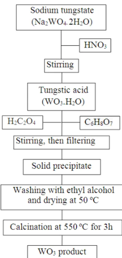
Synthesis of WO3 NPs
Li2TiO3 powder was synthesized via sol-gel route
(Poulter and Pryde 1968; Maruyama et al. 1994; Han et al. 1995; Sofian et al. 2007; Ilıcan et al.
2008; Ghodsi and Absalan 2010) WO3
nanopowders (<100 nm) were prepared by sol-gel process. The experimental details are shown in
Figure 1. Firstly, nitric acid (HNO3) solution was
added drop-by-drop to sodium tungstate
(Na2WO4.2H2O). Oxalic acid (H2C2O4) and citric
acid (C6H8O7) were used as complex agents at sol
solution. The precipitate obtained was washed several times with absolute ethyl alcohol and dried
at 50 ºC. The yellow precipitate (WO3) powder
was produced with calcination at 550ºC for 3h.
Figure 1 - Schematic diagrams of steps involved in obtaining WO3 powders.
Animals
Male rats of Sprague-Dawley strain (from Medical
Experimental Research Center, Atatürk
University, Erzurum, Turkey) of 200-300 g body weight, were used throughout the present studies. They were allowed water and standard laboratory chow ad libitum and were maintained under the standard light, temperature, and relative humidity conditions. The study protocol was approved by the local ethical committee. All the experiments were performed in accordance with the Guide for the Care and Use of Laboratory Animals.
Hepatocyte isolation and cultivation
Rats were sacrificed by CO2 overdose and the
livers were removed immediately. Isolated hepatocytes from the rats were prepared by the collagenase perfusion technique. The liver was perfused through the hepatic portal vein with calcium-free Hanks balanced salt solution to remove the blood for about 10 min at a flow rate
of 2.5 mL min-1. As soon as the liver became
grayish brown in color, a second buffer solution containing collagenase (Hank’s balanced salt supplemented with 4 mM calcium chloride and 0.5
mg collagenase mL-1) was perfused at the same
rate until the liver appeared to have broken up. Then the liver was into 3-4 mm pieces with a
sterile scalpel. Following the mechanical
dissociation, the cells were filtered through the gauze and centrifuged at 250 xg for 5 min. Then, the hepatocytes were collected in the medium containing bovine serum albumin and bovine insulin. The cell suspension was filtered through the gauze again and allowed to sediment for 20 min to eliminate cell debris, blood, and sinusoidal cells. The cells were then washed three times by centrifugation at 50 g and tested by Trypan blue dye exclusion for the viability (always in the range of 82-93%). The hepatocytes were then suspended in a mixture of 75% Eagle’s minimum essential medium and 25% medium 199, supplemented with 10% fetal calf serum containing streptomycin, penicillin, bovine insulin, bovine serum albumin
and NaHCO3 (2.2 mg). The hepatocytes were
plated in multi-well tissue culture plates (3×10⁵
cells in a well area of 3.8 cm 2; 8×10⁵ cells in a
well area of 9.6 cm2). The medium was changed
3-4 h later. The effect of WO3 NPs was studied after
72 h of exposure in the cultures maintained with a medium deprived of fetal calf serum but supplemented with hydrocortisone hemisuccinate
(7x10⁻⁷ M). Hepatocytes were cultured for an
additional 8 h before the treatment.
Treatments
After 8 h of plating, when primary hepatocytes were adhered and attained their epithelial morphology, culture medium was aspirated and replaced with an equal volume of the medium supplemented with different concentrations of
aqueous WO3 NPs (5, 10, 20, 50, 75, 100, 150,
300, 500 and 1000 ppm) followed by incubation in
CO2 incubator for 72 h (n=6). Mitomycin C
(MMC; C15H18N4O5; Sigma®, at 10
-7
M) added group was considered as positive control
(Control+).
MTT assay
The viability of the cells was assessed by measuring the formation of a formazan from 3-(4,5-dimethylthiazol-2-yl)-2,5-diphenyl
tetrazolium bromide (MTT)
spectrophotometrically test, modified after
Mosmann (1983). Hepatocytes were incubated
with 0.7 mg mL-1 MTT for 30 min at 37ºC at the
photometrically determined at 560 nm (Lewerenz et al. 2003).
LDH assay
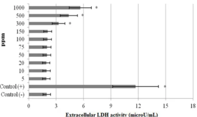
Lactate dehydrogenase (LDH) activity was measured in the culture medium as an index of cytotoxicity, employing an LDH kit (Bayer Diagnostics®, France) adapted to the auto analyzer (ADVIA 1650, USA). Enzyme activity was expressed as the extracellular LDH activity percentage of the total activity on the plates.
Total antioxidant capacity and total oxidant status assays
The automated Trolox equivalent total antioxidant capacity (TAC) and total oxidant status (TOS) assays were carried out in the culture medium by
commercially available kits (Rel Assay
Diagnostics®, Gaziantep, Turkey).
LMN assay
Liver micronucleus assay (LMN) assay was done by using the method of Suzuki et al. (2009).
Immediately prior to the evaluation, 10-20 µL of
hepatocyte suspension was mixed with an equal volume of acridine orange (AO)–DAPI (4´,6-diamidino-2-phenylindole dihydrochloride) stain
solution (AO, 0.5 mg mL-1; DAPI, 10 µg mL-1) for
fluorescent staining. Approximately 10-20 µL of
the mixture was dropped onto a glass slide and covered with a cover glass. Samples of well-isolated hepatocytes were evaluated with the aid of a fluorescence microscope counting the number of MNHEPs in 2000 hepatocytes for each animal. MNHEPs were defined as hepatocytes with round or distinct MNs that stained like the nucleus, with a diameter 1/4 or less than that of the nucleus, and confirmed by focusing up and down, taking into account hepatocyte thickness by one observer.
Nucleic acid oxidation
DNA oxidation was determined by measuring the amount of 8-OH-dG adducts. DNA was digested by incubation with DNAase I, endonuclease, and alkaline phosphatase (Schneider et al. 1993). The amount of 8-OH-dG was measured by high-performance liquid chromatography (HPLC) with electrochemical detection as described previously (Floyd et al. 1993).
Statistical analysis
The experimental data were analyzed using one-way analysis of variance (ANOVA) and Fischer’s least significant difference (LSD) tests to
determine whether any treatment significantly differed from the controls or each other’s. Results were presented as the mean ± SD values and the level of 0.05 was regarded as statistically significant.
RESULTS
The results of cell viability measured by the MTT
assay are shown in Figure 2. When assayed in
vitro on the hepatocyte cells using the MTT assay,
the value for the MMC-treated cells (as control+)
was approximately 2.4-fold lower than that for the control. Similarly, the higher concentrations of
WO3 NPs (300, 500 and 1000 ppm) caused
significant (p<0.05) decreases of the cell viability. However, the hepatocyte cells exposed to lower
doses than 300 ppm of WO3 NPs did not show any
significant change in cell viability during 72 h as determined by the MTT assay. No cytotoxicity
was observed for the control- cells. MMC-induced
hepatic damage was clearly evidenced by 6-fold increases in the activity of LDH compared with the observations of the negative controls (Fig. 3). Although LDH was not affected by the low doses
of WO3 NPs alone, but the increases of the levels
of enzyme reached statistical significances at 300, 500 and 1000 ppm.
Figure 2 - MTT reduction [3-(4,5-dimethyl-thiazol-2-yl) 2,5-diphenyltetrazolium bromide] in cultured rat hepatocytes maintained in the presence of different WO3 NPs
concentrations for 72 h.
Figure 3 - Extracellular level of lactate dehydrogenase (LDH) in cultured rat hepatocytes maintained in the presence of different WO3 NPs concentrations for 72 h. Each individual hepatocyte culture without NPs was studied as a negative control group (Control -). MMC alone added group was considered as a positive control (Control +). Values are means ± standard deviation (n = 6). * symbol presents significant differences at the p<0.05 level from the control - group).
The levels of 8-oxo-2-deoxyguanosine (8-OH-dG) in the cultured rat hepatocytes of controls and experimental groups are shown in Figure 4. Firstly, the levels of 8-OH-dG, a sensitive marker of oxidative DNA damage, were quantified with regard to MMC treatment. It was observed that
MMC significantly increased 8-OH-dG
concentrations in the hepatocyte cultures after 72 h. Similarly, 8-OH-dG levels increased in the hepatocyte cells that were treated with 500 and
1000 ppm of WO3 NPs.
The results of the observed LMN rates in the
primary rat hepatocyte cells after 72 h WO3 NPs
treatment are presented in Figure 5. LMN analyses did not show statistically significant differences
(p>0.05) between the control- and tested NPs
applied cultures.
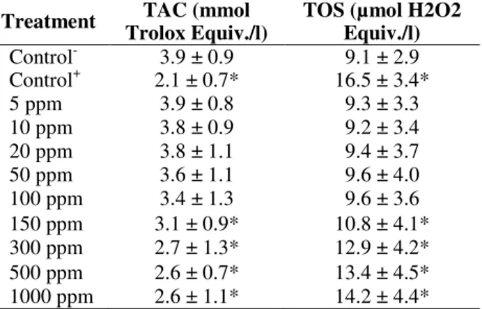
Table 1 showed the effects of WO3 NPs on oxidant
status in the cultured rat hepatocytes, which were determined by the TAC and TOS analysis. The TAC value decreased with the addition of MMC while TOS value increased. In contrast to the treatments with 5, 10, 20, 50, 75 and 100 ppm of
WO3, NPs did not alter the TAC and TOS levels.
However, nanomaterials applications at higher doses (150, 300, 500 and 1000 ppm) changed the
TAC and TOS levels. Thus, WO3 NPs had dose
dependent effects on the oxidative damage in hepatocytes in vitro.
Figure 4 - 8-oxo-2-deoxyguanosine (8-OH-dG) adducts in cultured rat hepatocytes maintained in the presence of different WO3 NPs concentrations for 72 h. Each individual hepatocyte culture without NPs was studied as a negative control group (Control -). MMC alone added group was considered as a positive control (Control +). Values are means ± standard deviation (n = 6). * symbol presents significant differences at the p<0.05 level from the control - group).
Table 1 - Extracellular TAC and TOS levels in cultured rat hepatocytes maintained with WO3 NPs for 72 h.
Each individual hepatocyte culture without NPs was studied as a negative control group (Control -). Ascorbic acid (10 ?M) and hydrogen peroxide (25 ?M) added groups were also used as the control+ in TAC and TOS analysis, respectively. Values are means ± standard deviation (n = 6). * symbol presents significant differences at the p<0.05 level from the control - group).
Treatment TAC (mmol
Trolox Equiv./l)
TOS (µmol H2O2 Equiv./l)
Control- 3.9 ± 0.9 9.1 ± 2.9 Control+ 2.1 ± 0.7* 16.5 ± 3.4*
5 ppm 3.9 ± 0.8 9.3 ± 3.3
10 ppm 3.8 ± 0.9 9.2 ± 3.4
20 ppm 3.8 ± 1.1 9.4 ± 3.7
50 ppm 3.6 ± 1.1 9.6 ± 4.0
100 ppm 3.4 ± 1.3 9.6 ± 3.6 150 ppm 3.1 ± 0.9* 10.8 ± 4.1* 300 ppm 2.7 ± 1.3* 12.9 ± 4.2* 500 ppm 2.6 ± 0.7* 13.4 ± 4.5* 1000 ppm 2.6 ± 1.1* 14.2 ± 4.4*
DISCUSSION
In the present investigation, it was aimed to evaluate the cytotoxicity, genotoxicity and oxidative damage in the cultured rat healthy
hepatocyte cells in response to different
concentrations of WO3 NPs. Trends in the
cytotoxicity as observed for the different concentrations of these NPs were overall highly similar using the two independent assays (MTT and LDH) in the cultured rat hepatocytes. A particular contrast was observed for the highest
concentrations of WO3 NPs, which induced a
pronounced LDH release and decrease of MTT. As a matter of fact, tungsten carbide nanoparticles (WC NPs) caused reduction in cell viability between 10 and 50% compared to controls upon
particle exposure in the rainbow trout
(Oncorhynchus mykiss) gill cell line, RTgill-W1 (Kühnel et al. 2009). Rothen-Rutishauser et al. (2007) reported that the size of nano-particles or their aggregates could be considered a potential determinant for the uptake and subsequent macrophage responses, which could explain the observed differences on LDH release at the highest concentration of NPs (Albrecht et al. 2009). Particles in the fine size range may induce more pronounced responses than in the nano-range (Rothen-Rutishauser et al. 2007), and the ‘‘ultrafine hypothesis” has been challenged
previously by the experimental studies (Schins et al. 2004). Albrecht et al. (2009) found that cytotoxicity as observed at high concentrations did not necessarily represent a compound-related effect, and could at least partly be due to physical coverage of the cells by the test NPs.
The fully understanding of how WO3 NPs interact
with hepatocytes, especially at the molecular level, is still unclear. Present findings demonstrated that
direct exposure of the hepatocytes to WO3 NPs
induced the intracellular ROS generation, reduced the total antioxidant capacity (TAC), increased
8-OH-dG levels, and subsequently caused
dysfunction of these cells. In accordance with the present results, it has been reported that engineered nanomaterials, either metals (like carbon and silver) or metal oxides (like zinc oxide, magnesium oxide, and titanium oxide), induce toxicity and oxidative stress by generating free radicals (Turkez and Geyikoglu 2007; Anreddy et al. 2013). Mechanisms involved in the reduction of
cell viability through oxidative stress by WO3 NPs
phosphorylation), P53 activation, Bax up regulation and Bcl-2 down regulation.
Previous studies showed that higher dose exposure to nanomaterials, including silica nanoparticles,
titanium dioxide, zinc oxide, alumina
nanoparticles led to proinflammatory and
procoagulant responses in endothelial cells. It is an accepted view point that ROS are involved in many of the processes underlying endothelial activation (e.g., the up regulation of adhesion molecules and chemokines, increased expression of tissue factor). Many of the key signal transduction molecules involved in the endothelial activation, such as various MAPKs and the transcription factors NF-kB, are known to be redox sensitive (Alom-Ruiz et al. 2008). To assess the biological effect of different concentrations of
WO3 NPs on rat hepatocyte cells, cell viability was
determined. In the present study, MTT and LDH assays showed that the higher concentrations of
WO3 NPs decreased cell viability. Also, Liu and
Sun (2010) found that exposure to high
concentrations (100-200 ppm) of silica
nanoparticles caused an increase of cytotoxicity in HUVECs. The LDH release was also increased by
silica nanoparticles only at the highest
concentration, indicating that exposure to high concentrations of silica nanoparticles could affect cell-membrane integrity and lead to cell death. They also reported the induction of apoptosis or necrosis by silica nanoparticles and oxidative stress as silica nanoparticles induced toxicity mechanism. Liu and Sun (2010) showed that intracellular ROS generation of HUVECs by silica nanoparticles was gradually increased in a time- and dose-dependent manner, suggesting that oxidative stress occurred not at once, but continuously during the cell culture. ROS played a central role in silica nanoparticles-mediated apoptosis. Disturbance of membrane integrity has been recently suggested as possibly one of the
mechanisms for cytotoxic action in the
nanoparticles (Kim et al. 2009). In accordance to the present findings, the results by Ding et al. (2009) had demonstrated that nano-WC-Co generated a higher level of hydroxyl radicals, induced greater oxidative stress, as evidenced by a decrease of glutathione (GSH) levels.
Present results indicated that WO3 NPs did not
induce the LMN formations in in vitro conditions. The study of DNA damage at the chromosome level is an essential part of genotoxicity testing because chromosomal mutation is an important
event in carcinogenesis. LMN assays have emerged as one of the preferred methods for assessing the chromosome damage because they enable both chromosome loss and chromosome breakage to be measured reliably. The NPs, which were located in the cytosol near the nucleus but not were found inside the nucleus, in mitochondria or ribosomes did not induce DNA-breakage
(Bhattacharya et al. 2009). But WO3 NPs led to
oxidative DNA damages as determined by the increases of 8-OH-dG levels, which were the major products of DNA oxidation in present study.
Thus, apparently WO3 NPs were not clastogenic
but were able to generate elevated amounts of free radicals, which induced indirect genotoxicity, mainly by DNA-adduct formation. In contrast to
our in vitro findings, WO3 NPs showed positive
mutagenic response in TA1537 and TA98
bacterial strains of Salmonella typhimurium by
using Ames test (Hasegawa et al. 2012). WO3 NPs
did not cause increase of the incidence of chromosome aberrations in rat bone marrow cells but led to increases of MN formation after chronic exposure for 30 days (Turkez et al. 2013a). These
conflicting evidences on genotoxicity by WO3 NPs
could be explained by the differences in conditions
(in vivo or in vitro), exposuring time (acute,
chronic or sub chronic) and chemical features of tested NPs (exact size, shape, composition, and aggregation). It is known that studies dealing with
nanotoxicity have focused on in vitro cell culture
studies. However, data obtained from such studies
might not correspond to in vivo results. Hence, a
full in vivo life cycle characterization framework
would be necessary for systematic evaluation of the size, shape, and surface chemistry of NPs, and
their correlation to in vivo behavior. In vivo
systems were extremely complicated and the interactions of NPs with biological components, such as proteins and cells could lead to unique bio-distribution, clearance, immune response, and metabolism (Fischer and Chan 2007; Berger 2008).
In summary, data from the current study showed
that exposure to WO3 NPs at high concentrations
caused ROS generation and decreased the total antioxidant capacity (TAC) in cultured primary rat hepatocytes. NPs caused decreased cell viability, which was detected by the increased MTT and LDH release. The present findings also showed
that WO3 NPs had weak genotoxic potential in
exposure to WO3 NPs could pose environmental
and human health risk.
REFERENCES
Albrecht C, Scherbart AM, van Berlo D, Braunbarth CM, Schins RPF, Scheel J. Evaluation of cytotoxic effects and oxidative stress with hydroxyapatite dispersions of different physicochemical properties in rat NR8383 cells and primary macrophages. Toxicol In Vitro. 2009; 23: 520-530.
Alom-Ruiz SP, Anilkumar N, Shah AM. Reactive oxygen species and endothelial activation. Antioxid Redox Signal. 2008; 10: 1089-1100.
Anreddy RN, Yellu NR, Devarakonda KR. Oxidative biomarkers to assess the nanoparticle-induced oxidative stress. Methods Mol Biol. 2013; 1028: 205-219.
Berger M. Nanotechnology and toxicity: the growing need for in vivo study. http://www.nanowerk.com. 2008.
Bhattacharya K, Davoren M, Boertz J, Schins RP, Hoffmann E, Dopp E. Titanium dioxide nanoparticles induce oxidative stress and DNA-adduct formation but not DNA-breakage in human lung cells. Part Fibre Toxicol. 2009; 6: 17.
Bupesh G, Chinnaiah A, Sakthivel V, Natesan M, Ramalingam SR, Raju K, Periyasamy S. Hepatoprotective efficacy of Hypnea muciformis
ethanolic extract on CCl4 induced toxicity in rats.
Braz Arch Biol Technol. 2012; 55: 857-863.
Ding M, Kisin ER, Zhao J, Bowman L, Lu Y, Jiang B, Leonard S, Vallyathan V, Castranova V, Murray AR, Fadeel B, Shvedova AA. Size-dependent effects of tungsten carbide-cobalt particles on oxygen radical production and activation of cell signaling pathways in murine epidermal cells. Toxicol Appl Pharmacol. 2009; 241: 260-268.
ECETOC. Workshop on Testing Strategies to Establish the Safety of Nanomaterials. November 2005, Barcelona, Workshop Report No: 7.
Erik L, Wolf-Dieter S. Tungsten: properties, chemistry, technology of the element, alloys, and chemical compounds. 1999; Kluwer Academic, USA.
Fischer HC, Chan WCW. Nanotoxicity: the growing need for in vivo study. Curr Opinion Biotechnol. 2007; 18: 565-571.
Floyd RA, Watson JJ, Wong PK, Altmiller DH, Rickard RC. Hydroxyl free radical adduct of deoxyguanosine: Sensitive detection and mechanisms of formation. Free Radic Res Commun. 1993; 1: 163-172.
Geyikoglu F, Turkez H. Protective effect of sodium selenite on genotoxicity to human whole blood cultures induced by aflatoxin B-1. Braz Arch Biol Technol. 2005; 48: 905-910.
Ghodsi FE, Absalan H. Comparative study of ZNO thin films prepared by different sol-gel route. Acta Phys Pol A. 2010; 118: 659-664.
Grandjean-Laquerriere A, Laquerriere P, Guenounou M, Laurent-Maquin D, Phillips TM. Importance of the surface area ratio on cytokines production by human monocytes in vitro induced by various hydroxyapatite particles. Biomater. 2005; 26: 2361-2369.
Han SD, Campet G, Huang SY, Shasrty MCR, Portier J. A new method for the preparation of fine-grained SnO2 and WO3 powders: influence of the crystallite size on the electrochemical insertion of Li + in SnO2 and WO3 electrodes. Active Passive Electron Component. 1995; 18: 39-51.
Hasegawa G, Shimonaka M, Ishihara Y. Differential genotoxicity of chemical properties and particle size of rare metal and metal oxide nanoparticles. J Appl Toxicol. 2012; 32: 72-80.
Hsin YH, Chen CF, Huang S, Shih TS, Lai PS, Chueh PJ. The apoptotic effect of nanosilver is mediated by a ROS- and JNK-dependent mechanism involving the mitochondrial pathway in NIH3T3 cells. Toxicol Lett. 2008; 179: 130-139.
Hoshino T, Tanaka K, Makita J, Hashimoto T. Investigation of phase transition in Li2TiO3 by high temperature X-ray diffraction. J Nuclear Mater. 2007; 367: 1052-1056.
Ilıcan S, Caglar Y, Caglar M. Preparation and characterization of ZnO thin films deposited by sol-gel spin coating method. J Optoelectron Adv Mater.
2008; 10: 2578-2583.
Jian-wen Y, Hui Z, Haryun Z, Yarryang D, Jian L, Xuan Z. Synthesis and electrochemical properties of nanocrystalline Li[Li1/3Ti3/5O4] by complex sol-gel method. Trans Nonferrous Met Soc China. 2004; 14: 1012-1016.
Kim S, Choi JE, Choi J, Chung KH, Park K, Yi J. Oxidative stress-dependent toxicity of silver nanoparticles in human hepatoma cells. Toxicol In Vitro. 2009; 23: 1076-1084.
Knaapen AM, Borm PJ, Albrecht C, Schins RP. Inhaled particles and lung cancer. Part A: Mechanisms. Int J Cancer. 2004; 109:799-809.
Kühnel D, Busch W, Meissner T, Springer A, Potthoff A, Richter V. Agglomeration of tungsten carbide nanoparticles in exposure medium does not prevent uptake and toxicity toward a rainbow trout gill cell line. Aquat Toxicol. 2009; 93: 91-99.
Lanone S, Rogerieux F, Geys J, Dupont A, Maillot-Marechal E, Boczkowski J. Comparative toxicity of 24 manufactured nanoparticles in human alveolar epithelial and macrophage cell lines. Part Fibre Toxicol. 2009; 6: 14-19.
Lewerenz V, Hanelt S, Nastevska C, El-Bahay C, Rohrdanz E, Kahl R. Antioxidants protect primary rat hepatocyte cultures against acetaminophen-induced DNA strand breaks but not against acetaminophen-induced cytotoxicity. Toxicol. 2003; 191: 179-187. Lewinski N, Colvin V, Drezek R. Cytotoxicity of
nanoparticles. Small. 2008; 4: 26-49.
Li SQ, Zhu RR, Zhu H, Xue M, Sun XY, Yao SD, Wang SL. Nanotoxicity of TiO(2) nanoparticles to erythrocyte in vitro. Food Chem Toxicol. 2008; 46: 3626-3631.
Liu X, Sun J. Endothelial cells dysfunction induced by silica nanoparticles through oxidative stress via JNK/P53 and NF-kB pathways. Biomater. 2010; 31: 8198-8209.
McInturf SM, Bekkedal MYV, Wilfong E, Arfsten D, Chapman G, Gunasekar PG. The potential reproductive, neurobehavioral and systemic effects of soluble sodium tungstate exposure in Sprague-Dawley rats. Toxicol Appl Pharmacol. 2011; 254: 133-137.
Maruyama T, Arai S. Electrochromic properties of tungsten trioxide thin films prepared by chemical vapor deposition. Phys Inorg Chem. 1994; 25: 31005. Merck Index. Tungsten trioxide. 2006; Vol. 14.
Mosmann T. Rapid colorimetric assay for cellular growth and survival: application to proliferation and cytotoxicity assay. J Immunol Meth. 1983; 65: 55-63. Pan Y, Leifert A, Ruau D, Neuss S, Bornemann J,
Schmid G. Gold nanoparticles of diameter 1.4 nm trigger necrosis by oxidative stress and mitochondrial damage. Small. 2009; 5: 2067-2076.
Patel DJ and Patel JK. Design and evaluation of famotidine mucoadhesive nanoparticles for aspirin induced ulcer treatment. Braz Arch Biol Technol.
2013; 56: 223-236.
Poulter KF, Pryde JA. Chemisorption of hydrogen on rhenium. Brit J Appl Phys Ser. 1968; 1: 169.
Pradyot P. Handbook of inorganic chemical compounds. 2003; ISBN. 0-07-049439-8. McGraw-Hill, USA, p. 1086.
Ramesh M, Turner LF, Yadav R, Rajan TV, Vella AT, Kuhn LT. Effects of the physico-chemical nature of two biomimetic crystals on the innate immune response. Int Immunopharmacol. 2007; 7: 1617-1629. Riehemann K, Schneider SW, Luger TA, Godin B, Ferrari M, Fuchs H. Nanomedicine-challenge and perspectives. Angew Chem Int Ed Engl. 2009; 48: 872-897.
Rothen-Rutishauser B, Mühlfeld C, Blank F, Musso C, Gehr P. Translocation of particles and inflammatory responses after exposure to fine particles and nanoparticles in an epithelial airway model. Part Fibre Toxicol. 2007; 4: 9.
Rodgher S, Espindole ELG, Sımoes FCF, Tonietto AE. Cadmium and chromium toxicity to
Pseudokirchneriella subcapitata and Microcystis
aeruginosa. Braz Arch Biol Technol. 2012; 55: 161-169.
Schins RP, Lightbody JH, Borm PJ, Shi T, Donaldson K, Stone V. Inflammatory effects of coarse and fine particulate matter in relation to chemical and biological constituents. Toxicol Appl Pharmacol.
2004; 195: 1-11.
Schmid O, Moller W, Semmler-Behnke M, Ferron GA, Karg E, Lipka J. Dosimetry and toxicology of inhaled ultrafine particles. Biomarkers. 2009; 14: 67-73. Schneider JE, Jr Phillips JR, Pye Q, Maidt ML, Price S,
Floyd RA. Methylene blue and rose bengala photoinactivation of RNA bacteriophages: Comparative studies of 8-oxoguanine formation in isolated RNA. Arch Biochem Biophys. 1993; 301: 91-97.
Shan D, Xie Y, Ren G, Yang Z. Inhibitory effect of tungsten carbide nanoparticles on voltage-gated potassium currents of hippocampal CA1 neurons.
Toxicol Lett. 2012; 209: 129-135.
Simbula G, Columbano A, Ledda-Columbano GM, Sanna L. Increased ROS generation and p53 activation in alpha-lipoic acidinduced apoptosis of hepatoma cells. Apoptosis. 2007; 12: 113-123. Sisman T, Turkez H. Toxicologic evaluation of imazalil
with particular reference to genotoxic and teratogenic potentials. Toxicol Ind Health. 2010; 26: 641-648. Sofian MK, Carl PT. Synthesis, FTIR studies and
sensor properties of WO3 powders. Curr Opin Solid State Mater Sci. 2007; 11: 19-27.
Song Y, Li X, Du X. Exposure to nanoparticles is related to pleural effusion, pulmonary fibrosis and granuloma. Eur Respir J. 2009; 34: 559-567.
Tsuchiya K, Nakamichi M, Nagao Y, Enoeda M. In-situ tritium recovery experiments of blanket in-pile mockup with Li2TiO3 pebble bed in Japan. J Nuclear Sci Technol. 2001; 38: 996-999.
Turkez H, Cakmak B, Celik K. Evaluation of the potential in vivo genotoxicity of tungsten (VI) oxide nanopowder for human health. Key Engineering Mater. 2013a; 543: 89-92.
Turkez H, Celik K, Cakmak B. Biosafety evaluation of nanoparticles in view of genotoxicity and carcinogenicity studies: A systematic review. Key Engineering Mater. 2013b; 543: 200-203.
Turkez H, Geyikoglu F, Tatar A, Keles MS, Kaplan I. The effects of some boron compounds against heavy metal toxicity in human blood. Exp Toxicol Pathol.
2012; 64: 93-101.
Turkez H. The role of ascorbic acid on titanium dioxide-induced genetic damage assessed by the comet assay and cytogenetic tests. Exp Toxicol Pathol. 2011; 63: 453-457.
Turkez H, Togar B. The genotoxic and oxidative damage potential of olanzapine in vitro. Toxicol Ind Health. 2010; 26: 583-588.
Turkez H, Geyikoglu F. Boric acid: a potential chemoprotective agent against aflatoxin b(1) toxicity in human blood. Cytotechnol. 2010; 62: 157-165. Turkez H. Effects of boric acid and borax on titanium
dioxide genotoxicity. J Appl Toxicol. 2008; 28: 658-664.
Turkez H, Geyikoglu F, Tatar A, Keleş S, Ozkan A. Effects of some boron compounds on peripheral human blood. Z Naturforsch C. 2007; 62: 889-896. Turkez H, Sisman T. Anti-genotoxic effect of hydrated
sodium calcium aluminosilicate on genotoxicity to human lymphocytes induced by aflatoxin B1. Toxicol Ind Health. 2007; 23: 83-89.
Turkez H, Geyikoglu F, Keles MS. Biochemical response to colloidal bismuth subcitrate: dose-time effect. Biol Trace Elem Res. 2005; 105: 151-158. Turkez H, Geyikoglu F. An in vitro blood culture for
evaluating the genotoxicity of titanium dioxide: the responses of antioxidant enzymes. Toxicol Ind Health. 2007; 23:19-23.
Unfried K, Albrecht C, Klotz LO, Mikecz von A, Grether-Beck S, Schins RPF. Cellular responses to nanoparticles: target structures and mechanisms.
Nanotoxicol. 2007; 1: 52-71.
Wang Y, Aker WG, Hwang HM, Yedjou CG, Yu H, Tchounwou PB. A study of the mechanism of in vitro
cytotoxicity of metal oxide nanoparticles using catfish primary hepatocytes and human HepG2 cells.
Sci Total Environ. 2011; 409: 4753-4762.
Witten ML, Sheppard PR, Witten BL. Tungsten toxicity. Chem Biol Interact. 2012; 196: 87-88. Xu Z, Zhang YL, Song C, Wu LL, Gao HW.
Interactions of hydroxyapatite with proteins and its toxicological effect to zebrafish embryos development. PLoS One. 2012; 7: e32818.
Zhang XD, Zhao J, Bowman L, Shi X, Castranova V, Ding M. Tungsten carbide-cobalt particles activate Nrf2 and its downstream target genes in JB6 cells possibly by ROS generation. J Environ Pathol Toxicol Oncol. 2010; 29: 31-40.
Zhou G, Hou Y, Liu L, Liu H, Liu C, Liu J, Qiao H, Liu W, Fan Y, Shen S, Rong L. Preparation and characterization of NiW-nHA composite catalyst for hydrocracking. Nanoscale. 2012; 4: 7698-7703.
![Figure 2 - MTT reduction [3-(4,5-dimethyl-thiazol-2- [3-(4,5-dimethyl-thiazol-2-yl) 2,5-diphenyltetrazolium bromide] in cultured rat hepatocytes maintained in the presence of different WO 3 NPs concentrations for 72 h](https://thumb-eu.123doks.com/thumbv2/123dok_br/15960560.684066/4.892.472.810.680.891/reduction-dimethyl-dimethyl-diphenyltetrazolium-hepatocytes-maintained-different-concentrations.webp)