http://dx.doi.org/10.190/1678-4324-2017160409 ISSN 1678-4324 Online Edition
BRAZILIAN ARCHIVES OF BIOLOGY AND TECHNOLOGY
A N I N T E R N A T I O N A L J O U R N A L
Phenolic Content and Biomolecule Oxidation Protective
Activity of
Globularia alypum
Extracts
Hamama Bouriche
1*, Seoussen Kada
1, Abderrahmane Senator
1, Ibrahim demirtas
2, Tevfik
Ozen
3, Bircan Çeken Toptanci
4, Goksel Kizil
4, Murat Kizil
4.
1Laboratory of Applied Biochemistry, Faculty of SNV, University Ferhat Abbas, Sétif 1, Algeria. 2Department of
Chemistry, Faculty of Science, Cankırı Karatekin University, Cankırı, Turkey. 3Department of Chemistry, Faculty of
Science and Arts, Ondokuz Mayis University, Samsun, Turkey. 4Department of Chemistry, Faculty of Science and Arts, University of Dicle, Diyarbakir, Turkey
ABSTRACT
The protective activity of methanolic (Met E) and aqueous (Aq E) extracts of Globularia alypum L. (G. alypum) against DNA, lipid and protein oxidative damage was investigated. Moreover, the scavenging, chelating, and reducing power activities of the extracts were also evaluated. Phytochemical analysis was performed to determine phenolic compounds. Results showed that Met E and Aq E were rich in phenolic compounds, and were able to scavenge DPPH˙ with IC50 values of48.61 µg/mL and 51.97 µg/mL, respectively. In addition, both extracts were
able to chelate ferrous ions. At 300 μg/mL, the chelating activity was 97.53% and 91.02%, respectively. The reducing power of these extracts was also remarkable and concentration dependent. At 100 µg/mL, both extracts inhibited lipid peroxidatin by only 42.45% and 4.03%. However, the DNA oxidation damage was inhibited dose-dependently in the presence of G. alypum extracts. At 1 mg/mL, both extracts suppressed DNA cleavage by 83%-84%. The protein oxidation was also inhibited by G. alypum extracts. At 1 mg/mL, Aq E and Met E protected BSA fragmentation by 77%-99%. The overall results suggest that G. alypum extracts exerted antioxidant activity and protect biomolecules against oxidative damage; hence it may serve as a potential source of natural antioxidants.
Key words: antioxidants, DNA damage, Globularia alypum, lipid peroxidation, protein oxidation
*
INTRODUCTION
Reactive oxygen species (ROS) generation, results from a number of endogenous metabolic pathways, as well as from exposure to chemical and physical factors in the ambient environment. In small amounts, ROS play important roles in a number of biological processes and regulate cell physiology and functions. They act as signal
transducers1,2, growth factor regulators 3,4, second messengers and regulators of
vascular homeostasis 5. However, the overproduction of ROS leads to several cell
damaging effects. Cells have a variety of defense mechanisms that intercept ROS to prevent or limit intracellular damages and ameliorate their harmful effects. When the equilibrium between pro-oxidant and antioxidant systems within the cell is failed, the overproduced ROS, which could not be fully neutralized can cause oxidative
damage to a broad range of cell components, leading to many diseases6. ROS can
interact and modify different classes of biological macromolecules including DNA, lipids and proteins.
ROS can react with the polyunsaturated fatty acids of lipid membranes and induce lipid peroxidation. Lipid peroxidation is a source of secondary free radicals, which
can directly react with other biomolecule, enhancing biochemical lesions 7. Increased
formation of oxidized lipid products contribute to several pathologies such as
atherosclerosis, ischemia-reperfusion, heart failure, Alzheimer’s disease, rheumatic
arthritis, cancer, and other immunological disorders 8. Moreover, lipid peroxidation
is one of the major factors causing deterioration of foods during storage and
processing, and could have harmful effects on human health 9.
DNA is continually attacked by ROS that can affect its structure and function severely. Excess oxidative stress results in DNA base modifications, single- and double-strand breaks, and the formation of apurinic/apyrimidinic lesions, which may
be toxic and/or mutagenic 10. Mutagenic lesions are a critical contributor to the
development of aging, some degenerative diseases 11, 12, and many cancers 13,14
Proteins are also major targets for ROS attacks in biological systems due to their abundance and high rate constants for reaction. During oxidation, several amino acid residues irreversibly form carbonyl products, which can further lead to the formation
of high-molecular-mass aggregates 15. Protein aggregates can be highly cytotoxic,
altering cell functions and leading to cell necrosis or apoptosis 16. The accumulation
of oxidized proteins was observed in several pathological states such as diabetes,
neurodegenerative diseases and aging 12,17,18.
Medicinal plants constitute one of the main sources of safe antioxidants, healthcare supplements and phytochemicals that assist in maintaining good health and
combating diseases. Globularia alypum L. is an aromatic medicinal plant belongs to
Globulariaceae family, found throughout the Mediterranean area and largely used in Algerian folk medicine to treat some inflammatory disorders. The leaves of the plant are reported to be used in the treatment of diabetes, renal disorders and
cardiovascular diseases 19,20. However, few studies are reported on the phytochemical
constituents and the biological activities of this plant. Moreover, the literature survey did not reveal any reference to previous work comparing the antioxidant activities of this plant by reducing biomacromolecule oxidative damages. Therefore, this study is aimed to investigate the antioxidant activity and the potential role of methanolic and
aqueous extracts of G. alypum L. to prevent lipid, DNA and proteins against
oxidative damages.
MATERIALS AND METHODS
Ascorbic acid, bovine serum albumin (BSA), 1,1-Diphenyl-2-picrylhydrazyl
(DPPH), ethidium bromide, FeCl2, FeCl3, FeSO4, folin-ciocalteu reagent, gallic acid,
H2O2, linoleic acid, sodium carbonate (Na2CO3), thiobarbituric acid (TBA),
trichloroacetic acid(TCA), tween-20, potassium ferricyanide (K3FeCN6) and
quercetin, were purchased from Sigma chemical (St. Louis, MO). Plasmid miniprep kit was obtained from Quiagen (Valencia, CA). All other chemicals and solvents were of analytic grade and are from Panreac (Spain), Fluka (French), Riedel-de Haén (Germany).
Plant Material
Globularia alypum L.was collected in June 2010 from Sétif, in Eastern Algeria. The
plant was identified and authenticated taxonomically by Dr. N. Boulaacheb, Univesity of Sétif 1, Algeria. A voucher specimen (No. G.A. 2010-1) was preserved at the local Herbarium of Botany, Department of Botany, University of Sétif, Algeria. Leaves were air-dried at room temperature and then reduced to powder.
Preparation of the Plant Extracts
Globularia alypum methanolic extract (Met E) was prepared by maceration of 100 g
of powdered aerial part of the plant material twice with 80% methanol and then with methanol 50% at room temperature for 48 h with frequent agitation. After filtration, the filtrate was concentrated under reduced pressure at 40 °C. The residue was lyophilized to give a brown powder (yield: 37.8%) which was stored at -32 °C until use.
Aqueous extract (Aq E) was prepared by boiling 50 g of powdered plant in 500 mL of distillated water for 20 min. After filtration, the filtrate was lyophilized to give a brown powder (yield: 30%).
Determination of Total Polyphenols
The content of total polyphenolic compounds in the extracts was determined using
Folin-Ciocalteu’s reagent according to Li et al. 21. Sample of 40 μL of crude extract
(1 mg/ml) were mixed with 200 μL of Folin-Ciocalteu reagent and 1160 μL of
distilled water, followed by the addition of 600 μL of 7.5% Na2CO3. The mixtures
were shaken for 2 h at room temperature and the absorbance was measured at 765 nm. Gallic acid was used as a standard. The concentration of total polyphenol compounds was determined as mg of Gallic acid equivalents per 1 g of extract (GAE/mg extract).
Determination of Total Flavonoids
The method of Bahorun et al. 22 was used to determine the total flavonoid content of
G. alypum extracts. Briefly, 1 mL of 2% AlCl3 (in ethanol) was added to 1 mL of
each extract (1 mg/mL). After 10 min of incubation at room temperature, the absorbance was measured at 430 nm. Quercetin was used as a standard. Total flavonoid content was expressed as mg quercetin equivalent per g of extract (QAE/g extract).
Determination of Total Condensed Tannins
Tannin content of the extracts was determined using the hemoglobin precipitation
assay according to Bate-Smith 23, using tannic acid as standard. An aliquot of 0.5 mL
the absorbance was measured at 578 nm. Tannin content was expressed as mg tannic acid equivalent per g of extract (TAE/g extract).
HPLC-TOF/MS Analysis
Phenolic acids and flavonoids in the extracts were analyzed by e HPLC-TOF/MS
using Agilent Technology of 1260 Infinity HPLC System coupled with 6210 Time
Of Flight (TOF) LC/MS detector and ZORBAX SB-C18 (4.6 x 100 mm, 3.5 μm)
column as described by Abay et al. 24. Mobile phases A and B were ultra-pure water
with 0.1% formic acid and acetonitrile, respectively. Flow rate was 0.6 mL min-1 and
column temperature was 35 °C. Injection volume was 10 μL. The solvent program was as follow: 0-1 min 10% B; 1-20 min 50% B; 20-23 min 80% B; 23-25 min 10% B; 25-30 min 10% B. Ionization mode of HPLC-TOF/MS instrument was negative and operated with a nitrogen gas temperature of 325°C, nitrogen gas flow of 10.0 L
min-1, nebulizer of 40 psi, capillary voltage of 4000 V and finally, fragmentor
voltage of 175 V. For sample analysis, dried crude extracts (200 ppm) were dissolved in methanol at room temperature. Samples were filtered through a PTFE
(0.45 μm) filter by an injector to remove particulates.
Free Radical Scavenging Activity
Free radical scavenging activity of the extracts was measured according to the
method of Que et al. 25. The solution of the free DPPH radical in ethanol (0.1 mM)
was prepared and 0.5 mL of aqueous or methanolic extracts at different concentrations (10 - 400 µg/mL) was added. The mixture was shaken vigorously and left standing at room temperature for 30 min. before measuring the absorbance at 517 nm. Butylated hydroxytoluene (BHT) was used as a standard antioxidant. The
antiradical activity was expressed as IC50 (mg.mL−
1
). A lower IC50 value corresponds
to a higher antioxidant activity of extracts. The ability to scavenge the DPPH radicals was calculated using the following equation:
DPPH˙ scavenging activity (%) = [(A0− A1) / A0] × 100
Where A0 is the absorbance of the control and A1 is the absorbance of the sample at
30 min.
Ferrous Ions Chelating Activity
The chelating of ferrous ions by the methanolic and aqueous extracts was estimated
by the method of Le et al. 26. Briefly the extract samples (20 – 300 µg/mL) were
added to 50 µl of 0.6 mmol/l FeCl2. The reaction was initiated by the addition of 50
µL of ferrozine (5 mM) and the mixture was shaken vigorously and left standing at room temperature for 10 min before measuring the absorbance at 562 nm. EDTA
was used as a standard chelator. The percentage of inhibition of ferrozine–Fe2+
complex formation was calculated using the following formula:
Ferrous ions chelating activity (%) = [(A0 − A1) / A0] × 100
Where A0 is the absorbance of the control (contained complex formation molecules;
FeCl2 and ferrozine) and A1 is the absorbance of the test compound.
Reducing Power
The reducing power of G. alypum extracts was determined according to the method
of Oyaizu 27. A volume of 2,5 mL of each each extract at different concentration (20
– 350 µg/mL) was mixed with 2.5 mL of 200 mM sodium phosphate buffer (pH 6.6)
and 2.5 mL of 1% potassium ferricyanide. After the incubation of the mixture at 50°C for 20 min, 2.5 mL of 10% TCA were added and the mixture was centrifuged at 200 × g for 10 min. The upper layer (2.5 mL) was mixed with 2.5 mL of deionized
water and 0.5 mL of 0.1% FeCl3 and the absorbance was measured at 700 nm.
triplicate and the results are expressed as mean values ± standard deviations. BHT was used as a standard antioxidant.
Linoleic Acid Peroxidation Assay
The protective effect of G. alypum extracts against lipid peroxidation was tested by
assessing the peroxidation of linoleic acid in an oxidation system catalysed by Fe+2
-ascorbate 28.Different concentrations (50 –500 μg/mL) of extracts were mixed with
linoleic acid solution (0.28 mg linoleic acid and 0.28 mg tween-20) in 500 mL of
100 μM of phosphate buffer ( pH 7.4) and 150 μL of 10 μM ascorbic acid solution.
The mixture was mixed and sonicated to obtain a homogeneous emulsion. The
linoleic acid peroxidation was initiated by the addition of 0.1 mL FeSO4 (10 μM).
After incubation at 37Co for 60 min, the mixture was cooled and 1.5 mL of TCA
(10% in 0.5% HCl) was added, followed by the addition of 3 mL of TBA (1%, in 50
mM NaOH). The mixture was then heated in a water bath at 90 Co for 60 min. After
cooling, aliquots of 2 mL were taken from each sample and mixed with 2 mL of butanol and centrifuged at 1000 x g for 30 min. The upper layer solution was separated for measuring the absorbance at 532 nm. The percentage of linoleic acid peroxidation inhibition was calculated according to the following equation:
Linoleic acid peroxidation inhibition (%) = [(Ao-A1)/Ao] x 100
Where Ao is the absorbance of control reaction (containing all reagents except the
extracts) and A1 is the absorbance of the sample with extracts or standard.
DNA Oxidation Assay
Protective activity of methanolic and aqueous extracts of G. alypum against DNA
damage was tested by photolysing H2O2 with UV radiation in the presence of
pBluescript M13+ plasmid DNA. Plasmid DNA was isolated by Qiagene plasmid
miniprep (and oxidized with H2O2+UV treatment in presence of different
concentrations of G. alypum extracts (100, 250, 350 and 500 µg/mL) and checked on
1% agarose gel electrophoresis according to a modified method 29. Briefly, the
experiments were performed in a volume of 10 µ L in a microcentrifuge tube containing 200 ng of plasmid DNA in phospate buffer (7.14 mmol phospate and
14.29 mmol NaCl, pH 7.4) and H2O2 was added at a final concentration of 2.5
mmol/L with or without 1μL of extracts. The reactions were initiated by UV
irradiation and continued for 5 min on the surface of a UV transilluminator with
intensity 8000 μW/cm2 at 300 nm under room temperature. After irradiation, the
reaction mixture (10 μL) with gel loading dye was placed on 1% agarose gel for
electrophoresis. Electrophoresis was performed at 40 V for 3 h in the presence of
ethidium bromide (10 mg/mL). Untreated pBluescript M13+ plasmid DNA was used
as a control in each run of gel electrophoresis along with partial treatments (i.e. only
UV treatment and only H2O2). Percent inhibition of the DNA strand scission was
calculated as follows:
Inhibition (%) = I − (Sm+a − Sc)/(Sm− Sc).
Where Sm+a is percentage remaining supercoiled DNA after treatment with UV +
H2O2 in the presence of the extracts, Sc is percentage remaining supercoiled DNA in
the control untreated plasmid, and Sm is percent remaining supercoiled DNA with
UV + H2O2 without extracts.
Protein Oxidation Assay
The protective ability of methanolic and aqueous extracts of G. alypum against
protein oxidation was evaluated according to Kizil et al. 29. BSA (1 mg/mL), used as
a model protein, was dissolved in 20 mM potassium phosphate buffer (pH 7.4) and
then 50 μM FeCl3, 1 mM H2O2, and 100 μM ascorbic acid were added in the reaction
mixture. This mixture was incubated in the presence or absence of methanol and
aqueous extracts of G. alypum (50 − 1000 μg/mL) in a final volume of 1.2 mL for 3
h at 37 oC, and then the reaction mixture was analyzed by electrophoresis in 10%
SDS polyacrylamide gel (SDS-PAGE) according to Laemmli (1970). Samples were mixed with equal volumes of sample buffer (Tris HCl pH 6.8, 2% SDS, 5% of 2-mercaptoethanol, 10% sucrose, and 0.002% bromophenol blue). The mixture was
then boiled for 5 min, and then 5 μL of each sample was electrophoresed by
SDS-PAGE. The gel was run in a BioRad tank in running buffer (25 mM Tris pH 8.3, 190 mM glycine, and 0.1% SDS) at a maximum voltage and a constant current of 25 mA for a mini gel, using a BioRad 1000/500 power supply. Gels were stained with 0.15% Coomassie Brillant Blue R-250 for 2 h and then distained and digitally photographed.
The density of each band, corresponding to the protein fragments was estimated using the Gel Documentation System (Gel-Doc-XR; BioRad, Hercules, CA, USA). Bands on the gels were quantified by Discovery Series Quantity One program (version 4. 5. 2, BioRad Co.).
Statistical Analysis
Data were analyzed by analysis of variance (ANOVA). IC50 values were calculated
by regression analysis. Results were expressed as the mean ± standard deviation
(SD). Difference was considered significant when P < 0.05.
RESULS
Total Polyphenol, Flavonoid and Tannin Content
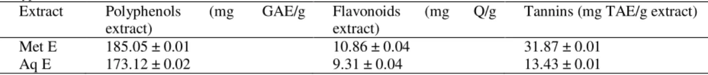
Total content of phenolic compounds of G. alypum extracts are presented in Table 1.
Methanolic extract contains the highest amount of polyphenols, flavonoids and tannins compared to aqueous one.
Table 1. Polyphenol, tannin and flavonoid content of methanolic extract (Met E) and aqueous extract (Aq E) of G. alypum.
Extract Polyphenols (mg GAE/g extract)
Flavonoids (mg Q/g extract)
Tannins (mg TAE/g extract)
Met E 185.05 ± 0.01 10.86 ± 0.04 31.87 ± 0.01
Aq E 173.12 ± 0.02 9.31 ± 0.04 13.43 ± 0.01
Values are mean of triplicate determination (n=3) ± SD.
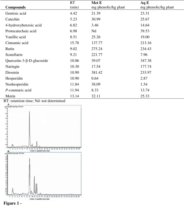
HPLC-TOF/MS Analysis
The HPLC-TOF/MS analysis revealed the presence of phenolic acids and flavonoids in G. alypum methanolic and aqueous extracts (Table 2 and Fig. 1). Both extracts contain high quantity of glycoside flavonoids such as diosmin, rutin and scutellarin.
A highest amount of naringin and quercetin-3-β-D-glucoside was observed in
Table 2. Phenolic compounds identified by HPLC-TOF/MS analysis in methanolic extract (Met E) and aqueous extract (Aq E) of G. alypum.
Compounds
RT (min)
MetE
mg phenolic/kg plant
Aq E
mg phenolic/kg plant
Gentisic acid 4.42 21.39 23.31
Catechin 5.23 30.99 25.67
4-hydroxybenzoic acid 6.82 3.46 14.64
Protocatechuic acid 6.98 Nd 39.53
Vanillic acid 8.51 25.26 19.00
Cinnamic acid 15.78 137.77 213.16
Rutin 9.02 275.24 234.43
Scutellarin 9.21 221.77 7.96
Quercetin-3-β-D-glucoside 10.06 39.07 347.38
Naringin 10.30 17.54 177.74
Diosmin 10.90 381.42 233.97
Hesperidin 10.90 0.64 2.87
Neohesperidin 11.84 38.09 1.54
P-coumaric acid 11.94 8.33 13.74
Morin 13.14 32.11 25.33
RT: retention time; Nd: not determined
Figure 1 -
Free Radical Scavenging Activity
Globularia alypum extracts showed a concentration-dependent scavenging activity
of DPPH free radicals (Fig. 2). However, methanolic extract was more potent than
the aqueous one. The recorded IC50 were 48.61 µg/mL and 51.97 µg/mL,
respectively. The activity of methanolic extract was close to that of the standard
Figure 2 -
Ferrous Ions Chelating Activity
The methanolic and aqueous extracts of G. alypum were able to chelate ferrous ions
in a concentration-dependent manner (Fig. 3). However, the aqueous extract showed
more activity than the methanolic extract. The IC50 values were 52.69 μg/mL and
148.15 μg/mL, respectively. This activity was less important than that obtained with
the standard chelator, EDTA, which gave an IC50 value of 5.97μg/mL.
Figure 3 -
Reducing Power
The reductive activity of both extracts of G. alypum compared with BHT have been
illustrate in Figure 4. The methanolic extract exerted a remarkable reducing power
with IC50 of 50.4 μg/mL, followed by the aqueous extract with an IC50 of 61 μg/mL.
This reductive activity is significant but less than that observed with BHT which
showed an IC50 value of 17 μg/mL.
Figure 4 -
Protective Effect of G. alypum Extracts Against Lipid Peroxidation
Both extracts of G. alypum exerted a weak inhibition on linoleic acid peroxidation
Figure 5 -
Protective Effect of G. alypum Extracts Against DNA Damage
Electrophoretic patterns of DNA after UV-photolysis of H2O2 in the presence or
absence of G. alypum methanolic and aqueous extracts on agarose gel
electrophoresis is shown in Figure 6. The conversion of supercoiled circular DNA (sc-DNA) to open circular (oc-DNA) derived from pBluescript M13+ DNA plasmid showed two bands on agarose gel electrophoresis (lane 1); the faster moving band corresponds to the native form of supercoiled circular DNA and the slower moving band corresponds to the open circular form. The UV irradiation of DNA in the
presence of H2O2 (lane 2) resulted in the cleavage of sc-DNA to oc-DNA form and
linear form (lin-DNA). It was noted that only UV treatment (lane 3), only H2O2
treatment (lane 4) and only UV treatment with 250 of extract (lane 5) could not
induce damage to DNA, as observed with the mixture treatment; UV + DNA + H2O2
(lane 2). The addition of the extracts (lanes 6 - 9) to this reaction mixture induced a partial recovery of sc-DNA. In fact, the intensity of sc-DNA bands scanned from the
agarose gel electrophoretic patterns at 100, 250, 350 and 500 µg/mL of G. alypum
Met E and Aq E was 45.88%, 53.77%, 70.91%, and 84.24% and 37.75%, 70.88%, 72.79% and 83.08%, respectively compared with the untreated plasmid DNA.
Figure 6 -
Protective Effect of G. alypum Extracts Against Protein Oxidation
Electrophoretic patterns of BSA after incubation for 3 h with Fe3+/H2O2/ascorbic
acid system in the absence or presence of different concentrations of methanolic and
aqueous extracts of G. alypum, and the density of the corresponding bands were
presented in Figure 7. The density of BSA band of the control induced by
Fe3+/H2O2/ascorbic acid decreased to 49.59%. The treatment by G. alypum extracts
exerted protective effect on BSA degradation induced by Fe3+/H2O2/ascorbic acid
system. Indeed, methanolic and aqueous extracts of G. alypums showed a
1000 μg/mL, the aqueous extract protected BSA very efficiently and restored the protein band intensity by 78.06%, 80.75, 85.41%, 98.16% and 98.83% compared to the control level, respectively. At the same concentrations, the methanolic extract restored the BSA band intensity by 53.18%, 56.2%, 57.9%, 74.29 and 76.97%, respectively.
Figure 7 -
DISCUSSION
The identification of new natural products, which can provide protection against the generation of oxidative stress, may have important human health implications. In this
study, we investigated whether G. alypum methanolic and aqueous extracts prevent
ROS mediated damages to lipids, DNA and proteins, employing a variety of in vitro
methods. Firstly, free radical scavenging activity, metal ion chelating, and reducing power of the extracts were tested. Results showed that both extracts were able to quench DPPH free radicals, and this activity is highly related to the amount of phenolic compounds (polyphenols, flavonoids and tannins). These compounds are electron donors and can react with DPPH free radicals to convert them to more
stable products 30, 31. In addition, G. alypum extracts showed metal chelating activity
and may serve as secondary antioxidants, by reducing the redox potential, thereby stabilizing the oxidized form of the ferrous ions. Ferrous ions are the most effective
pro-oxidants, which lead to the hydroxyl ions (HO˙) production via Fenton reaction.
Hydroxyl radicals are highly toxic and react with all kinds of biological macromolecules. Furthermore, both extracts exerted a remarkable reducing capacity, indicating that these extracts contain bioactive compounds that exhibit electron-donating capacity. The reducing capacity may serve as a significant indicator of the
potent antioxidant activity of phenolics and flavonoids 32.
Since both studied extracts showed significant anti-radical, chelating and reducing activities, so their protective activity against lipid, DNA and protein oxidation was evaluated in the current study.
Lipid peroxidation is preceded by radical mediated abstraction of the hydrogen atom from a methylene carbon on a polyunsaturated fatty acid side chain and it further proceeds with radical chain reaction. During this process, a number of compounds are formed such as malanoaldehyde (MDA), which used as a marker in the lipid
peroxidation assay 7. In this study, the lipid peroxidation was conducted using
linoleic acid catalyzed by Fe2+-ascorbate. The transition of ferrous ions can stimulate
lipid oxidation via Fenton reaction, and accelerate the reaction by decomposing the lipid hydroperoxides into peroxyl and alkoxyl radicals that can propagate the chain
of lipid peroxidation 33. Thus, minimizing ferrous ions may afford protection against
oxidative damage by inhibiting the production of hydroxyl radicals (·OH), which is
able to initiate free radical chain reactions 30. This study showed that despite both
were less effective on lipid peroxidation. This result may be due to the experimental conditions, in particular the time of measuring of MDA, since this assay is time consuming and therefore may not give accurate results due to degradation of the
hydroperoxidesbefore they converted to MDA in vitro during sample preparation 34.
Indeed, in another study, aqueous and methanolic extracts of G. alypum showed a
significant potential to protect the unsaturated β-carotene molecules (data not shown)
against free radicals attacks, by inhibiting strongly the linoleic acid peroxidation and then neutralizing free radicals formed in the system.
Similarly, the generation of oxidative damaged DNA is hypothesized to occur via
production of ROS. The UV irradiation photolysis of H2O2 generates OH radicals,
which attack DNA and resulting in the cleavage of the DNA strand, which is clearly
visible in the control DNA (treated with UV + H2O2). The scission of DNA strand
was assessed by measuring the conversion of supercoiled circular DNA to open circular DNA and further to linear form derived. The OH radicals can damage all components of DNA molecules such as purine and pyrimidine bases and also the
deoxyribose 35, inhibiting the normal functions of the cell. Among different types of
damages, DNA double strand breaks are the most deleterious, since they can lead to
the loss of genetic material 36. The treatment with G. alypum extracts protected the
supercoiled double strand DNA from hydroxyl radical-induced strand scission. Indeed, in the presence of increasing concentration of these extracts, the proportion of both open circular DNA and linear DNA were significantly decreased, while the amount of the residual supercoiled DNA was recovered. This protective activity against DNA damage could be assigned to the presence of potent antioxidants in the
extracts. It has been reported that the oxidative damage caused by exposure to H2O2
can be prevented by metal sequestering agents via chain-breaking antioxidants that
scavenge the free radicals generated by metal ion-catalyzed decomposition of H2O2
30
. In fact, the studied extracts were found to be rich in phenolic acids and flavonoids, which are potent antioxidants. Phenolic compounds possess ideal chemical structure for reducing free radicals because they have phenolic hydroxyl groups that are prone to donate a hydrogen atom or an electron to a free radical, and
extended conjugated aromatic system to delocalize an unpaired electron 30. Sevgi et
al.37 reported that phenolic acids possessed protective effects on pBR322 plasmid
DNA against the mutagenic and toxic effects of UV and H2O2.
The most common mechanism for inducing protein oxidation is metal-catalysed
reaction. Metal ions react with H2O2 in the so called Fenton reaction and produces
hydroxyl radicals that attack neighboring amino acid residues 38. ROS induce the
cleavage of peptide bonds and then leads to different peptide fragmentation or
protein-protein cross-linkages 39. Moreover, amino acids can be modified directly via
side chain reaction with ROS, methionine and cysteine are the most susceptible to oxidative changes due to high reaction susceptibility of the sulfur group in those
amino acids 39. In the present study, the metal-catalysed protein oxidation caused
different peptide fragmentations. This oxidative damage was assessed using a common method (SDS-PAGE) for determination of protein fragments. Densitometric analysis of each protein band showed clearly that both aqueous and
methanolic extracts of G. alypum protected significantly BSA against ROS attack
and restored the protein band intensity. This ability to protect protein fragmentations is mainly due to the strong antioxidant activities exerted by both extracts. Generally, phenolic compounds are responsible for the antioxidant activities of plant extracts, and they have been reported as scavengers of ROS and chelators of metal cations
that participate in hydroxyl radical formation 30. Therefore, these compounds are
CONCLUSION
The overall results revealed that G. alypum possess high antioxidant activity. The
presence of bioactive compounds such as polyphenols and flavonoids might be responsible for this activity. So, this plant can be considered as a good source of natural products that may be used in the treatment of different diseases associated to the oxidative stress.
CONFLICT OF INTEREST
Authors declare no conflict of interest in this study.
ACKNOWLEDGEMENTS
Authors acknowledge the Algerian Ministry of High Education for the financial support.
REFERENCES
1- Finkel T. Signal transduction by reactive oxygen species. J Cell Biol. 2012; 194: 17-15. 2- Reczek CR, Chandel NS. ROS-dependent signal transduction. Curr Opin Cell Biol. 2015;
33:8-13.
3- Jain M, Rivera S, Monclus EA, Synenki L, Zirk A, Eisenbart J, Feghali-Bostwick C, Mutlu, GM, Budinger GRS, Chandel NS. Mitochondrial reactive oxygens regulate transforming growth factor-β signaling. J Biol Chem. 2013; 288: 770-777.
4- Krstic J, Trivanović D, Mojsilovic S, Santibanez JF. Transforming growth factor-Beta and oxidative stress interplay: implications in tumorigenesis and cancer progression. Oxid Med Cell Longev [internet]. 2015. Available from: http://dx.doi.org/10.1155/2015/654594.
5- Vara D, Pula G. Reactive oxygen species: physiological roles in the regulation of vascular cells. Curr Mol Med. 2014; 14: 1103-25.
6- Kayama Y , Raaz U, Jagger A, Adam M, Schellinger IN, Sakamoto M , Suzuki H , oyama K, Spin JM, Tsao PS. Diabetic Cardiovascular Disease Induced by Oxidative Stress. Int J Mol Sci. 2015; 16: 25234-25263.
7- Lobo V, Patil A, Phatak A, Chandra N. Free radicals, antioxidants and functional foods: Impact on human health. Pharmacogn Rev. 2010; 4: 118-126.
8- Ramana KV, Srivastava S, Singhal SS. Lipid peroxidation products in human health and disease. Oxid Med Cell Longev [internet]. 2013. Available from: http://dx.doi.org/10.1155/2013/583438.
9- Laguerre, M, Lecomte J, Villeneuve P. Evaluation of the ability of antioxidants to counteract lipid oxidation: Existing methods, new trends and challenges. Prog Lipid Res. 2007; 46: 244-282.
10- Jena NR. DNA damage by reactive species: Mechanisms, mutation and repair. J Biosci. 2012; 37: 503-517.
11- Chao M-R, Rossner P, Haghdoost JrS, JengHA, Hu C-W. Nucleic acid oxidation in human health and disease. Oxid Med Cell Longev [internet]. 2013. Published online 2013 Dec 25. Available from: http://doi.org/10.1155/2013/368651.
12- Davalli P, Mitic T, Caporali A, Lauriola A, D’Arca D. ROS, cell senescence, and novel molecular mechanisms in aging and age-related diseases. Oxid Med Cell Longev [internet]. Published online 2016 Avr 16. Available from: http://dx.doi.org/10.1155/2016/3565127.
14- Schumacker PT. Reactive Oxygen species in cancer: A dance with the devil. Cancer Cell. 2015; 27 :156-157.
15- Xu Y-J, Qiang M, Zhang J-L, Liu Y, He R-Q. Reactive carbonyl compounds (RCCs) cause aggregation and dysfunction of fibrinogen. Protein Cell. 2012 3: 627- 640. 16- Dasgupta A, Zheng J, Bizzozero OA. Protein carbonylation and aggregation precede
neuronal apoptosis induced by partial glutathione depletion. ASN Neuro. 2012; 4: e00084.
17- Chen X, Chunyan Guo C, Kong J. Oxidative stress in neurodegenerative diseases. Neural Regen Res. 2012; 7: 376-385.
18- Rahman T, Hosen I, Towhidul Islam MM, Shekhar HU. Oxidative stress and human health. Adv Biosci
Biotechnol. 2012, 3: 997-1019.
19- Jouad H, Haloumi M, Rhiouani H, El Hilaly J, Eddoukes M. Ethnobotanical survey of medicinal plants used for the treatment of diabetes; cardiac and renal diseases in North centre region of Morocco (Fez-Boulmane). J Ethnopharmacol. 2001,77: 175-182.
20- Taleb-Dida N, Krouf D, Bouchenak M. Globularia alypum aqueous extract decreases hypertriglyceridemia and ameliorates oxidative status of the muscle, kidney, and heart in rats fed a high-fructose diet. Nutr Res. 2011; 31: 488-495.
21- Li HB, Cheng KW, Wong CC, Fan KW, Chen F, Jiang Y. Evaluation of antioxidant capacity and total phenolic content of different fractions of selected microalgae. Food Chem. 102: 2007; 771-776.
22- Bahorun T, Gressier B, Trotin F, Brunete C, Dine T, Vasseur J, et al. Oxygen species scavenging activity of phenolic extracts from hawthorn fresh plant organs and pharmaceutical preparations. Arzneimittel-Forsch. 1996; 46: 1086-1089.
23- Bate-Smith EC. Haemanalysis of tannins, the concept of relative astringency. Phytochemstry. 1973; 12: 907-912.
24- Abay G, Altun M, Koldas¸ S, Tüfekçi AR, Demirtas I. Determination of antiproliferative activities of volatile contents and HPLC profiles of Dicranum scoparium (Dicranaceae, Bryophyta). Comb Chem High Throughput Screen. 2015; 18:453-563.
25- Que F, Mao L, Pan X, Antioxidant activities of five Chinese rice wines and the involvement of phenolic compounds. Food Res Int. 2006; 39: 581-587.
26- Le K, Chiu F, Ng K. Identification and quantification of antioxidants in Fructus lycii. Food Chem. 2007; 105: 353-363.
27- Oyaizu M. Studies on products of browning reactions: antioxidative activities of browning reaction prepared from glucosamine. Jpn J Nutr. 1986; 4: 307-315.
28- Choi Y, Jeong HS, Lee J. Antioxidant activity of methanol extracts from some grains consumed in Korea. Food Chem. 2007; 103: 130-138.
29- Kizil G, kizil M, Ceken B,Yavuz M, Demir H. Protective activity of ethanol extracts of Hypericum scabrum L. and Hypericum retusum Aucher against the protein oxidation and DNA damage. Int J Food Prop. 2011; 14: 926-940.
30- Dai J, Mumper RJ. Plant phenolics: Extraction, analysis and their antioxidant and anticancer properties.
Molecules. 2010; 15: 7313-7352.
31- Mathew S, Abraham TE, Zakaria ZA. Reactivity of phenolic compounds towards free radicals under in vitro conditions. J Food Sci Technol. 2015; 52: 5790 - 5798.
32- Jing L, Ma H, Fan P, Gao R, Jia Z. Antioxidant potential, total phenolic and total flavonoid contents of Rhododendron anthopogonoides and its protective effect on hypoxia-induced injury in PC12 cells. BMC. Complement Altern Med. 2015; 15: 287. 33- Ayala A, Munoz, MF, Arguelles S. Lipid peroxidation: production, metabolism, and
signaling mechanisms of malondialdehyde and 4-Hydroxy-2-Nonenal. Oxid Med Cell Longev [internet]. 2014. Available from: http://dx.doi.org/10.1155/2014/360438.
34- Yamaguchi Y, Kunitomo M, Haginaka J. Assay methods of modified lipoproteins in plasma. J Chromatogr B. 2002; 781: 313-330.
36- Klaunig JE, Kamendulis LM, Hocevar BA. Oxidative stress and oxidative damage in carcinogenesis. Toxicol Pathol. 2010; 38: 96-109.
37- Sevgi K, Tepe B, Sarikurkcu C. Antioxidant and DNA damage protection potentials of selected phenolic acids. Food Chem Toxicol. 2015; 77: 200-204.
38- Davies MJ. Protein oxidation and peroxidation. Biochem J. 2016; 473: 805-825.
39- Zhang W, Xiao S, Ahn DU. Protein oxidation: basic principles and implications for meat quality. Crit Rev Food Sci Nutr. 2013; 53: 1191-201.
40- Carocho M, Ferreir IC. The role of phenolic compounds in the fight against cancer. Anticancer Agents Med Chem. 2013; 1236-1258.
41- Scapagnini G, Caruso C, Calabrese V. Therapeutic potential of dietary polyphenols against brain ageing and neurodegenerative disorders. Adv Exp Med Biol. 2010; 698: 27-35.
42- Yamada M, Ono K, Hamaguchi T, Noguchi-Shinohara M. Natural phenolic compounds as therapeutic and preventive agents for cerebral amyloidosis. Ad Exp Med Biol. 2015; 63: 79-94.