UNIVERSIDADE DA BEIRA INTERIOR
Ciências
Purification of Recombinant Human Membrane
COMT by Ionic Exchange Chromatography
Filipa Inês Feliciano Correia
Dissertação para obtenção do Grau de Mestre em
Biotecnologia
(2
ºCiclo de estudos)
Orientador: Prof. Dr. Luís António Paulino Passarinha
Para os Meus Queridos Pais
São a Razão da Minha Vida
Acknowledgments
A realização desta dissertação marca o fim de uma importante etapa da minha vida. Embora a tese seja, pela sua finalidade académica, um trabalho individual, há contributos de natureza diversa que não podem e nem devem deixar de ser realçados. Por essa razão, gostaria de agradecer a todos aqueles que contribuíram de alguma forma para a sua concretização. Em primeiro lugar, agradeço ao professor Doutor Luís António Paulino Passarinha, meu orientador, pelo excelente acompanhamento, competência científica, disponibilidade e generosidade reveladas ao longo deste ano, assim como pelas críticas, correções e sugestões relevantes feitas durante a orientação. E ainda, agradecer pelo simples facto de nunca ter virado costas aos meus problemas no trabalho, mostrando eterna amizade e apoio para me ajudar a ultrapassá-los. Consegui em parte finalizar este meu projeto graças à motivação e confiança depositadas, que me deu força e vontade para conquistar todos os passos para estar aqui hoje a escrever palavra a palavra a minha dissertação.
Ao professor Doutor João Queiroz por todo o apoio científico e bibliográfico no decorrer no trabalho.
Agradecer do fundo do coração à minha família, por quem tenho o maior respeito e amor do mundo, o facto de terem estado presentes em toda a minha vida. Em especial, à minha mãe e ao meu pai que me deram ao longo da vida, com muito esforço, o possível e o impossível, para chegar onde cheguei e mostrar com o orgulho a pessoa que sou hoje. Não me cansarei de lhes agradecer porque aos meus pais devo tudo, são o meu pilar nesta vida. Um agradecimento também muito especial à Tia Gabriela, Tio Tó-Zé, Ana e Álvaro pelo apoio e
ajuda prestado. Adoro-vos!
Ao João Farias, o meu mais que tudo, por todo o amor dado nestes últimos dois anos e por nunca ter desistido de me apoiar nos momentos mais difíceis. Se há coisa que vou guardar sempre é o teu apoio e confiança que não me permitiram em momento algum desistir dos meus sonhos. Realmente revelaste ter muita paciência, João. Obrigado meu amor por me permitires atingir o auge da Felicidade!
À Fátima Santos, minha “pseudo-co-orientadora”. Digo isto com toda a satisfação do mundo porque para além de me orientar no trabalho ao longo do ano, mostrou ser uma amiga incondicional que me ajudou em tudo o que precisei e sem hesitar partilhou os seus inestimáveis conhecimentos para que eu pudesse concretizar mais uma etapa da minha vida. O meu muito obrigado Fatucha!
A todos os meus colegas e amigos que me acompanharam e contribuíram para a minha chegada até aqui. Em especial, agradeço à Marta Esteves, Claúdia Mouro, Joana Vaz, Ana
Afonso e Filipa Pires pela amizade e paciência em aturar os meus devaneios, as minhas palermices e sobretudo pelo companheirismo e carinho prestado.
A todos os membros do CICS que graças a eles esteve sempre presente um bom ambiente de trabalho que proporcionou realizar as tarefas da melhor maneira possível. Em particular, ao Augusto, Margarida Grilo, Luís Miguel, Guilherme, Armanda, Luís Rocha e Sílvia Rocha, pelo apoio, companheirismo, espírito de equipa e amizade prestadas ao longo deste ano.
Resumo
A catecol-O-methyltransferase (COMT) é um alvo importante na engenharia das proteínas devido à sua função na actividade normal do cérebro e ao seu possível papel em desordens neurológicas humanas, como a doença de Parkinson’s, esquizofrenia, depressão e Alzheimer’s. Assim, o desenvolvimento de novos inibidores da COMT proporcionou uma melhoria no tratamento da doença de Parkinson’s. No entanto, apesar do facto da SCOMT humana ter sido extensivamente estudada, poucos estudos têm sido publicados relativamente à recuperação cromatográfica da MBCOMT e respectivos níveis de bioactividade. Portanto, neste trabalho foi comparado o perfil cromatográfico da hSCOMT e hMBCOMT usando a Q-Sepharose como ligando, de forma a avaliar os seus níveis de actividade e taxas de recuperação. Os resultados obtidos mostram que ambas as isoformas requerem diferentes condições de adsorção; A adsorção da isoforma solúvel é alcançada com a aplicação de baixa força iónica, enquanto que para a hMBCOMT torna-se necessário um aumento linear do gradiente de sal. Porém, a aplicação de 0,5% de Triton X-100 na fase móvel permitiu a adsorção da hMBCOMT a uma baixa força iónica. Aparentemente, o uso de detergentes aproxima os comportamentos das duas isoformas em termos de condições cromatográficas. Especificamente, as estratégias cromatográficas com e sem detergentes resultaram num índice de purificação respectivamente de 4,3 e 7, o que corresponde a valores de actividade específica de 331nmol/h/mg e 496noml/h/mg. Em relação ao valor de actividade específica da hSCOMT, este corresponde a 250nmol/h/mg e um grau de purificação de 3,6. Assim, apesar da instabilidade da COMT durante o seu manuseamento laboratorial, a aplicação da Q-Sepharose como permutador aniónico revelou ser eficiente na recuperação de ambas as isoformas, tornando-se um requisito transversal de aplicação cromatográfica para estudos cinéticos e farmacológicos.
Palavras-chave
Q-Sepharose; COMT solúvel; COMT membranar; Purificação; Cromatografia de Interacção Aniónica.
Abstract
Catechol-O-methyltransferase (COMT) is an important target in protein engineering due to their role in normal brain and their possible role in human neurological disorders such as Parkinson's, schizophrenia, depression and Alzheimer's disease. So, the development of new COMT inhibitors led to an improvement of Parkinson disease treatment. However, despite the fact that human SCOMT has been extensively studied, few studies have been published in relation MBCOMT chromatographic recovery and respective bioactivity levels. Therefore, in this work we compare the chromatographic profile of hSCOMT and hMBCOMT using Q-Sepharose as ligand, in order to evaluate activity levels and COMT recovery rates. Results show that both isoforms required different conditions for its adsorption; soluble isoform adsorption is perform at low ionic strength while for MBCOMT it becomes necessary an increasing linear salt gradient. However, the application of 0,5% Triton X-100 in mobile phase promotes hMBCOMT’s adsorption at low ionic strength. Apparently, the use of detergents approaches both isoforms behavior in terms of chromatographic conditions. Specifically, the chromatographic strategies with and without detergents resulted in a 4,3 and 7-fold purification respectively, corresponding to specific activity values of 331 nmol/h/mg and 496 nmol/h/mg. Regarding to specific activity value of hSCOMT, this corresponding to 250 nmol/h/mg and a 3,6-fold purification. Thus, despite the instability of COMT during their handling laboratory, the application of Q-Sepharose as anion exchanger shown to be effective in recovery of both isoforms, it becomes a transversal requirement of chromatographic application for kinetic and pharmacological trials.
Keywords
Q-Sepharose; Soluble COMT; Membrane bound COMT; Purification; Anion Interaction Chromatography
Table of Contents
Chapter I ... 1
Introduction ... 1
1.1
Membrane proteins ... 1
1.2
Purification of membrane proteins ... 6
1.3
The enzyme catechol-O-methyltransferase ... 15
1.4
Properties of COMT isoforms ... 23
Chapter II ... 27
Materials and Methods ... 27
2.1 Materials ... 27
2.2 Plasmids, bacterial strains and media ... 27
2.3 Recombinant COMT isoforms biosynthesis and recuperation ... 28
2.4 Anionic Chromatography ... 29
2.5 Total protein quantification ... 30
2.6 COMT isoforms enzymatic assays ... 30
2.7 SDS-PAGE, Western and Dot Blotting ... 30
Chapter III ... 32
Results and Discussion ... 32
3.1. hSCOMT recovery assays on Q-Sepharose... 32
3.2 hMBCOMT recovery assays on Q-Sepharose ... 39
3.3 Comparison of hMBCOMT and hSCOMT recovery ... 52
Chapter IV ... 54
Conclusions ... 54
Chapter V ... 55
Future perspectives ... 55
Chapter VI ... 56
References... 56
Chapter VII ... 61
Appendices ... 61
List of Figures
Figure 1– Structure of an ionic detergent type, Sodium Dodecyl Sulfate (SDS) ... 3
Figure 2 – Structure of bile acid salts ... 4
Figure 3 – Structure of non-ionic detergents ... 4
Figure 4- Structure of zwitterionic detergents ... 5
Figure 5 - Phases of a typical chromatographic procedure ... 8
Figure 6 - Anionic Exchange Chromatography ... 12
Figure 7 - Scale of counter ions with high or low-affinity relatively to anion/cation exchanger applied ... 12
Figure 8 - Typical chromatographic profile obtained from anion exchange chromatography .. 13
Figure 9 - Effect of pH on IEC elution strategies of various compounds ... 14
Figure 10 – A typical reaction catalyzed by catechol-O-methyltransferase ... 15
Figure 11 - Schematic representation of a three-dimensional structure of COMT ... 16
Figure 12 - The COMT enzyme gene and their transcripts ... 18
Figure 13 - Inhibitors of type I of class COMT inhibitors - Tolcapone and Entacapone ... 20
Figure 14 - Structure of S-adenosyl-L-homocysteine (SAH)... 21
Figure 15 - Examples of Class III COMT inhibitors ... 21
Figure 16 - Structure of two duplicates substructures - an example of Class IV inhibitors ... 22
Figure 17 - Structure of the first substrate, inhibitor class of type V ... 22
Figure 18 - Constitution of tropolone, an inhibitor of type VI ... 23
Figure 19 - The soluble COMT chromatographic profile on Q-Sepharose and respective SDS-PAGE/Western blot analysis of collected fractions ... 34
Figure 20 - Agarose electrophoresis analysis of recovered fractions from chromatographic assay on Q-sepharose ... 35
Figure 21 - Chromatographic profiles obtained by HPLC analysis corresponding to: SCOMT lysate - Control (A) and peak IV of SCOMT purified fraction (B) ... 37 Figure 22 - Initial purification trials on Q-Sepharose ... 40 Figure 23 - Dot blotting and SDS - PAGE analysis of samples collected on chromatographic profiles of figure 22 ... 41 Figure 24 - Dot blot analysis of colleted fractions with no retained species on Q- Sepharose using several adsorption strategies ... 43 Figure 25 - Chromatographic profile for hMB-COMT isolation on Q-Sepharose (IEC) with respective dot blot analysis ... 44 Figure 26 - hMBCOMT chromatographic profile on Q-Sepharose by AEC and respective SDS-PAGE analysis ... 45 Figure 27 - Chromatographic profiles obtained by HPLC analysis corresponding to: MBCOMT lysate without Triton X-100 - Control (A) and peak I of MBCOMT purified fraction (B) ... 46 Figure 28 - hMBCOMT chromatographic profile on Q-Sepharose by AEC and respective dot blot analysis ... 48 Figure 29 - Chromatographic profiles obtained by HPLC analysis corresponding to: MBCOMT lysate with Triton X-100 - Control (A) and peak II of MBCOMT purified fraction ... 50
List of Tables
Table 1 - Groups of ion exchangers used for the isolation of proteins ... 11 Table 2 - Summary of salt concentrations used in adsorption and elution SCOMT's ... 33 Table 3 - Recombinant hSCOMT activity levels after recovery by AEC, using Q-Sepharose as anion exchanger ... 38 Table 4 - Summary of salt concentrations used in adsorption and elution MBCOMT's ... 42 Table 5 - Recombinant hMBCOMT activity levels after recovery by AEC using Q-Sepharose as anion exchanger ... 48
List of Acronyms
A Adenine
AEC Anion exchange chomatography
Ala Alanine
AS Ammonium sulphate
B. choshinensis Brevibacillus choshinensis
BSA Bovine serum albumin
C Cytosine
CAPE Caffeic acid phenethyl ester
CHAPS 3-[(3-cholamidopropyl)dimethylammonio]-1-propane-sulfonate
Cl- Chloride ion
CMC Critical micellar concentration COMT catechol-O-methyltransferase
CV Column volume
DAGK E.coli diacylglycerolkinase DDAO N-dodecyldimethyl amineoxide DDM N-dodecyl-β-D-maltoside
DEAE Diethylaminoethyl
DFP Diisopropylfluoro-phosphate
DHPC L-a-1,2-dihexanoylphosphatidyl choline DMPC 1,2-dimyristolysn-glycero-3-phosphocholine
DNA Deoxyribonucleic acid
DNAse Deoxyribonuclease
DTT Dithiothreitol
E.coli Escherichia coli
ECG (-)-epicatechin-3-gallate
EDTA Ethylenediamine tetraacetic acid
EGC Epigallocatechin
EGCG Epigallocatechin gallate
FPLC Fast performance liquid chromatography
G Guanine
HCl Hydrogen chloride
HIC Hydrophobic Interaction Chromatography
His Histidine aminoacid
hMBCOMT Human membrane bound catechol-O-methyltransferase HPLC High performance liquid chromatography
hSCOMT Human soluble catechol-O-methyltransferase
IEC Ion Exchange chromatography
IMAC Immobilized metal-affinity chromatography IPTG Isopropylthiogalactosidase
Km Michaelis- Menten constant
L-Dopa Levodopa
Leu Leucine
Lys Lisine
MBCOMT Membrane bound catechol-O-methyltransferase
Met Methionine
Mg (II)/Mg2+ Magnesium ion
MP’s Membrane proteins
nA Nanoamperes
NaCl Chloride sodium
NMR Nuclear magnetic resonance
OD660 Optical density at 660nm
OG N-octyl-β-D-glucopyranoside
PD Parkinson’s Disease
PI Isoelectric point
PIS Isoeletric points
Pro Proline aminoacid
PVDF Polyvinyl difluoride
SAH S-adenosyll-homocysteine
SAM S-adenosyll-L-methionine
SCOMT Soluble catechol-O-methyltransferase
SDS Sodium dodecyl sulphate
SDS-PAGE Reducing sodium dodecyl sulphate-polyacrylamide gel electrophoresis
Ser Serine SH- Sulfhydryl group T Thymine Tris Tris(hydroxymethyl)aminomethane Thr Threonine Trp Tryptophan UV Ultra Violet Val Valine
Chapter I
Introduction
1.1 Membrane proteins
The membrane system is one of the most important interfaces in biological systems and contain many types of receptor proteins, transport proteins and channel proteins that play critical roles in biological activity [1]. Membrane proteins (MPs) play a fundamental role in biological processes such as transport of molecules, cellular signaling, energy production and use and maintenance of cells and tissue structures [2, 3].
MPs represent 20-30% of all proteins encoded by the genomes of various organisms [2, 4] and can be classified as peripheral or integral [2, 5]. Peripheral MPs are poorly associated with the membrane and are usually water-soluble after being released from the lipid environment [2, 5]. On the other hand, the integral MPs are water insoluble and contain one or more transmembrane segments comprising polypeptide stretches that cross the membrane [2, 5]. The transmembrane fraction may be constituted by a single beam or multiple polypeptide stretches [2, 5].
Nowadays, the demand for therapeutic proteins has increased significantly due to scientific developments and continued growth of biotechnology and biopharmaceutical industries [3]. Specifically, the investigation of MPs has become a focus of interest for biochemical, biotechnological and biomedical researchers [6]. However most of experimental approaches require large quantities of proteins [6]. The main difficulty in handling MPs is to obtain sufficient quantities of the target protein since its level of expression is low in their native environment [4, 6, 7]. Thus, attempts are made to overexpress the protein of interest in order to achieve high cell concentrations of this protein [2, 4, 6, 7]. However, MPs aggregation has been reported in cytoplasm after its overexpression in heterologous expression systems such as E. coli [6–8]. As matter of fact, high yields of functional and stable proteins are rarely obtained [7]. MPs possess a great tendency to form aggregates even in the presence of detergents, which decreases its biological activity and reduces the efficiency of separation techniques [2, 6].
So, in order to improve MPs solublization several variables such as the type and concentration of detergents, ionic strength and pH should be considered. The accurate combination of this inputs contributes to achieve a homogeneously dissolved state of the protein, to increase its stability and to avoid the dissociation of labile protein complex structures [6]. Specifically, the use of detergents ensures the homogeneity and integrity of MPs interacting directly with its hydrophobic regions and allowing it solubilization by interacting with aqueous phase [6, 9]. The ionic strength should be adjusted to the minimum that allows a homogeneous
solubilization and to a maximum which avoid the dissociation of labile protein structures [6]. Indeed, MPs solubilization can lead to a complex procedure which must be carefully optimized in order to avoid protein and activity losses [2].
1.1.1
Membrane proteins environment mimetization – Solubilization
The in vitro studies such as crystallization are dependent of the successful solubilization or MPs reconstitution [7]. The use of detergent maintains the stability and function of the protein [4, 7]. A major difficulty in handling with MPs is that they are naturally incorporated into the lipid bilayer, which requires their removal [7]. This fact limits the application of conventional techniques used to determine and identify structure-function relationships such as NMR, X-ray crystallography and circular dichroism [6, 7]. Then, regarding the complexity of lipid bilayer is highly desirable to transfer proteins from the cellular membrane to an environment easier to handle, for further experimental studies [7]. So, the application of general methodologies are frequently restricted since they require the protein extraction from its native membrane and its subsequent solubilization with detergents or in a lipid native environment [7].
An effective solubilization with detergents or other hydrophobic compounds is necessary due to MPs requirement of be surrounded by a hydrophobic environment [7]. Indeed, solubilization is one of the most critical steps in the preparation of MPs and is effective only when occurs the dissociation of most lipid-protein and protein-protein interactions, separating the protein from its native environment [2]. In order to promotes this separation, the transference solution should include solubilizing components to satisfy the hydrophobic nature of transmembranar segments and to remove the MP of interest from their natural environment a aqueous medium [2, 6, 7].
Several approaches have been developed in order to fulfill these requirements [7]. The methods currently available to for solubilize and reconstitute MPs included typically the use of detergents. The detergents are amphipathic compounds that consist in a polar head group (hydrophilic) and a non-polar tail (hydrophobic). The polar part may present different ionic characters, providing specific classifications, namely, non-ionic, anionic, cationic or zwitterionic detergents [2, 7]. The hydrophobic surface areas of membrane proteins, and "tails" of the lipid are incorporated into the hydrophobic interior of the detergent micellar structures, while the hydrophilic parts of the protein are in contact with the aqueous medium [2, 7]. Thus, detergents act in order to disrupt the lipid bilayer, incorporating lipids and proteins into detergent micelles, mimicking its natural environment [2]. In general, the physical properties and behavior of detergents in solution can be described by several particular proprieties but the most relevant is the critical micellar concentration (CMC). The
CMC is defined as the concentration of detergent in aqueous medium in which detergent molecules are associated to form multimolecular complexes and micelles, with hydrophobic cores and hydrophilic surfaces [2, 7]. The CMC differs between detergents and also varies with temperature, pH and ionic strength [7]. So, the individual CMC values can be used to choose the optimum (minimum) concentration of a specific detergent [6]. In fact, all buffers used for solubilization, purification and storage of MPs should have a detergent concentration above the CMC [2]. Detergents can be divided in four major categories, the nonionic detergents, bile acid salts, ionic detergents and zwitterionic surfactants [2, 7, 10]. The ionic detergents contain a hydrophobic hydrocarbon chain and a polar head group, which has a net charge that can present a cationic or an anionic character [7]. Ionic detergents such as sodium dodecyl sulfate (SDS), represented in Figure 1, are highly effective in solubilization of membrane proteins but generally led to denaturation a great extension [6, 7, 11]. However, some proteins can be renatured after SDS step [7]. For example E. coli diacylglycerolkinase (DAGK) [11] was renatured by transfer the protein from SDS solution to a less denaturant buffer with others detergents or lipids [7]. However, occasionally the removal of SDS can often lead to an irreversible aggregation and precipitation of the protein under study [7].
Figure 1– Structure of an ionic detergent type, Sodium Dodecyl Sulfate (SDS) (adapted from [7]).
The salts of bile acids, represented in Figure 2, are another ionic detergent but differ from SDS structure, once its backbone consists in rigid steroidal groups [7, 10]. As a result, bile acid salts form small kidney-shaped aggregates unlike the spherical micelles traditionally formed by detergents with ionic linear chains [7]. The bile acids are mild and, generally, non-deactivating detergents, offering a suitable alternative to ionic detergents with the same head group [7].
Figure 2 – Structure of bile acid salts (adapted from [7]).
The non-ionic detergents (Figure 3) contain an uncharged hydrophilic head group instead of a charged group [2, 7]. They are generally considered mild detergents and relatively non-denaturing because they break lipid–lipid and lipid–protein interactions rather than protein– protein interactions [7]. Thus, non-ionic detergents improve MPs solubilization without affecting its structural characteristics, allowing biomolecule isolation in its biologically active form [7, 10]. However, the nonionic detergents with short chains (C7-C10) such as n-octyl-β-D-glucopyranoside (OG) can often lead to deactivation of MPs, unlike their intermediate (C12-C14), a chain derivative [7]. On the other hand, n-dodecyl-β-D-maltoside (DDM) has become the non-ionic detergent more widely used for the solubilization of MPs, presenting good results in maintenance of MPs , namely, functional and structural properties [7].
The zwitterionic detergents (Figure 4) combine the properties of ionic and non-ionic detergents [6]. Despite zwitterionic detergents being more deactivating than non-ionic detergents, its application is common in MPs structural studies [6, 7]. Few examples include the use of N-dodecyldimethyl amineoxide (DDAO) in the crystallization of the reaction centre of Rhodopseudomonas sphaeroides [7].
Figure 4- Structure of zwitterionic detergents (adapted from [7]).
Instead of the application of traditional detergent in MPs solubilization, new solubilizing agents, with less destabilizing effects have been developed [7]. For this purpose, a new class of compounds such as tripod amphiphiles and amphipols or some mixed lipid–detergent systems have been tested for MPs solubilization. The tripod amphiphiles consist in a tetrasubstituted carbon atom carrying three hydrophobic tails and a polar head group [7, 12]. These compounds limit the length and flexibility of hydrophobic portions of MPs, which may be involved in its inactivation [7]. However, tripod amphiphiles has been successfully used in the solubilization of bacteriorhodopsin and bovine rhodops [12], maintaining the protein in its native monomer form for several weeks [7]. The amphipols, another class of new solubilizing agents, was designed to involve the hydrophobic portion of proteins and expose their hydrophilic backbone to the aqueous environment [7, 12, 13].
Also, MPs may be solubilized into detergent micelles/lipid mixtures [7, 14]. In these systems, hydrophobic regions of the protein are solvated by non-polar groups available in lipid dispersed solution [7,14]. When certain detergents, such as 3-[(3-Cholamidopropyl)dimethylammonio]-1-propane-sulfonate (CHAPS) or l-a-1,2- dihexanoylphosphatidylcholine (DHPC), are involved with short-chain lipids, such as 1,2-dimyristoylsn-glycero-3-phosphocholine (DMPC), with the right composition and temperature, bilayer structures known as bicelles can be formed [7, 15]. Bicelles formation required lower detergent concentrations than traditional mixed micelles and possess certain characteristics that makes it more advantageous for functional MPs solubilization [7]. However, this
An ideal environment, for MPs in vitro studies should be as similar as possible to natural lipid bilayer that surrounds MPs in vivo [6, 7]. Despite this, due to the complexity of the bilayer, this environment becomes impossible to mimics [6, 7]. However, a suitable solubilization strategy should be designed to stabilize MPs, through the use of new solubilizing agents, lipids or appropriate detergents [7]. In fact, a combination of detergent and lipid can often prove to be useful in NMR and crystallization experiments [7].
1.2 Purification of membrane proteins
After a successful solubilization, it is recommended to proceed immediately to the purification step in order to minimize the decrease and loss of membrane protein activity due to aggregation, complete loss of terciary/quaternary structure or proteolytic degradation [2]. The high purity of a specific protein is crucial to resolve its molecular structure and assess its bioactivity [16]. Normally, the range of separation techniques available is limited and in the majority of experiments attempts for the full MPs isolation failed and only a partial purification can be achieved [6]. So, in order to design a successful chromatographic technique, the biochemical properties of target protein must be exploited [17]. The molecular adsorption in the surface of a solid is the basis of the adsorptive chromatography used for bioseparation [18].
Proteins adsorption/desorption is a complex process, generally constituted by five sequential steps: diffusion of the protein from the mobile phase into the ligands surface, protein adsorption on the surface transition, conformational changes of the adsorbed protein, protein desorption and diffusion of the protein back into mobile phase [18]. The orientation of the protein also has significant effects in its interaction with the surface, once protein surface is usually quite heterogeneous, with hydrophobic and hydrophilic regions [18, 19]. For example, when the hydrophobic region, located on protein surface, is close to the hydrophobic ligands, the protein is attracted to chromatographic support [18]. However, if the hydrophilic region of protein, is nears hydrophobic ligands the protein will be repelled [18].
1.2.1 Preparative Chromatography
The chromatography is a powerful separation method that was initially developed for extraction and purification of complex mixtures of vegetal origin [20]. Nowadays, many chromatographic techniques are available, such as Ion Exchange Chromatography (IEC), Hydrophobic Interaction Chromatography (HIC), Affinity Chromatography (AC), Reverse Chromatography and Gel Filtration [2, 20–23].
Currently, chromatographic processes can be differentiated in two types, the analytical and the preparative chromatography [1]. In analytical chromatography complex mixtures are
separated in order to identify and quantify one or more specific components that after signals acquisition are discarded [20]. This purpose may involve the use of simple sensors (e.g. UV) or complex detectors (e.g. mass spectrometry) [20]. So, the principal aim of analytical chromatography is the rapid detection of specific components through the direct signal acquisition in order to calculate its concentration by a calibration curve [20] . On the other hand, in preparative chromatography a specific component is isolated from a complex mixture [20]. The preparative separation requires the injection of large quantities of sample in the column in order to obtain the maximum performance [20].
So, independently of the type of chromatography used, components of a complex mixture are preferentially distributed in stationary phase or in mobile phase, according to their physic-chemical properties [2, 24]. Typically, an insoluble matrix is used as stationary phase is packed into a column and in mobile phase is pumped through the system [24]. Generally, the chromatographic step compromise five major stages: phase equilibrium, injection of sample, washing of non-retained species, elution of molecules adsorbed in matrix and regeneration (Figure 5) [2, 24]. The equilibrium of the column ensures that initially all the stationary phase remains in same optimal conditions for binding the target biomolecule [24]. The sample injection consists in insertion of a certain quantity of a complex mixture, which contains the target protein, into a stationary phase [24]. After injection, sample crosses the column by the continuous addition of mobile phase and, according their affinity to each phase, the constituents may remain in mobile phase or be distributed in stationary phase [24]. In the washing stage, impurities that not interact within stationary phase are removed from the column using the same buffer of column equilibrium [24]. The elution of the retained species is achieved using a buffer that decline the strength of interactions established between the matrix and the target biomolecule [2, 24]. During this stage, the proteins strongly adsorbed move more slowly through the column than the weakly bound biomolecules, so their elution takes more time [24]. The regeneration step is a very important cleaning process that maintains the binding capacity, selectivity and lifetime of the chromatographic support [24]. After each chromatographic step, precipitated and denatured proteins and other biomolecules or salts may remain in matrix. So these molecules must be removed in order to maintain the native characteristics of the support [24]. The cleaning procedure depends on matrix type but in general are used highly acid or basic solutions, low or high-salt concentrated solutions, organic solvents or detergents [24].
Figure 5 - Phases of a typical chromatographic procedure (adapted from [2]).
1.2.2 Chromatographic methods for MP’s purification
The permanent requirement to understand the structure, function and properties of MPs encourages the design of new separation strategies [25]. The choice of a suitable purification technique depends on location, characteristics and desired purity of the target protein [26]. As is well known, each protein has unique physicochemical properties which make possible their isolation from complex mixtures if a suitable chromatographic purification process was designed [26]. However, the MPs purification is quite difficult, due to a great tendency to form aggregates even in the presence of detergents, reducing the efficacy of all separation techniques [6]. Moreover, remove MPs from their lipid environment can led to losses of catalytic activity during purification process [6]. Thus, it becomes important to maintain a minimum number of steps during a purification procedure to maximize the yields of the process, quality, purity and to avoid irreversible losses of activity [26].
Usually, various chromatographic procedures can be applied in MPs purification. The AC is a high-resolution technique commonly used in MPs isolation, separating proteins based in highly specific biological interactions between the protein and an affinity ligand, providing elevated selectivity [2, 6, 23]. These methods require often the addition of an affinity tag to protein during production step, which facilitates target protein binding to chromatographic matrix [26]. Consequently, affinity tag removal is necessary after the purification step, which usually implies a significant reduction in process yield and irreversible activity losses [26]. Nevertheless, there are characteristics that make AC an optimum bioseparation method for
purifying MPs such as; allows use of a wide variety of buffers in order to obtain the stability of the protein, provides an optimal separation at moderate concentrations (NaCl), reducing the MPs aggregation MPs and permits the use of neutral detergent which does not interfere with the binding and elution of the protein [6].
The Immobilized metal-affinity chromatography (IMAC) is an affinity technique of chromatographic separation based in affinity between the immobilized metal ions on a solid matrix and the biomolecule in solution [2, 27, 28]. This affinity results of reversible linkages formed between a metal ion chelated and certain groups in amino acids naturally presented or in residues of tags incorporated biotechnologically in target protein [27, 29].
Recently, a highly active membrane multiprotein complex was purified after use of Histidine-tagged [6]. Also, the purification of chloroplast ATP synthase (membrane protein complex) [6] and Mammalian Serine Palmitoyltransferase [8] was achieved through affinity techniques. The HIC is a widely applied technique in the purification of biomolecules [26]. The HIC separates proteins with differences in hydrophobicity and is based on the reversible interaction between a protein and hydrophobic surface in a chromatographic medium [2, 16]. However, HIC applications to MPs isolation are uncommon, since detergents, binds very strongly to the matrix, preventing target protein binding [6]. However, some membrane proteins have been isolated by this technique, such as cytochrome F-450 [6] and hMBCOMT, evaluating the performance of different hydrophobic ligands [30].
In addition, size exclusion chromatography or gel filtration is also applied to MPs purification, in which fractionation is entirely based on molecular size [2, 16, 25]. The size exclusion chromatography is considered appropriate for a final purification step, such as polishing and final desalting in a downstream bioprocess [2, 6, 16].
This technique was the main advantage that can be employed for the purification of any kind of protein and allowed that target protein retains its bioactivity since no molecular interactions are established [16]. But, the resolution is very low, and is able to distinguish proteins with small differences in their molecular weight [6, 16]. In literature, there are references to the application of size exclusion chromatography to cytochrome bc1 complex as
1.2.3 Anionic Interaction Chromatography
The IEC is one of the most powerful and common techniques used in the purification of proteins, both for soluble proteins and MPs [1, 31]. The IEC allows the separation of biomolecules with high degree of resolution according to differences in their surface charge at specific pH value [2, 21, 31, 32]. This chromatographic procedure has revealed to be quite efficient, separating selectively proteins with minor differences in its charge, amino acids composition and with transactional modifications [16, 31]. Indeed, IEC is frequently a method of choice for isolating both soluble proteins and MPs for several reasons [6]. Firstly, a wide range of chromatography mediums, with different binding properties, are available at relatively low cost [6]. Additionally, high flow rates can often be achieved with mediums, allowing purifications in large scale [6]. Secondly, this chromatographic technique allows the use of any neutral detergents, which reduces the risk of MPs instability during the
chromatographic step [6].
In general, one of the main advantages of this technique is the capacity to purify biomolecules with positive or negative charge [6, 21]. As most chromatographic techniques, IEC, requires a stationary phase, usually composed by hydrated insoluble polymers such as cellulose, dextran or Sephadex, to which is coupled an ion exchanger group, that can be cationic or anionic [21] . Some of ion exchanger groups commonly used IEC and respective chemical structures are shown in Table 1 [21].
Table 1 - Groups of ion exchangers used for the isolation of proteins (adapted from [21]).
Cation exchangers interact with positively charged molecules, due to the presence of anionic ligands in its surface. Anion exchangers are coupled ligands with positive charge capable to establish reversible interactions with negatively charged biomolecules and negatively charged counter ions (Figure 6) [6]. For example, counter ion Cl- of the anion exchanger DEAE has a
weak binding and may be substituted by anions with high affinity, such as negatively charged amino acids on protein surface (Figure 6) [6]. So, the basis of IEC is that charged ions can be freely exchanged by others ions with the same charge [21]. Other types of counter ions can be used depending of it level of affinity with anion or cation exchanger (Figure 7). The counter ions with high-affinity may be used for tightly binding proteins [6].
Group
Chemical structure
Cation Exchangers S (Methyl Sulphonate) SP (Sulphopropyl) CM (Carboxymethyl)
Anion Exchangers
QAE (Quartenary Aminoethyl)
Q (Quarternary Ammonium)
DEAE (diethylaminoethyl)
Figure 6 - Anionic Exchange Chromatography: a) Anionic proteins (negatively charged) state exchange
with chloride ions; b) the protein establishes electrostatic bonds with groups DEAE - cellulose (adapted from [21]).
Figure 7 - Scale of counter ions with high affinity or low-affinity relatively to anion/cation exchanger
The ion exchangers can be classified as strong and weak exchangers based on the difference between the functional groups [6]. The ion exchangers are strong due the fact that they are completely ionized at a specific pH, while the weak exchangers have a degree of ionization dependent of pH [6]. An example of a weak anion exchanger is usually diethylaminoethyl (DEAE), whereas a strong anion exchanger is, for example, the diethyl-2-hydroxypropylaminoethyl (QAE). Weak cation exchangers are constituted by carboxymethyl (CM) groups, while strong cation exchangers are characterized by sulfo, sulfomethyl, or sulfopropyl groups [6, 21].
Typically, DEAE has been used for purification of MPs such as the cytochrome bc1 complex using moderate salt concentrations [6]. Moreover, porins from Mycobacterium smegmatis [8], quinol: fumarate reductase [6], rat zinc transporter [8] and a rat MBCOMT (only partially) [8] were isolated using IEC.
Generally, in IEC the samples are loaded into column at low ionic strength and proteins charged interact with the oppositely charged ligands, while molecules with similar charge are eluted (Figure 8) [2]. Then, for elution of the adsorbed proteins, chemical conditions of mobile phase must be changed [2]. The elution is usually performed by increasing the salt concentration or suitable modification of pH [2], being salt application the most common [24]. This changes can be made gradually using a gradient as elution step, a stepwise strategy or combination of both [2].
An increased ionic strength reduces the degree of ionization of ion exchanger and protein, resulting in a lower effective binding. This effect is dependent of the counter ion applied [6]. For example, using NaCl, the counter ion Cl- will compete with the proteinfor binding in the
matrixand willprogressively replace it, allowing protein elution [6]. In elution step, weakly bound proteins are remove firstly, followed by the strongly bound proteins, the proteins with greater net negative charge [21].
Figure 8 - Typical chromatographic profile obtained from anion exchange chromatography (adapted from
Typically in IEC the net surface charge of proteins vary with the operational pH [2]. When operating pH is above its isoelectric point (pI), the target protein will assume a negative net charge and will bind to an anion exchanger (figure 9) [2]. Consequently, the protein that has a more negatively net charge will be stay more time adsorbed on the column and will elute at last. On the other hand, if the pH is below of pI, the target protein will assume a positively net charge and will bind to a cation exchanger (Figure 9) [2]. Thus, the protein that has a more positively net charge in a protein mixture will be eluted at last, since that presents more affinity with the cation exchanger. So, different pH strategies can be applied to separate proteins with different pI values, as shown in Figure 9 [2].
1.3 The enzyme catechol-O-methyltransferase
The enzyme catechol-O-methyltransferase (COMT, EC 2.1.1.6) was firstly discovered in rat liver extracts, and since then, it has been found in plants, fungi, invertebrates and vertebrates [8, 33, 34]. In mammals, the higher COMT activity levels have been found in liver, kidney and gut wall [33, 35].
COMT is a magnesium-dependent enzyme that catalyzes the transference of a methyl group from S-adenosyl-l-methionine (SAM) to a hydroxyl group on a catechol substrate, having as reaction products an O-methylated catechol and S-adenosyl-L- homocysteine (SAH) (Figure 10) [4, 8, 35–39].
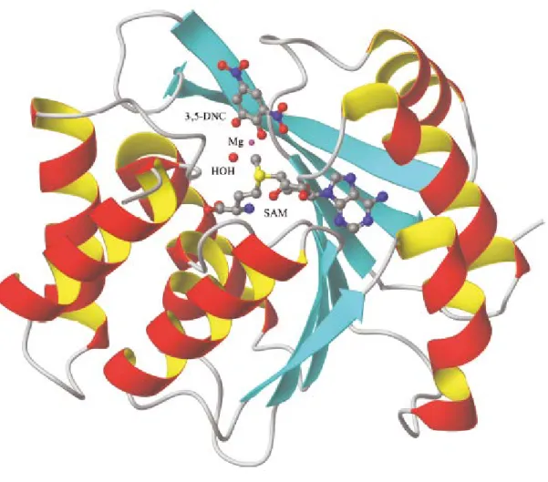
According to Figure 10, in the first step of reaction SAM binds to COMT, forming a stable complex. In the second step, the divalent metal cation bind to the complex, specifically by coordination of various acid residues in COMT, no prevailing a direct interaction between the magnesium and cofactor SAM [36]. In the last step, catechol substrate coordinates with the metal and establishes a network of hydrogen bonds with the active site residues [36]. After the formation of this complex, the methyl group of the cofactor SAM is ready to be transferred towards the hydroxyl group of the substrate [36]. This reaction mechanism has been reinforced when the structure of soluble COMT (Figure 11) in rats and humans was crystallized in the presence of SAM, Mg (II) and the substrate analogue, 3,5-dinitrocatechol [34, 36].
Figure 10 – A typical reaction catalyzed by catechol-O-methyltransferase (COMT). SAM -
Figure 11 - Schematic representation of the three-dimensional structure of COMT. The S-adenosyl-L-methionine co-substrate (SAM), the 3,5-dinitrocatechol (3,5-DNC), the magnesium ion, and coordinated water molecules are depicted. SAM - S-adenosyl-L-methionine; Mg – magnesium; DNC - 3,5-dinitrocatechol (adapted from [34]).
1.3.1 Physiological Functions
COMT is an important target for biotechnological engineering due to their role not only in normal brain function but also to their possible role in human neurological disorders such as Parkinson's disease (PD), schizophrenia, depression [26, 35, 38] and Alzheimer's disease [38]. In general, COMT physiological function is the inactivation of biologically active or toxic catechols [8]. Specifically, a major role of COMT includes its participation in the inactivation of the neurotransmitters in the central nervous system and regulation of both dopaminergic and noradrenergic systems. Additionally, COMT acts as enzymatic barrier, limiting the access of toxic catecholamines to certain biological compartments [33, 40]. The enzyme also acquires an important role in the metabolism of catecholestrogens and melanin and in the inactivation of toxic and carcinogenic compounds [8, 33]. The metabolic role of COMT is not
restricted to the inactivation of neurotransmitters [18] but also includes the inactivation of neuroactive xenobiotics that have a catechol group, and regulation of its quantity in brain and other organs [36]. Indeed, catecholamines regulation is quite important to the survival and well-being of the body [36].
The malfunction of COMT and its polymorphisms has been associated to several medical conditions, such as Parkinson disease, anxiety, substance abuse and schizophrenia [26, 35, 36, 38]. Thus, COMT role in regulation of catecholamines levels of in tissues, becomes an important target for various pharmaceutical drugs [36].
1.3.2 Isoforms: SCOMT and MBCOMT
In nature, COMT can be found in two molecular forms: a soluble form (SCOMT) and a membrane-bound form (MBCOMT) [26, 35]. In humans, SCOMT, the predominant isoform, is a non-glycosylated protein that contains 221 amino acid residues and has a molecular weight of 24.7 kDa. The membrane isoform, associated to endoplasmic reticulum, has an additional peptide of 50 amino acids in its amino terminus besides of 221 residues of the soluble isoform, corresponding to a final molecular weight of 30 kDa [4, 35, 38, 41]. This additional peptide contains a stretch of 21 hydrophobic residues, which comprises a membrane anchor region of the enzyme [4, 35].
Despite of similar characteristics of both isoforms, MBCOMT has a higher affinity for catecholamines substrates and a lower reaction rate, when compared with soluble isoform [4]. Indeed, MBCOMT has a 100-fold higher affinity for catechol substrates than SCOMT [8]. Therefore, this suggests that MBCOMT plays an important role in metabolism of catecholamines at physiological concentrations in vivo [4]. However, for the catechol estrogens, both isoforms present similar km values [4].
Concerning its structure, COMT is composed by a β chain of seven sheets, trapped between two sets of helices α, as seen in Figure 11 [23, 34]. The enzyme active site consists in a S-adenosyl-l-methionine binding domain and some amino acids extremely relevant for the binding of substrate, water and Mg2+ [23, 34]. For example, amino acid residues such as
Lys144 accept a proton of the hydroxyl group and residues such as Trp38, Trp143 and Pro174 form hydrophobic walls that define COMT selectivity for the substrate [23, 34].
1.3.3 Gene and localization of COMT isoforms
Both COMT isoforms are coded on a single gene but present different subcellular localizations [8, 33, 38]. Regarding its subcellular localization, SCOMT is present in cytoplasm while MBCOMT is associated to rough endoplasmic reticulum [8, 33, 38]. The MBCOMT, as an integral membrane protein, is associated to a membrane by its anchor region and with its catalytic portion oriented toward the cytoplasmic side of the membrane [8].
In humans and rats, a single gene with two distinct promoters encodes both COMT isoforms of COMT, and performs a translational regulation (Figure 12) [8, 33, 42]. The COMT gene is located on the long arm of chromosome 22 and is highly polymorphic [35, 38, 39, 42].
Through a translational regulation mechanism, COMT gene origins two primary transcripts using the two distinct promoters: an 1.5 kb transcript relative to MBCOMT and 1.3 kb transcript relative to SCOMT, from promoters P1 and P2, respectively.
Figure 12 - The COMT enzyme gene and their transcripts (adapted from [42]).
1.3.4 Genetic polymorphisms
Genetic polymorphisms, both in upstream and downstream regions or in COMT gene itself, are been associated to a variety of neuropsychiatric phenotypes [38, 39]. The most studied and relevant is Val108Met polymorphism, associated to Parkinson's disease [41, 43], Alzheimer's disease [38] and others [35]. However, there are other polymorphisms in the COMT gene, such as Ala22Ser (G/T), His12His (C/T), Leu86Leu (C/G and C/T) and Ala52Thr (G/A) in which COMT activity is not affected by mutation [38].
Recent studies have reported the relation of the brain structure with COMT polymorphisms. It suggests that functional variants of COMT may affect the volume of the brain in patients with schizophrenia and even among healthy individuals [44].
Another COMT functional polymorphism is located at codon 158 causing a substitution of amino acid valine (Val) by methionine (Met) (Val158Met) [39, 40, 44–46]. Results in three genotypes (Val/Val, Val/Met and Met/Met) shows that polymorphism can affect differentially COMT stability and cognitive flexibility [40, 46]. The COMT-Met allele had an enzymatic activity 40% lower than the COMT-Val allele, which is associated to elevated dopamine levels [40]. Thus, the variant Val/Val developed higher enzymatic activity, increasing the efficiency of elimination of dopamine from synaptic cleft and, as result, decreasing it levels [40]. Therefore, COMT enzyme appears to play an important role in the metabolism of dopamine in prefrontal cortex and in other brain regions [39, 40].
1.3.5 Stability of COMT
The protein COMT is highly unstable and loses rapidly its activity during isolation and storage [22, 33, 35]. For example, the recombinant hSCOMT can lose 80% of its activity after 30 minutes at physiological temperature [47] and 50-70% when kept at 4ºC during 24 h [33]. Therefore, low COMT activities are associated with its thermal stability [48, 49]. In addition to the temperature, the pH is also essential to ensure maintenance of catalytic activity and specifically for COMT optimum pH is achieved between 7.5 and 8.0 [6]. Moreover, differences between both isoforms in terms of stability were been found in a variety of tissues [47]. The enzyme contains few cysteine residues in its primary structure and is suggested that in secondary structure hydrogen bonding with the serine hydroxyl group makes cysteine’s particularly prone to oxidation [35]. Thus, a reasonable reason for COMT poor stability is the oxidation of thiol (-SH free groups of cysteine) and the consequent formation of intra- or intermolecular disulfide bridges [22, 35].
Some studies show that cofactor SAM and magnesium chloride (MgCl2) reduce cysteine
oxidation, preventing COMT inactivation, since these residues are essential to catalytic activity [35]. Additionally, experimental stability studies show that the combination of ethylenediaminetetraacetic acid (EDTA), β-mercaptoethanol and MgCl2 with a reducing agent
such as dithiothreitol (DTT) have a stabilizing effect in all enzyme preparations [6, 22, 35]. Frequently, the protein stabilization is also achieved by the use of protease inhibitors such as EDTA and diisopropyl fluorophosphate (DFP), which are currently applied in a lab scale, since host proteases activity is a current problem during the purification of proteins [6]. Other types of stabilizers may have a stabilizing effect on proteins, such as sugars, divalent metals, glycerol and some amino acids (glycine and proline), protecting them against loss of activity and thermal denaturation [50, 51].
1.3.6 COMT inhibitors
The design of inhibitors has been investigated since COMT discovery in 1958 [52]. Nowadays, COMT inhibitors are used in Parkinson’s disease therapy [26].
The first generation of COMT inhibitors such as tropolone, N-butylgallate and 2-hydroxylated estrogens presented a low efficacy and selectivity in vivo, little lasting effects and some toxicity [38]. The second-generation inhibitors include nitrocatechols as entacapone (OR-611), nitecapone (OR-462) and tolcapone (Ro 40-7592) [38]. The tolcapone and entacapone are currently used for the treatment of Parkinson's disease [26, 34]. These COMT inhibitors have beneficial effects in increasing the half-life of levodopa (L-Dopa), a drug used as substitute of dopamine [34, 38].
Since COMT discovery, many enzyme inhibitors have been developed and, nowadays, they are grouped as catechol-based COMT inhibitors (Type I), SAM-based COMT inhibitors (Type II), natural-products COMT inhibitors (Type III), bifunctional COMT inhibitors (Type IV), bisubstrate type of COMT inhibitors (Type V) and other types of COMT inhibitors (Type VI), according to similarities in its structure [38].
The type I class of inhibitors are based on catechol substructure and specific structural modifications resulted in a diverse range of compounds, including nitrocatechols, pyrogallol, salsolinol and polychlorinated biphenyl, among others [34, 38]. As mentioned above, tolcapone and entacapone (Figure 13) are clinically used for the treatment of Parkinson's disease [26, 53].
Figure 13 - Inhibitors of type I of class COMT inhibitors - Tolcapone and Entacapone, inhibitors currently
used in the treatment of Parkinson's disease (adapted from [38]).
The inhibitors of type II are analogous to SAH, the demethylated product of SAM, which is a potent inhibitor of several methyltransferases (Figure 14) [38].
Figure 14 - Structure of S-adenosyl-L-homocysteine (SAH) (adapted from [38]).
The type III class of inhibitors (Figure 15) are natural product derivatives, including flavonoids that possesses great inhibitory action against COMT [38]. Also, four catechins found in green tea have showed inhibitory activity: epigallocatechin gallate (EGCG), (-)-epicatechin-3-gallate (ECG), epigallocatechin (EGC), (-)-epicatechin [38]. Also, coffee constituents such as chlorogenic acid, caffeic acid and caffeic acid phenethyl ester (CAPE) are effective inhibitors of COMT [38]. Additionally, natural products extracted from various plants, such as Cistus parviflorus, Vitex agnus-cactus and seeds of Peganum harmala have been reported due to their inhibition effects in COMT activity [54].
The type IV class of inhibitors (Figure 16) are duplicated substructures with capacity to interact with binding domains of the enzyme [38]. Some of this inhibitors are d-catechin, desmethylpapaverine and nordihydroxyguaiaretic acid [38]. Recently, bifunctional inhibitors were designed with dual substituted catechols and were mostly catechol derivatives such as 3,4- dihydroxybenzamide or 3,4,5-trihydroxybenzamide, linked by a spacer section consisting of various methylene units [34, 38].
Figure 16 - Structure of two duplicates substructures - an example of Class IV inhibitors (adapted from
[38]).
The type V class of inhibitors includes bisubstrate inhibitors, which are designed by replacing both the cofactor SAM and the catechol substrate (Figure 17) [34, 38].
Figure 17 - Structure of the first substrate, inhibitor class of type V (adapted from [38]).
Finally, type VI of COMT inhibitors comprise all inhibitors that structurally are not related to any of the classes referred above [38]. These class includes tropolones (Figure 18), benzotropolones and many others inhibitors [38].
Figure 18 - Constitution of tropolone, an inhibitor of type VI (adapted from [38]).
1.4 Properties of COMT isoforms
The COMT has become an interesting biopharmaceutical target due to it association to some human disorders, particularly in Parkinson disease where the development of new COMT inhibitors lead to significant improvements in diseases prophylaxis [34, 53].
Initially, it was thought that MBCOMT was only an artifact, resulting from nonspecific binding of soluble form to cellular membrane [55]. However, MB-COMT represents a biochemically distinct molecular entity despite both isoforms share some similarities [55]. As referred above, MB-COMT has much lower capacity and Km value than the soluble form but a higher affinity to catecholamines [48, 55]. Thus, at low substrate concentrations, MB-COMT, a high affinity isoform, is probably the predominant isoform [55]. On the other hand, when substrate levels are raised above saturation, S-COMT activity increases and possibly prevails over MBCOMT activity [55].
Furthermore, there are other differences between the two isoforms such as pI values, which is 6.2 for MBCOMT and 5.2 for soluble form [55]. Although both COMT isoforms display considerable similarities specific differences in it kinetic and biochemical properties are evident [55].
1.4.1 Biosynthesis of hSCOMT and MBCOMT
The bacterial expression system is a challenging research area, although it has as advantages the relatively low cost and safety, when compared to eukaryotic systems such as mammalian cell cultures [26]. Indeed, expression systems such as E. coli have been extensively used as hosts for the expression of foreign proteins due to its rapid growth rate and capacity for continuous fermentation [26, 33].
As matter of fact, the recombinant hSCOMT was successfully produced in its active catalytically form in E. coli SG 13009 [33], DH-5 [33] and BL21-(DE3) Gold strains using
Isopropylthiogalactosidase (IPTG) as an inducible promoter [26, 56]. This recombinant system is an adequate host for expression of recombinant SCOMT since it is completely lacks endogenous COMT enzyme [33]. Indeed, with an optimized heterologous expression system in E.coli for recombinant hSCOMT production it was possible to produce and recover the active monomeric enzyme directly from cell crude culture using as lysis step freeze/thaw cycles or ultrasonication [26].
Furthermore, the expression of recombinant hSCOMT was also possible in insect cells using the baculovirus expression system [33].
Regarding hMBCOMT recombinant, several expression systems have been used to produce considerable amounts of the membrane enzyme for structural, function and pharmacological studies [4]. However, in some systems is impossible to express this recombinant protein, since its hydrophobic sequence can be toxic to bacteria [4]. Nevertheless, recombinant hMBCOMT was successfully produced in a catalytically active form and at relatively high levels in strains E. coli BL21 and SG 13009 [4].
Also other systems, have been described for recombinant expression of hMBCOMT, eukaryotic expression systems such as Sf9 insect cells [33], transfected human embryonic kidney fibroblast cell lines and hamster BHK-cells [4]. Despite of advantages associated, a drawback is the fact that hMBCOMT was expressed with low levels of biological activity [4].
The expression system based in Brevibacillus choshinensis cells is suitable to the production and secretion of heterologous proteins with high efficiency and presents a single membrane and low levels of protease activity [4]. Brevibacillus choshinensis is a gram-positive microorganism that shows an excellent capacity to produce several extracellular proteins [57]. In addition, this expression system allows the secretion of heterologous proteins with high efficiency and, as it produces a negligible amount of extracellular proteases, target bioproducts remain unscathed in the culture medium [57]. In sum, this system presents several advantages, becoming a system amenable to genetic engineering [57].
Nowadays, a rapid grow of knowledge in area of biogenesis of MPs has created new possibilities to design new strategies for improving yields in terms of host performance and protein production levels [4]. Brevibacillus recombinant expression system allows the production and recovery of human recombinant MBCOMT in a single procedure with a biological and immunological active form [4].
1.4.2 Purification of hSCOMT and MBCOMT
Over the past years, the human SCOMT has extensively studied and subjected to numerous purification procedures [22, 23, 26, 33, 47, 58, 59]. The SCOMT was subject to chromatographic processes such as hydrophobic interaction chromatography [22, 26, 47], bioaffinity chromatography [23, 33], size exclusion, anion and cation exchange chromatography [33, 58, 59].
Anionic exchange chromatography (AEC), a well-established technique widely used in isolation of soluble and membrane proteins (MPs) [6, 31], was one of the most usual chromatographic techniques applied to SCOMT isolation [33, 58, 59]. Although some of these purification procedures presented as drawback significant COMT activity and yield losses, some of their led to the successful resolving of SCOMT atomic structure [33]. Additionally, usual combination of AEC with others purification steps contributed to reinforce these issues [26, 33].
Regarding to hMBCOMT, some through kinetic and biochemical characterization has been carried out [34, 55], but few studies have been published concerning it isolation and so its structure remains unknown [4, 30, 34, 48, 60].
Recently, some work was developed on MBCOMT purification by AEC, where the authors describe MBCOMT recovery from microssomal fraction of rat liver homogenates [8, 60]. Specifically, hMBCOMT was partially isolated from the microsomal fraction of rat liver homogenates using a Resource Q column [8]. This strategy involved hMBCOMT solubilization with Triton X-100 and its purification was also performed using Triton X-100 and an increased sodium chloride concentration gradient for target protein elution [8].
Also, a new purification strategy were studied for recombinant human COMT (hMBCOMT) isolation from crude Brevibacillus choshinensis cell lysates, comparing different hydrophobic ligands such as octyl, butyl and epoxy [30]. In case of octyl and butyl ligands, the hMBCOMT’s adsorption was performed at moderates salt concentrations, whereas on epoxy was used higher concentrations for their adsorption [30]. In addition, the hMBCOMT elution in octyl and epoxy was promoted using Triton X-100 and in butyl media besides the use of Triton X-100 was used a monosodium phosphatedecrease [30].
In this work, chromatographic conditions required for each isoform isolation were compared and the results show that, using the same hydrophobic resins, lower ionic strength is required for MBCOMT’s adsorption than for SCOMT [22, 30].
So, similarly to hMBCOMT’s behavior on chromatographic hydrophobic adsorbents [30], it’s likely that the additional amino acid residues in membrane anchor region domain, may contribute for the reinforcement of electrostatic interactions establish between MBCOMT and anion exchangers when compared with soluble isoform. Thus, it may be interesting to study the differences between both isoforms in terms of ionic interactions that can be established between each COMT isoform and an ionic exchanger, in a chromatographic procedure.
In this work it is intended to study, compare and assess the chromatographic behavior of both COMT isoforms using quaternary ammonium (Q-sepharose) as anion exchanger, in order to analyze Q-Sepharose performance in terms of binding and elution conditions for hSCOMT and hMBCOMT recovery. Furthermore, is also intended to determine enzyme recovery, activity and procedure yield by HPLC analysis.
Chapter II
Materials and Methods
2.1 Materials
Ultrapure reagent-grade water for Fast Performance Liquid Chromatography system (FPLC) and ÄKTA™ avant was obtained with a Mili-Q system (Milipore/Waters). Neomycin (trisulphate salt hydrate), Carbenicillin disodium salt, isopropylthiogalactosidase (IPTG), tryptone, Bacto yeast extract, Glucose, Calcium chloride dehydrate, lysozyme, Deoxyribonuclease (DNase), dithiothreitol (DTT), Cysteine (L-), Sucrose, DL-Metanephrine Hydrochloride (MN), citric acid monohydrate, 1-Octanesulfonic acid and bovine serum albumin (BSA) were obtained from Sigma Chemical Co. (St. Louis, MO). Potassium chloride and sodium acetate (anhydrous) were supplied by Fluka (Buchs, Switzerland). Bacto soytone and polypeptone were obtained from Becton Dickinson (NJ, USA). Tris(hydroxymethyl)aminomethane (Tris) and Acetonitrile (HPLC grade) was obtained from Fisher scientific (Epson, United Kingdom). Sodium dihydrogen phosphate (NaH2PO4) and Glycerol were obtained from Riedel-de-Haën (Hanover, Germany) and Himedia (Mumbai, India), respectively. Sodium chloride (NaCl) and perchloric acid was purchased from Panreac (Barcelona, Spain). NZYcolour Protein Marker II used for estimation of subunit molecular weight was purchased in NZYTech (Lisboa, Portugal). Anti-rabbit IgG alkaline phosphatase secondary antibody were purchased on GE Healthcare Biosciences (Uppsalla, Sweden). Monoclonal rabbit anti-COMT antibody was produced in BIAL (S. Mamede do Coronado, Portugal) using purified recombinant rat COMT. All chemicals used were of analytical grade commercially available and used without further purification.
2.2 Plasmids, bacterial strains and media
The Champion pET101 Directional TOPO expression kit (Invitrogen Corporation, Carlsbad, CA, U.S.A.) was used for the expression of hSCOMT on E. coli BL21 (DE3) strain according to the manufacturer’s instructions and as previously described [26]. The SOB medium [20 g/L triptone, 5 g/L yeast extract, 0.5 g/L NaCl, 2.5 mM KCl, 10 mM MgCl2 with 50 µg/mL
cabenicillin] was used for E. coli growth.
Plasmid pNCMO2-hMBCOMT was used for hMBCOMT expression on Brevibacillus expression system [Takara Bio Inc. (Otsu, Japan)] according to the manufacturer’s instructions and as previously described [4]. Typically, B. choshinensis cells were grown in 2SYNM medium (20.0 g/L glucose, 40.0 g/L Bacto Soytone, 5.0 g/L Bacto Yeast Extract, 0.15 g/L CaCl2.2H2O and
Bacto Yeast extract, 10.0 mg/L FeSO4.7H2O, 10.0 mg/L MnSO4.H2O, 1.0 mg/L ZnSO4.7H2O, 4.1
g/L MgCl2 and 50 μg/mL Neomycin for MTNm liquid medium; 3.75 g/L Agar and 10.0 μg/mL
Neomycin).
2.3 Recombinant COMT isoforms biosynthesis and recuperation
The biosynthesis of recombinant hSCOMT was performed according to the following protocol. E. coli cells transformed with the expression construct were grown overnight at 37°C in LB medium plates containing 50 µg/mL cabenicillin. A single colony was precultivated in 62.5 mL of SOB medium in 250 mL shake flasks. Cells were grown at 37ºC and 250 rpm until the optical density at 600 nm (OD600) reached 2.6, corresponding to middle of log phase. This bacterial preculture was then diluted using fresh SOB medium (250 mL) in a 1 L erlenmeyer flask, in order to achieve an initial OD600 nm of 0.2–0.3 units. When the OD600 nm of culture reached 0.7, the production of recombinant hSCOMT was induced by the addition of IPTG in a final concentration of 1 mM. After a 4 h growth at 37ºC, cells were harvested by centrifugation (7000×g, 15 min, 4ºC) and stored frozen at−80ºC until use [26, 56].
The supernatant obtained from centrifugation were resuspended in 5 mL of lysis buffer (150 mM NaCl, 10 mM DTT, 50 mM Tris pH 8.0) with protease inhibitors (5 µg/mL leupeptin and 0.7 µg/mL pepstatin) and COMT stabilizers (75 mM DTT, 250 mM sucrose, 150 mM cysteine and 20% glycerol). In cellular lysis, lysozyme (10 mg/ml) was added to extract for 15 minutes at room temperature, followed by six freeze (-196 ºC in liquid nitrogen)/thaw (42ºC) cycles. Then, DNAse (1,0 mg/ml) was added to the lysate and the supernatant resulting from centrifugation (16000xg, 20 min, 4ºC) was stored until use [26, 56].
Unless otherwise stated, recombinant hMBCOMT was carried out according to the following protocol. The B. choshinensis cells transformed with the expression construct were grown overnight at 37ºC in MTNm plates. A single colony of transformed cells was precultivated in 62.5 mL of 2SYNM medium in 250 mL shake flasks. Cells were grown at 30ºC and 120 rpm until cell density at 660 nm (OD660) reached 2.6. After grown time, an aliquot of pre-inoculum was diluted using 2SYNm medium on 500 mL shake flasks, corresponding to an initial OD660 of 0.2 units in a final volume of 125 ml. After a 48h growth at 30ºC and 120 rpm, cells were harvested by centrifugation (5000xg, 25 min, 4ºC) and stored frozen at -20.0ºC until use [4]. The bacterial cell pellet obtained was incubated with 5 mL of lysis buffer (150 mM NaCl, 10 mM DTT, 50 mM Tris, 1 mM MgCl2, pH 8.0) complemented with protease inhibitors (5.0 μg/ml leupeptin and 0.7 μg/ml pepstatin) and COMT stabilizers (75 mM DTT, 250 mM sucrose, 150 mM cysteine and 20% glycerol), disrupted by lysozyme treatment (10 mg/ml) for 15 minutes at room temperature and followed by six freeze (-196 ºC in liquid nitrogen)/thaw (42ºC) cycles. Then, DNAse (1,0 mg/ml) was added to the lysate and the soluble material removed by centrifugation (16000xg, 20 min, 4ºC). Full solubilization was carried out by pellet incubation with lysis buffer at 4ºC, overnight.
2.4 Anionic Chromatography
The chromatographic assays were performed at room temperature (25oC) in an ÄKTA Avant
system with UNICORN 6Software (GE Healthcare, Uppsala, Sweden) equipped with a 2 mL injection loop and in a FPLC system (GE Healthcare Biosciences, Uppsalla, Sweden) consisting of a double-piston pumps P500, an MV 7 injection valve equipped with a 2 mL injection loop, a fraction collector FRAC 100, a dual path monitor UV-1 and a data processor 112, controlled by a LCC 501 Plus controller. All buffers pumped into the system were prepared with Mili-Q system water, filtered through a 0.20 µm pore size membrane (Schleicher Schuell, Dassel, Germany) and degassed ultrasonically.
Q-Sepharose (GE Healthcare Biosciences) was packed according to company guide-lines (20 mL of gel volume) into a C 16/20 [16 mm (diameter) x 200 mm (length)] glass column purchased from GE Healthcare Biosciences. Screening experiments were performed at different salt concentrations in order to assess sodium chloride concentration required for hSCOMT and hMBCOMT’s retention. Unless otherwise stated, column was initially equilibrated with 10 mM Tris HCl.
Aliquots of recombinant hSCOMT containing supernatant (2 mL with a protein concentration 3,65mg/mL) were injected into column at 1 ml/min with 10 mM Tris-HCl. After elution of unretained species, the concentration of sodium chloride was increased to 350 mM in a step mode for 2.5 column volume (CV). Subsequently, sodium chloride concentration in mobile phase was increased to 1M (2.5 CV).
Solubilized pellet containing hMBCOMT (2 mL with a protein concentration 2.7mg/mL) was injected into the column at 1 ml/min with 10 mM Tris-HCl. The elution of unretained species occurred with an increasing sodium chloride gradient from 0 mM to 100 mM (2 CV). After elution of unretained species, sodium chloride concentration was increased to 300 mM in a step mode (2.5 CV). Subsequently sodium chloride concentration in mobile phase was gradually increased from 350 mM to 1M NaCl in 10 mM Tris buffer (2,5CV). Finally, a washing step was applied with 1M of sodium chloride in Tris buffer 10 mM (1CV).
Otherwise, in an alternative chromatographic procedure was used 0.5 % Triton X-100 in 10 mM Tris HCl in adsorption step, followed by a step at 300 mM NaCl in 0.5% of detergent. Thereafter, a final salt increasing gradient from 350 mM to 1M NaCl and 0.5% Triton X-100 in 10 mM Tris buffer was applied.
In all chromatographic runs, conductivity was continuously monitored, as well as absorbance at 280 nm. Fractions volumes of 3 mL were collected and pooled according to the chromatograms profile obtained and stabilized in a suitable solution composed by 75 mM DTT, 150 mM of cysteine, 250 mM sucrose and 20% of glycerol in an adequate buffer. Finally, samples were concentrated and desalted with Macrosep® Advance centrifugal devices with Omega™ membrane from VWR (Carnaxide, Portugal) and conserved at 4oC until further
![Figure 1 – Structure of an ionic detergent type, Sodium Dodecyl Sulfate (SDS) (adapted from [7])](https://thumb-eu.123doks.com/thumbv2/123dok_br/18901545.935345/16.892.314.649.581.735/figure-structure-ionic-detergent-sodium-dodecyl-sulfate-adapted.webp)
![Figure 4 - Structure of zwitterionic detergents (adapted from [7]).](https://thumb-eu.123doks.com/thumbv2/123dok_br/18901545.935345/18.892.219.746.305.497/figure-structure-zwitterionic-detergents-adapted.webp)
![Figure 5 - Phases of a typical chromatographic procedure (adapted from [2]).](https://thumb-eu.123doks.com/thumbv2/123dok_br/18901545.935345/21.892.159.815.133.497/figure-phases-typical-chromatographic-procedure-adapted.webp)
![Table 1 - Groups of ion exchangers used for the isolation of proteins (adapted from [21])](https://thumb-eu.123doks.com/thumbv2/123dok_br/18901545.935345/24.892.136.793.198.742/table-groups-ion-exchangers-used-isolation-proteins-adapted.webp)
![Figure 7 - Scale of counter ions with high affinity or low-affinity relatively to anion/cation exchanger applied (adapted from [6])](https://thumb-eu.123doks.com/thumbv2/123dok_br/18901545.935345/25.892.169.775.692.990/figure-counter-affinity-affinity-relatively-exchanger-applied-adapted.webp)
![Figure 8 - Typical chromatographic profile obtained from anion exchange chromatography (adapted from [2])](https://thumb-eu.123doks.com/thumbv2/123dok_br/18901545.935345/26.892.181.774.773.1082/figure-typical-chromatographic-profile-obtained-exchange-chromatography-adapted.webp)
![Figure 9 - Effect of pH on IEC elution strategies of various compounds (adapted from [2] )](https://thumb-eu.123doks.com/thumbv2/123dok_br/18901545.935345/27.892.299.693.394.909/figure-effect-iec-elution-strategies-various-compounds-adapted.webp)
![Figure 10 – A typical reaction catalyzed by catechol-O-methyltransferase (COMT). SAM - S-adenosyl-l- S-adenosyl-l-methionine; Mg 2+ - Magnesium (adapted from [38] )](https://thumb-eu.123doks.com/thumbv2/123dok_br/18901545.935345/28.892.143.820.721.917/reaction-catalyzed-catechol-methyltransferase-adenosyl-adenosyl-methionine-magnesium.webp)
![Figure 12 - The COMT enzyme gene and their transcripts (adapted from [42]).](https://thumb-eu.123doks.com/thumbv2/123dok_br/18901545.935345/31.892.189.789.512.782/figure-comt-enzyme-gene-transcripts-adapted.webp)