Ciências da Saúde
The protective effect of regucalcin against
radiation-induced testicular damage
Ana Manuela dos Santos Silva
Dissertação para obtenção do Grau de Mestre em
Ciências Biomédicas
(2º ciclo de estudos)
Orientador: Prof. Doutor Cláudio Maia
Co-orientador: Prof.ª Doutora Sílvia Socorro
iii
Agradecimentos
Antes de mais, começo por agradecer a todos aqueles que, de alguma forma, me transmitiram algum ensinamento ao longo da vida, contribuindo assim, em todos os sentidos, para a minha formação pessoal e profissional.
Ao Professor Doutor Cláudio Maia, meu orientador, e à Professora Doutora Sílvia Socorro, minha co-orientadora, que desde sempre se mostraram bastante acessíveis e disponíveis, por todo o interesse e dedicação, revelando-se incansáveis. Também por todo o conhecimento transmitido, e pelas sugestões e críticas construtivas, sem os quais seria impossível evoluir. Pelo contributo minucioso, bem como pela competência e rigor científico, motivando-me a tentar fazer sempre melhor.
À Fundação para a Ciência e Tecnologia, pelo financiamento através do programa COMPETE (Project PEst-OE/SAU/UI0709/2014).
Ao Instituto de Imagem Biomédica e Ciências da Vida (IBILI) e ao Centro Hospitalar e Universitário de Coimbra, nas pessoas da Professora Doutora Filomena Botelho, do Dr. João Casalta, e da Mestre Ana Mamede, cuja colaboração foi indispensável para tornar este projecto viável.
À Mestre Catarina Ferreira, Técnica de Anatomia Patológica, Citológica e Tanatológica, pela assistência no processamento dos tecidos para análise histológica, e também a todos os responsáveis pelo bom funcionamento do Biotério, do Centro de Investigação em Ciências da Saúde e da Faculdade de Ciências da Saúde da Universidade da Beira Interior, pela simpatia, boa disposição e disponibilidade sempre demonstradas.
Aos meus colegas e amigos de laboratório, Tânia, Luís, Inês, Margarida, Carlos, Rúben, Filipe, Ricardo, Cátia, Marília e Henrique por me terem recebido e acompanhado tão bem desde o primeiro dia. Pelo bom ambiente de trabalho, espírito de entreajuda, troca de opiniões, desabafos, conselhos, preocupação, e paciência para responderem às minhas infindáveis questões. Foi, sem dúvida, uma sorte e um prazer trabalhar convosco.
À Sara, que esteve activamente envolvida nas diversas fases e tarefas desta dissertação, não poderia deixar de atribuir o devido reconhecimento. Os meus primeiros passos na investigação, incluindo os mais variados e inúmeros aspectos implicados que não me são aqui possíveis descrever, devo-os, especialmente, a ti. Por ao longo deste projecto te teres tornado num exemplo, e por tudo o resto, estou extremamente grata.
iv
Aos meus amigos mais próximos, que ao longo destes anos se têm mostrado realmente verdadeiros, marcando presença quer nos bons quer nos maus momentos.
Ao Ricardo, por ter o dom de me acalmar, pela paciência, compreensão, motivação, e por acreditar em mim.
Aos meus pais e avós, pelo amor e apoio incondicional que nunca será passível de descrever, sendo a maior dádiva que poderia alguma vez receber.
A todos os que seguiram de perto esta dissertação, o meu profundo e sincero agradecimento. Este trabalho também é vosso, porque tudo se torna mais leve quando temos as pessoas certas do nosso lado.
v
Resumo
O cancro testicular é a malignidade masculina mais frequente nos jovens, e a radioterapia é normalmente usada no seu tratamento. No entanto, a exposição à radiação tem vários efeitos secundários na fertilidade masculina, tornando-se necessária a identificação de estratégias efectivas para a proteger do dano testicular provocado pela radioterapia. A regucalcina (RGN)
é uma proteína de ligação ao cálcio (Ca2+) que se encontra amplamente expressa no tracto
reprodutor masculino. Vários estudos demonstraram a capacidade supressora da RGN na morte celular de diferentes tipos de células. Anteriormente, o nosso grupo de investigação mostrou que a sobre-expressão de RGN teve efeitos benéficos na espermatogénese por suprimir a apoptose induzida quimicamente. Para além disso, a RGN é regulada positivamente em linhas celulares radiorresistentes, sugerindo que esta pode proteger de danos causados pela radiação. O presente trabalho visa avaliar se a RGN desempenha um papel benéfico na recuperação da espermatogénese após radioterapia. Ratos, quer transgénicos que sobre-expressam a RGN (Tg-RGN) quer os seus homólogos selvagens (Wt), foram expostos a raios-X. Às dez semanas de recuperação após a radioterapia, o estado testicular e os parâmetros espermáticos foram avaliados. A expressão da RGN, bem como de vários reguladores do ciclo celular e da apoptose foi também avaliada. Para além disso, a actividade enzimática da caspase-3 foi determinada. Às dez semanas de recuperação após a radioterapia, tanto o estado testicular como os parâmetros espermáticos parecem ter sido menos afectados pelos raios-X nos Tg-RGN. Verificou-se ainda uma diminuição da expressão de p53 e de p21, o que pode indicar a reiniciação da espermatogénese. Para além disso, a reduzida actividade da caspase-3 detectada nos Tg-RGN está de acordo com os baixos níveis de caspase-8 e com o elevado rácio Bcl-2/Bax, sugerindo que os Tg-RGN são mais resistentes à apoptose testicular em resposta à radiação. A expressão de RGN aumentou significativamente nos ratos Wt, suportando o seu envolvimento na resposta anti-apoptótica. De forma geral, estes resultados indicam que a sobre-expressão da RGN desempenhou um papel protector relativamente ao dano testicular induzido pela radiação.
Palavras-chave
Testículo, Epidídimo, Espermatogénese, Espermatozóides, Radioterapia, Preservação da fertilidade, Regucalcina, Dano testicular, Apoptose, Oncofertilidade masculina.
vii
Resumo Alargado
O cancro testicular é a malignidade masculina mais frequente nos jovens, apresentando mais de 10 novos casos por 100.000 homens por ano na Europa, e uma mortalidade de 0.3 casos por cada 100.000 homens por ano. A radioterapia é normalmente usada no tratamento do cancro testicular, mas a exposição à radiação tem vários efeitos secundários na fertilidade masculina porque os testículos são dos órgãos mais sensíveis à radiação devido à elevada taxa de divisão das células germinativas. Considerando a idade de incidência do cancro testicular e as suas elevadas taxas de sobrevivência, a maioria dos doentes ambiciona ainda ter filhos no futuro. Para além disso, os problemas reprodutivos são considerados pelos doentes uma das consequências mais frequentes e angustiantes do tratamento oncológico, tornando-se necessária a identificação de estratégias efectivas para os proteger do dano testicular
provocado pela radioterapia. A regucalcina (RGN) é uma proteína de ligação ao cálcio (Ca2+)
que se encontra amplamente expressa no tracto reprodutor masculino quer em rato quer em humano, incluindo nas vesículas seminais, epidídimos, e testículos. Vários estudos demonstraram a capacidade da RGN como supressora da morte celular em diferentes tipos de células. Anteriormente, o nosso grupo de investigação mostrou que a sobre-expressão de RGN teve efeitos benéficos na espermatogénese, nomeadamente, na supressão da apoptose induzida por fármacos como a tapsigargina ou actinomicina D. Para além disso, a RGN é regulada positivamente em linhas celulares radiorresistentes, sugerindo que esta proteína pode proteger de danos causados pela radiação. O presente trabalho visa avaliar se a RGN desempenha um papel benéfico na recuperação da espermatogénese após radioterapia. Com esse intuito, ratos com três meses, quer transgénicos que sobre-expressam a RGN (Tg-RGN) quer os seus homólogos selvagens (Wt), foram submetidos a uma dose única de 6 Gray (Gy) de raios-X. Às dez semanas de recuperação da radioterapia, o estado testicular e os parâmetros espermáticos da cauda do epidídimo foram analisados. Os animais e os seus testículos foram pesados, permitindo o cálculo do índice gonado-somático (GI). Os testículos foram processados histologicamente de modo a determinar o índice de diferenciação tubular (TDI), ou usados para análise da expressão de genes. A expressão da RGN, bem como de vários reguladores do ciclo celular e da apoptose foi avaliada por Western Blot (WB). Além disso, também a medição colorimétrica da actividade enzimática da caspase-3 foi incluída como um ponto final da apoptose.
Às dez semanas de recuperação da radioterapia, o GI estava diminuído tanto nos animais Tg-RGN como nos Wt, mas esta diminuição parece ser menor no Tg-Tg-RGN. Em resposta aos raios-X, também foi observada uma diminuição no TDI. Também a contagem, a motilidade, e a viabilidade dos espermatozóides parecem ter sido menos lesadas nos Tg-RGN. Quanto à morfologia dos espermatozóides, a percentagem de espermatozóides normais nos Tg-RGN parece ter sido menos afectada pela radiação, incluindo uma reduzida variação na
viii
percentagem de defeitos de pescoço e de cauda. Embora a diminuição da expressão de p53 e de p21 verificada às dez semanas de recuperação após a radioterapia seja provavelmente independente dos níveis de expressão de RGN, estes resultados sugerem a activação do ciclo celular e a reiniciação da espermatogénese. Considerando o reconhecido aumento da apoptose em resposta à radiação, a expressão do receptor de Fas (FasR) e do seu ligando (FasL) diminuiu inesperadamente. Contrariamente ao observado nos ratos Wt, a expressão de caspase-8 aumentou apenas moderadamente nos Tg-RGN, sugerindo uma menor susceptibilidade à apoptose. Em concordância, após a radioterapia, o rácio da expressão de proteína Bcl-2 (anti-apoptótica)/Bax (pró-apoptótica) foi significativamente mais alto nos ratos Tg-RGN, o que sugere também uma menor taxa de apoptose. Para além disso, ao contrário dos Wt, não foi observada diferença significativa na actividade da caspase-3 nos Tg-RGN, sugerindo menores níveis de apoptose testicular também nestes animais. Curiosamente, verificou-se ainda que a exposição à radiação aumentou significativamente a expressão de RGN nos ratos Wt, o que suporta o envolvimento desta proteína na resposta anti-apoptótica. De forma geral, os resultados obtidos indicam que a sobre-expressão da RGN desempenhou um papel protector relativamente ao dano testicular induzido pela radiação, provavelmente por suprimir a apoptose das células testiculares. Apesar de serem necessários mais estudos, estes resultados despertam a curiosidade acerca da manipulação dos níveis de RGN nos testículos, no sentido de poder ajudar a preservar a fertilidade de doentes do sexo masculino submetidos a tratamentos oncológicos.
ix
Abstract
Testicular cancer is the most common malignancy among young men, and radiation therapy is a generally used as treatment. However, the exposure to radiation has several adverse effects on male fertility. Considering the high survival rates and the age of incidence of this malignancy, it is mandatory to identify effective strategies protecting against
radiation-induced testicular damage. Regucalcin (RGN) is a calcium (Ca2+)-binding protein that is
broadly expressed in the male reproductive tract. Several studies have demonstrated RGN ability suppressing cell death in different cell types. Previously, our research group showed that RGN overexpression had beneficial effects on spermatogenesis by suppressing chemical-induced apoptosis. Moreover, RGN is upregulated in radioresistant cell lines, suggesting that it also can protect from radiation damage. The present work aimed to evaluate whether RGN may play a role in spermatogenesis recovery after radiation treatment. For this purpose, transgenic rats overexpressing RGN (Tg-RGN) and their wild-type (Wt) counterparts were exposed to X-rays. At ten weeks of recovery after irradiation, the testicular status and the epididymal sperm parameters were evaluated. The expression of RGN and several cell cycle and apoptosis regulators was also evaluated. In addition, the enzymatic activity of caspase-3 was measured. Upon radiation treatment and ten weeks of recovery, both the testicular status and sperm parameters seem to have been less affected by X-rays in Tg-RGN. We also found a diminished expression of p53 and p21, which may indicate the reinitiating of spermatogenesis. Moreover, the reduced activity of caspase‐3 detected in Tg‐RGN is in accordance with low levels of caspase-8 and increased Bcl‐2/Bax ratio, suggesting that these animals are more resistant to testicular apoptosis in response to radiation. RGN expression was significantly enhanced in Wt rats after irradiation, supporting its involvement in the anti-apoptotic response. Altogether, the present findings point out a protective role for RGN overexpression against radiation-induced testicular damage.
Keywords
Testis, Epididymis, Spermatogenesis, Sperm, Radiation therapy, Fertility preservation, Regucalcin, Testicular damage, Apoptosis, Male oncofertility.
xi
List of Contents
I. Introduction ... 1
1. Brief overview of the testicular, epididymal and sperm structure ... 3
2. Testicular and epididymal physiology ... 7
2.1 Spermatogenesis ... 7
2.2 Control of spermatogenesis... 8
2.3 Sperm maturation ... 11
3. Testicular damage and male (in)fertility ... 13
3.1 Testicular cancer ... 13
3.2 Cancer treatment ... 13
4. Preservation of male fertility and spermatogenesis recovery after testicular damage .. 14
4.1 Cryopreservation ... 14
4.2 Hormonal and non-hormonal factors ... 14
5. Regucalcin protein as a protective molecule in reproductive function ... 18
II. Aim of the thesis ... 21
III. Material and Methods ... 25
1. Animals ... 27
2. Radiation Treatment... 27
3. Tissue Collection ... 28
4. Epididymal Sperm Count and Motility ... 28
5. Epididymal Sperm Viability and Morphology Analysis ... 29
6. Testicular Histological Analysis ... 29
7. Total Protein Extraction and Quantification ... 30
8. Western Blot ... 30
9. Caspase-3 Activity Assay ... 30
10. Statistical Analysis ... 31
xii
1. Spermatogenic status at ten weeks of recovery after radiation treatment shows less
injury in Tg-RGN ... 35
2. Epididymal sperm parameters of Tg-RGN were less damaged by radiation treatment ... 37
2.1 Epididymal sperm count ... 37
2.2 Epididymal sperm motility and viability ... 37
2.3 Epididymal sperm morphology ... 39
3. Testicular expression of cell cycle and apoptosis regulators at ten weeks of recovery after radiation treatment ... 41
3.1 The expression of p53 and p21 was decreased in the testis in response to radiation treatment ... 41
3.2 The Bcl-2/Bax protein ratio is higher in rats exposed to radiation treatment ... 42
3.3 The expression of FasL and FasR was decreased in the testis of Wt and Tg-RGN rats after radiation treatment ... 44
3.4 The expression of caspase-8 was lower in the testis of Tg-RGN rats comparatively with Wt rats after radiation treatment ... 44
3.5 The enzymatic activity of caspase-3 was diminished in Tg-RGN after radiation treatment ... 45
3.6 The expression of RGN is enhanced in both experimental groups after radiation treatment ... 46
V. Discussion ... 49
VI. Conclusions and Future Perspectives ... 57
VII. References ... 61
VIII. Publications and Communications ... 79
1. Publication in International Peer-Reviewed Journal ... 81
xiii
List of Figures
Figure I-1. Schematic representation of the mammalian testis and its relationship with the
epididymis ... 4
Figure I-2. Schematic organization of the rat epididymis ... 5
Figure I-3. Schematic representation of the structure of mammalian spermatozoa ... 6
Figure I-4. Schematic representation comparing the human (A) and rat (B) spermatozoa ... 6
Figure I-5. Schematic representation of the testicular histology and mammalian spermatogenesis ... 8
Figure I-6. The endocrinology of the testes ... 9
Figure I-7. Extrinsic and intrinsic pathways of apoptosis ... 11
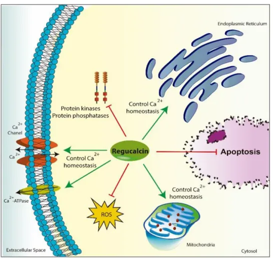
Figure I-8. The role of regucalcin (RGN) in cell biology ... 20
Figure III-1. Planning representation for testicular irradiation ... 28
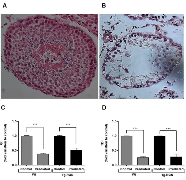
Figure IV-1. Representative photomicrographs of a differentiating (A) and non-differentiating (B) seminiferous tubule stained with H&E (400x magnification; Zeiss), and the effect of radiation treatment in GI (C) and TDI (D) both in Wt and Tg-RGN animals ... 36
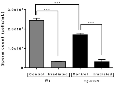
Figure IV-2. Effect of radiation treatment on sperm counts in Wt and Tg-RGN animals ... 37
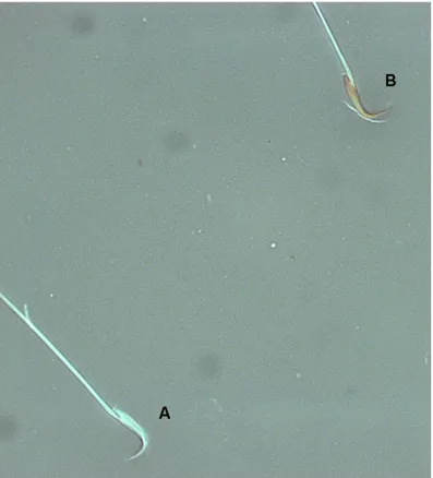
Figure IV-3. Representative photomicrographs of viable (A) and non-viable (B) rat sperm stained with Eosin-Nigrosin technique (1000x magnification; Zeiss). ... 38
Figure IV-4. Effect of radiation treatment on epididymal sperm motility (A) and viability (B) in Wt and Tg-RGN animals ... 39
Figure IV-5. Representative photomicrographs of a normal rat sperm (A) and different types of rat sperm abnormalities (B, C and D) stained with KwikTM-Diff stain kit ... 40
Figure IV-6. Mean distribution of the epididymal sperm morphology in Wt and Tg-RGN animals under control condition and ten weeks of recovery after radiation treatment ... 41
Figure IV-7. Effect of radiation treatment on p53 (A) and p21 (B) protein expression, determined by WB analysis, in the testis of Wt and Tg-RGN animals ... 42
Figure IV-8. Effect of radiation treatment on Bcl-2 (A) and Bax (B) expression and Bcl‐2/Bax protein ratio (E) in the testis of Wt and Tg-RGN animals determined by WB analysis ... 43
Figure IV-9. Effect of radiation treatment on FasL (A) and FasR (B) expression in the testis of Wt and Tg-RGN animals determined by WB analysis ... 44
Figure IV-10. Effect of radiation treatment on the protein expression of caspase-8 in the testis of Wt and Tg-RGN animals determined by WB analysis ... 45
xiv
Figure IV-11. Effect of radiation treatment on caspase‐3 activity in the testis of Wt and Tg-RGN animals, measured by spectrophotometric analysis ... 46 Figure IV-12. Effect of radiation treatment on the expression of RGN in the testis of Wt and Tg-RGN animals determined by WB analysis ... 47
xv
List of Tables
Table I-1. Hormonal protective endogenous factors in male reproductive function ... 16 Table I-2. Non-hormonal protective endogenous factors in male reproductive function ... 17
xvii
List of Abbreviations
[Ca2+]i Intracellular calcium concentration
Ac-DEVD-pNA Acetyl-Asp-Glu-Val-Asp p-nitroanilide ATP Adenosine Triphosphate
ABP Androgen-binding protein ART Assisted reproductive techniques Bcl-2 B-cell lymphoma 2
BSA Bovine Serum Albumin
Ca2+ Calcium
DTT Dithiothreitol
FasL Fas ligand
FasR Fas receptor
FSH Follicle‐stimulating hormone
GI Gonadosomatic index
GnRH Gonadotropin releasing hormone
Gy Gray
HBSSf Filtered Hank´s buffered salt solution H&E Hematoxylin and eosin
LH Luteinizing hormone
NIH National Institutes of Health NOS Nitric oxide synthase
PFA Paraformaldehyde
PMSF Phenylmethylsulfonyl fluoride pNA p‐nitro‐aniline
PVDF Polyvinylidene difluoride
RIPA Radioimmunoprecipitation assay ROS Reactive oxygen species
RGN Regucalcin
SDS-PAGE Sodium dodecyl sulfate-polyacrylamide gel electrophoresis STP Steroidogenesis stimulating protein
SOD Superoxide dismutase
T Testosterone
Tg-RGN Transgenic rats overexpressing RGN TDI Tubular differentiation index
WB Western Blot
1
I. Introduction
___________________________________
3
1. Brief overview of the testicular, epididymal and
sperm structure
Testes, the key organs of the male reproductive tract, are whitish and ovoid paired structures suspended outside of the abdomen in the scrotum, which is internally distributed into two sacs, one for each testis (Setchell et al. 1994).
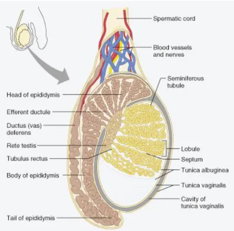
In mammals, the central functions of the testes are: 1) steroid synthesis and secretion; and 2) spermatozoa production (Cooke and Saunders 2002). The structural organization of testes determines the physical division of their dual function. Steroidogenesis takes place in the interstitium (vascularized region) whereas spermatogenesis occurs within the seminiferous tubules (avascular compartment), highly convoluted structures that are the functional units of the testis (Figure I-1). The testicular functions are coordinated by the communication between hormone and gamete-producing compartments (Schlatt et al. 1997). The interstitial area is coated by a tough fibrous membrane called tunica albuginea (Figure I-1) (Saladin 2003). There is also an outer tissue layer, named tunica vaginalis, a thin serous sac derived from the peritoneum during the descent of the testes, which covers both anterior and lateral surfaces of the testes but not their posterior surfaces (Setchell et al. 1994; Kent 2001). Fibrous internal septa, extending from the tunica albuginea, divide the testis in 250 to 300 wedge-shaped testicular lobules, each one enclosing 1 to 3 loop-shaped seminiferous tubules (Figure I-1) (Kent 2001; Rabbani et al. 2010).
The interior of each seminiferous tubule (Figure I-1) is limited by a basal membrane,
composed by germ cells that form numerous concentric layers penetrated by a single type of somatic cell, the Sertoli cell (de Rooij and Mizrak 2008). Externally, the seminiferous tubules are surrounded by mesenchymal cells, including the peritubular myoid cells whose contractile elements produce peristaltic waves along the tubules (Gaytan et al. 1994a; Gaytan et al. 1994b). The seminiferous tubules are connected with the rete testis by means of the tubulus
rectus, which in turn are linked to the efferent ductules (Figure I-1) (Rabbani et al. 2010).
The seminiferous tubules, which contain germ cells in different stages of development and Sertoli cells, represent about 80% of the testicular mass (Sharpe 1984; Colborn et al. 1993; Sikka and Wang 2008).
The interstitium contains blood and lymphatic vessels and various cell types, including fibroblasts, leukocytes, macrophages and endocrine cells, the Leydig (interstitial) cells (Sharpe 1984; Colborn et al. 1993). The major source of the testosterone (T) are the Leydig cells (Haider 2004), which play an important role in downstream masculinization events, descent of the human testes into the scrotum before birth and initiation and maintenance of spermatogenesis (Akingbemi 2005; Sikka and Wang 2008).
4
Figure I-1. Schematic representation of the mammalian testis and its relationship with the epididymis.The testis is encased by two tissue layers, from the inside to the outside, tunica albuginea and tunica vaginalis. Various septa extending from the tunica albuginea divide the testis in lobules, where the seminiferous tubules are located. The seminiferous tubules converge to the rete testis that is connected to the efferent ductules. The head of the epididymis receive testicular secretions by several efferent ductules (Marieb 2001).
The efferent ductules are mainly involved in fluid homeostasis and reabsorb more than 95% of the luminal fluid released from the seminiferous epithelium (Clulow et al. 1994). Moreover, these ductules carry spermatozoa from the rete testis and concentrate it before sperm maturation in the epididymis (Lee et al. 2009). The epididymis is a highly compartmentalized organ that is usually divided in three distinct regions (Figure I-1 and Figure I-2), the caput (head), corpus (body) and cauda (tail), which cooperate with different functions to reach the ultimate goal, sperm maturation and its fertilizing capacity (Robaire et al. 2006).
5 Figure I-2. Schematic organization of the rat epididymis. (A) Frontal view showing the three regions of the epididymis – caput, corpus, and cauda – as well as the initial segments. Oblique lines indicate sites where different regions were segmented. (B) Sagittal view evidencing the transit flow through the epididymis (adapted from (Guo et al. 2007) and (Shum et al. 2011)).
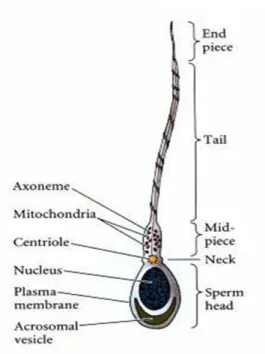
The sperm cell (Figure I-3) contains a haploid nucleus, with the acrosomal vesicle lying in front of it. The acrosomal vesicle, or acrosome, is derived from the Golgi apparatus and contains enzymes that digest proteins and complex sugars. These stored enzymes are used to lyse the outer coverings of the oocyte. Together, the acrosome and nucleus constitute the head of the sperm (Figure I-3) (Gilbert 2000). The flagellum of the mammalian spermatozoon consists of four distinct segments: the connecting piece (neck), the middle piece, the principal piece, and the end piece (Figure I-3) (Eddy 2006). The major motor portion of the flagellum is the axoneme, which is constituted by microtubules and responsible for sperm motility (Gilbert 2000; Rabbani et al. 2010). The flagellum is surrounded in turn by outer dense fibers extending from the neck into the principal piece of spermatozoa. The midpiece contains the mitochondrial sheath, a tightly wrapped helix of mitochondria surrounding the outer dense fibers and axoneme (Rabbani et al. 2010). The end piece is the thinnest portion of the sperm (Saladin 2003).
6
Figure I-3. Schematic representation of the structure of mammalian spermatozoa (adapted from (Gilbert 2000)).
There are some differences in the size and shape of the sperm head, in the length and relative amount of the different components of the flagellum among species (Rabbani et al. 2010). The head of human spermatozoa has a spatulate shaped whereas the rat sperm head is falciform-shaped (Figure I-4) (Eddy 2006).
Figure I-4. Schematic representation comparing the human (A) and rat (B) spermatozoa (adapted from (Frandson et al. 2009)).
7
2. Testicular and epididymal physiology
2.1 Spermatogenesis
Mammalian spermatogenesis is a complex biological process involving cell division and maturation of spermatogonial stem cells that culminates with the production of male gametes, the spermatozoa. It is a continuous and highly regulated process occurring in the seminiferous tubules within the testis (Hess and de Franca 2008). Sperm cells develop from the primordial germ cells and move towards the lumen of seminiferous tubule as they undergo a series of mitoses followed by the first and second meiotic division (Figure I-5) (Nussbaum et al. 2007; Sharma and Agarwal 2011).
Spermatogonial stem cells are localized at the basal membrane of the seminiferous tubules as single cells and upon division originate daughter cells, the spermatogonia (de Rooij and Mizrak 2008). The somatic Sertoli cells and germ cells, the only cell types within the seminiferous epithelium, are in close contact (Figure I-5) (Taylor et al. 2004). The cytoplasm of Sertoli cells extends as thin arms around the germ cells, spanning the thickness of the seminiferous epithelium. These cells supervise the several steps of spermatogenesis by providing structural and nutritional support to the germ cells (Griswold 1998; Taylor et al. 2004). The presence of tight junctions between neighbouring Sertoli cell forms the blood‐testis barrier, which divides the seminiferous tubule in basal and adluminal compartments (Figure I-5) (Griswold 1998). The somatic Leydig cells are located in the interstitial space between seminiferous tubules and play a key role in the regulation of spermatogenic process (Haider 2004).
The expression of a large number of genes is developmentally regulated during spermatogenesis (Lambard et al. 2004), with both transcriptional and translational control mechanisms being responsible for temporal and stage-specific expression pattern (Angelopoulou et al. 2007). Each spermatogenic cycle in the seminiferous tubules comprises three main phases: mitosis, meiosis, and the final stage of cell differentiation, spermiogenesis (Hess and de Franca 2008). Spermatogenesis (Figure I-5) begins with the proliferation of spermatogonia and, after a species‐specific fixed number of mitotic divisions, spermatogonia differentiate into primary spermatocytes (Clermont 1972). These proceed to the first division of meiosis resulting in secondary spermatocytes, which undergo the second meiotic division and become haploid spermatids. The cellular restructure in the spermiogenesis transforms round‐spermatids in elongated‐spermatids, and then, elongated‐spermatids into spermatozoa, which are finally released into the lumen of the seminiferous tubule (Hess and de Franca 2008).
The spermatogenesis process in rat is similar to another mammalian species, including primates (Ross et al. 1995). The total duration of spermatogenesis is about 50 days in rat and 64 days in man. The additional required period for maturation in epididymis is about a week for rat and 8 to 17 days for man (Adler 1996).
8
Figure I-5. Schematic representation of the testicular histology and mammalian spermatogenesis. The anatomic relationship between testis and epididymis, as well as the distinct functional regions of the epididymis are also shown (adapted from (Correia 2014)).
2.2 Control of spermatogenesis
The accomplishment of a successful spermatogenesis is dependent on various hormonal factors, which exert their actions via endocrine, paracrine, juxtacrine and autocrine signaling mechanisms. The central player in the hormonal control of spermatogenesis is the hypothalamic‐pituitary‐gonadal axis (Holdcraft and Braun 2004).
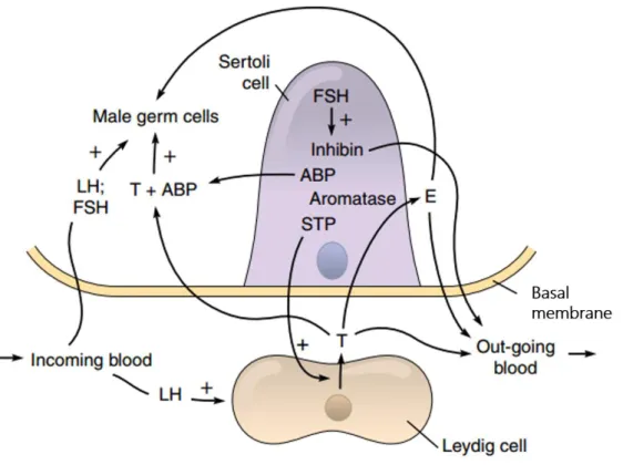
Generally, the hypothalamus releases gonadotropin releasing hormone (GnRH), which acts on the pituitary inducing the release of gonadotropins, namely luteinizing hormone (LH) and follicle‐stimulating hormone (FSH). In the testis, LH acts on Leydig cells stimulating the synthesis of T, while FSH acts on Sertoli cells inducing the production of several growth factors and other stimulatory factors required for spermatogenesis (Walker and Cheng 2005; Walker 2009), which includes the androgen-binding protein (ABP) and the steroidogenesis stimulating protein (STP) (Jones and Lopez 2006) (Figure I-6).
Androgens and ABP, produced by Sertoli cells in the seminiferous tubules, are released along with sperm and transported from testis to epididymis (Norris and Carr 2013). ABP has a high
9 affinity for androgens and binds specifically 5α‐dihydrotestosterone and T (Courot 1980). Through its binding activity, ABP may play a role in spermatogenesis and epididymal sperm maturation by enhancing the local concentration of androgens around the germinal cells and the male gametes (Courot 1980). STP enhances T secretion by Leydig cells (Figure I-6) (Jones and Lopez 2006).
Figure I-6. The endocrinology of the testes. Leydig cells secrete testosterone (T) in response to the circulating luteinizing hormone (LH). Then, T diffuses into the seminiferous tubules and in Sertoli cells can be converted to estrogens (E) by the aromatase enzyme. T is also present in the germ cell region, where may be bound to the androgen-binding protein (ABP) or may remain in free-form to stimulate the spermatogenesis. Follicle stimulating hormone (FSH) also acts on Sertoli cells to induce the production of inhibin, ABP, aromatase, and steroidogenesis stimulating protein (STP). The STP leaves the tubule and helps the LH to increase the production of T. Finally, inhibin, E, and T enter the outgoing blood and exert a negative feedback on gonadotropin secretion. Note that E produced by Sertoli cells can stimulate male germ cells directly (adapted from (Jones and Lopez 2006)).
Moreover, T diffuses into the seminiferous tubule where together with FSH exerts stimulatory effects on the activity of Sertoli cells, which is determinant for germ cells survival, maturation and sperm production (Erkkila et al. 1997; Walker and Cheng 2005; Walker 2009). Besides, T regulates the spermatogenic process by a negative feedback mechanism on the hypothalamus and pituitary inhibiting, respectively, the release of GnRH and LH (Holdcraft and Braun 2004). Other negative feedback regulatory mechanism is driven by inhibin, which is a member of the transforming growth factor β superfamily, produced by Sertoli cells in response to FSH (Figure I-6). Inhibin represses the production and release of FSH by the pituitary (Pierik et al. 2003; Bilezikjian et al. 2004) controlling the output of spermatogenesis. Even though androgens and FSH are considered the main regulators of spermatogenesis, in the last years, estrogens have also been recognized as important
10
modulators of spermatogenesis and male fertility (O'Donnell et al. 2001; Carreau and Hess 2010). The rodent and human testis express nuclear and membrane estrogen receptors, and also actively synthesize estrogens, including 17β‐estradiol, which is the most potent of estrogens (Carreau and Hess 2010). The synthesis of estrogens occurs through the aromatization of androgenic precursors by the cytochrome‐c P450 aromatase enzyme (Carreau et al. 2003; Carreau and Hess 2010). Estrogens can have a direct action on Leydig cells down-regulating the expression of steroidogenic enzymes involved in T biosynthesis (Sakaue et al. 2002). Since germ cells express estrogens receptors, estrogens produced by Sertoli cells are able to stimulate them directly (Correia et al. 2015). Estrogens also can act on the hypothalamus or pituitary exerting a negative feedback, suppressing the production of GnRH and LH, and consequently, decreasing the T levels (Hossaini et al. 2003; Chimento et al. 2014). Figure I-6 summarizes the hormonal modulation of spermatogenesis.
The output of spermatogenesis and the number of spermatozoa produced is regulated by an interaction between proliferation, differentiation and cell death (Pastor et al. 2011). Testicular germ cell apoptosis happens normally and continuously throughout the life (Wang et al. 2010). High rates of apoptosis, or programmed cell death, have been associated with the first waves of spermatogenesis (Aitken et al. 2011), and the germ cells that do not achieve the full maturity are more susceptible to die in response to numerous factors (Shaha et al. 2010). In fact, the quality control of spermatozoa is one of the most important aspects in spermatogenesis, and apoptosis is the best known quality control mechanism in testis (Shukla et al. 2012). Apoptosis occurs at the same time that spermatogonia undergo mitotic divisions and spermatocytes proceed through meiosis. Thus, apoptosis in the germ cell serves as a checkpoint to eliminate abnormal cells, as well as to provide an optimal germ/Sertoli cell ratio (Allan et al. 1992; Bartke 1995; Sinha Hikim et al. 2003). Spontaneous apoptosis provokes the loss of germ cells in the testis both in normal and pathological conditions. In the first case, it is estimated that up to 75% of potential spermatozoa degenerate in the testes of adult mammals (Huckins 1978). Regarding the pathological condition, the range of stimuli that trigger apoptosis is extraordinarily broad, including various forms of electromagnetic radiation, chemotherapeutic agents, environmental toxicants, heavy metals, heat exposure, growth factor depletion or hormonal alterations (Pastor et al. 2011; Aitken and Baker 2013). As for somatic cells, essentially two distinct pathways exist for the initiation of apoptosis of male germ cells: extrinsic or receptor-linked apoptosis and intrinsic or
11
Figure I-7. Extrinsic and intrinsic pathways of apoptosis. Extracellular ligand binding (FasL or tumor
necrosis factor, TNF) to death receptors (FasR and TNF receptor, TNFR) triggers the receptor-mediated (extrinsic) pathway resulting in the direct activation of initiator caspase-8. The mitochondrial (intrinsic) pathway is initiated in response to apoptotic stimuli (radiation, drugs, etc.) leading to the activation of proapoptotic members of the Bcl-2 protein family, such as the Bax. The Bax protein is translocated to the mitochondria allowing the permeabilization of mitochondrial membrane with consequent release of cytochrome c, which in turn, together with apoptotic protease activating factor 1 (Apaf-1), forms the apoptosome and activates caspase-9. Extrinsic and intrinsic pathways converge at the activation of the effector caspase-3. The transcription factor p53 is able to regulate downstream genes important in cell cycle arrest and apoptosis, including p21, Bax and Bcl-2. The cyclin-dependent kinase inhibitor p21 arrests the cell cycle at the G1 phase. Activation and inhibition are indicated by arrows and bar-headed arrows, respectively (adapted from (Correia et al. 2015).
2.3 Sperm maturation
Spermatozoa leaving the testis are non‐functional gametes and it is only during the passage through the long convoluted tubule of the epididymis that they undergo a maturation process (Cornwall 2009). The four main functions of the epididymis comprise transport of spermatozoa, acquirement of the ability to move progressively and to capacitate, eventually gaining the ability to fertilize, and the creation of a specialized luminal microenvironment which allows the maturation process through the absorptive and secretory activities of the epididymis epithelial cells (Robaire et al. 2006; Guyonnet et al. 2011).
12
The maturation process is androgen-dependent and conducts several biochemical and functional changes in spermatozoa (Vreeburg et al. 1992). Estrogens are also involved in sperm maturation by the regulation of fluid absorption in the efferent ducts and rete testis, which is a fundamental event for maintenance of the adequate osmolality in the epididymis and sperm concentration (Correia et al. 2015). The caput and corpus regions perform early and late sperm maturation events, respectively, while the cauda stores the functionally mature spermatozoa (Robaire et al. 2006). This regional compartmentalization is characteristically evident both in the number and quantity of proteins secreted, with the
caput as the most active, while the corpus and cauda possess a lower secretory activity
(Dacheux et al. 2009).
The epididymal lumen is rich in inorganic ions and organic molecules that create the appropriate ionic, oxidative and pH environment for sperm maturation throughout epididymis
transit (Cornwall 2009). The levels of calcium (Ca2+) in the epididymal fluid are quite low in
comparison with other ions, namely sodium, potassium, chloride, ammonium, and magnesium (Wales et al. 1966). Although the exact role of each component of the epididymal fluid needs to be deciphered, acidification has been shown to be essential for the alterations on sperm surface proteins required for sperm maturation and storage (Pholpramool et al. 2011). The acidification of epididymal fluid and water transport along epididymis are the critical events that ensure an appropriate environment. Acidification is implicated in sperm maturation and maintenance of its quiescent state during storage (Pholpramool et al. 2011) whereas water movement across the epididymis epithelium contributes to sperm concentration. This fact is crucial for proper sperm function, because sperm ability to reach maturation is enhanced by sperm concentration in the epididymal duct, achieved by water removal from the luminal fluid (Da Silva et al. 2006). In order to conserve energy and maintain structural integrity, sperm motility needs to be suppressed until it is required (Jones 1999). Some of the epididymal proteins contribute to the stabilization of the sperm plasma membrane preventing the occurrence of premature capacitation, whereas others proteins have been implicated in the acquisition of the sperm ability to bind and recognize the oocyte (Lefebvre et al. 2009; Cohen et al. 2011; Joseph et al. 2011). Physiological amounts of reactive oxygen species (ROS) are also involved in the regulation of some sperm functions, such as playing positive effects on maturation (Aitken and Baker 2004), capacitation (O'Flaherty et al. 2006), acrosome reaction (de Lamirande and O'Flaherty 2008), and sperm-oocyte fusion (Riffo and Parraga 1996), supporting the importance of preserving seminal ROS at low controlled levels through the delicate balance between ROS production and removal (Tremellen 2008).
In mammals, the normal duration of the transit through the epididymis cauda is in the range of 3 to 10 days, but spermatozoa can be stored in this segment for periods extending beyond 30 days (Rabbani et al. 2010).
13
3. Testicular damage and male (in)fertility
3.1 Testicular cancer
Testicular cancer is the most important malignancy in the young male, accouting >10 new cases per 100.000 males per year in Europe and a mortality of 0.3 cases per 100.000 males per year (McGlynn et al. 2003; Jemal et al. 2011). In fact, testicular germ cell tumors are the most common malignancy in males between 15 and 34 years old and also the most frequent cause of death from solid tumors (Chieffi and Chieffi 2013). Fortunately, the survival from cancer has been improved over the past few decades due to the advances in diagnostic tools, treatments and therapeutic modalities. As a result of early detection and successful adjuvant treatments, young cancer patients are living longer and subsequently the strategy of management has changed from cure with any cost to one in which quality of life has become increasingly important (Vassilakopoulou et al. 2015).
3.2 Cancer treatment
Depending on the type and stage of the testicular cancer and other factors, treatment options can include surgery, chemo- and/or radiotherapy (Albers et al. 2005; Brydoy et al. 2007). The testes are very sensitive organs and, thus, highly affected by exogenous damaging factors, such as chemo- and radiotherapy treatments (Trost and Brannigan 2012b). For this reason, the treatment of oncological diseases usually results in temporary or permanent arrest of spermatogenesis (Stahl et al. 2006), as well as in disrupted sex hormone production (Brauner et al. 1983). Taking into account that the majority of young patients wish to be parents, the reproductive problems are considered one of the most common and distressing consequences of cancer treatment (Schover et al. 2014).
Focusing on radiotherapy, the extent of testicular injury is directly related with the dose of radiation delivered as well as the underlying cell type (Zhang et al. 2007). The germinal epithelium is more sensitive than the Leydig cells as a result of its high mitotic rate (Osterberg et al. 2014). Radiation therapy negatively affects spermatogenesis either transiently or permanently, by directly inducing DNA damage (Lushbaugh and Casarett 1976; Apperley and Reddy 1995). Besides the dose, several variables can affect the deleterious effect of radiation on gonadal function, such as source of radiation, gonadal shielding, scatter radiation, and individual susceptibility (Colpi et al. 2004; Trottmann et al. 2007). Seminiferous tubules are particularly sensitive to radiation because energies as low as 0.1 gray (Gy) results in temporary arrest of spermatogenesis. Increasing doses have been shown to cause azoospermia at 0.65 Gy, and doses >0.65 Gy but <1 Gy, 2–3 Gy, and 4–6 Gy, result in azoospermia lasting 9–18 months, 30 months, and 5 years to permanent, respectively (Trost and Brannigan 2012a). Leydig cells are only affected when doses reach >15 Gy (Colpi et al. 2004). Also, the radiation delivered directly to the testes for treatment of testicular
14
leukemia, or as part of the total body irradiation prior to bone marrow transplant, involves doses that result in permanent sterility in most men (Leonard et al. 2004).
Although with some controversy, it is accepted that malignancy itself is associated with male infertility since numerous biological processes are affected in parallel (Petersen et al. 1999). Indeed, azoospermia is present in ≈ 3% to 18% of men at the moment of cancer diagnosis (Tournaye et al. 2004). In addition, it was shown that spermatogenesis is affected in oncological patients with lymphoma and leukemia without testicular pathology, even before the onset of gonadotoxic therapies (Rueffer et al. 2001; Howell and Shalet 2005; Hotaling et al. 2013; Katz et al. 2013; Bujan et al. 2014). Also, altered sperm production in patients with testicular cancer before orchiectomy has been detected (Rives et al. 2012).
4. Preservation of male fertility and spermatogenesis
recovery after testicular damage
4.1 Cryopreservation
Sperm cryopreservation preceding cancer treatments remains the only established method for fertility preservation in adult males (Hotaling et al. 2013), whereas in the pre-pubertal male the cryopreservation of testicular tissue is the adopted option for preserving fertility (Pennings and Mertes 2012). Other procedures are currently at experimental phase but are not devoid of ethical concerns (Pennings and Mertes 2012). Besides that, one of the major problems is related to the ability to avoid the oxidative damage that sperm cells and seminal plasma normally retain (Tremellen 2008). During the freeze–thawing practice, the antioxidant defenses could be insufficient (Bucak et al. 2010) to counteract the damage of that cryopreservation induces to the spermatozoa, which includes significantly decreased motility, viability, morphology, chromatin integrity, mitochondrial potential, in vivo fertilizing capacity, deterioration of acrosomal and plasma membrane integrity, and DNA damage (Bucak et al. 2010; Degl'Innocenti et al. 2013; Sharma et al. 2015). Furthermore, the use of cryopreserved sperm or tissue in assisted reproductive techniques (ART) has an economic obstacle due to the high costs associated (Linkeviciute et al. 2014).
The present challenge is to improve ART with cheaper and simpler alternatives with good acceptance by patients, and to develop effective strategies that would restrain the undesirable secondary effects of oncological treatments (Ko and Sabanegh 2014).
4.2 Hormonal and non-hormonal factors
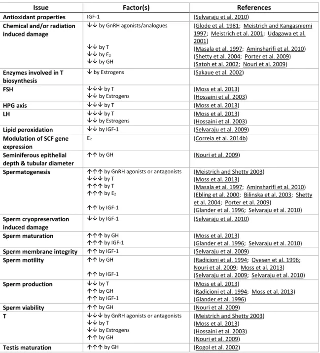
There are several endogenous agents, hormonal and non-hormonal, which have been described to have protective or advantageous properties on the recovery of male reproductive function after testicular injury and cancer, as summarized in Tables I-1 and I-2. The knowledge reviewed in Tables I-1 and I-2 identify promising factors, which might be able
15 to mitigate the male fertility problems arising either from oncological treatments or other gonadal damage, opening new possibilities to ameliorate the recovery of spermatogenesis or to preserve fertility. Furthermore, the perspective of the endogenous molecules that could act as cryoprotectants in order to improve the quality of cryopreserved semen samples was also included.
16
Table I-1. Hormonal protective endogenous factors in male reproductive function.
, up-regulated; , down-regulated; , increased; , reduced; , induced/stimulated; , suppressed; Testosterone (T), follicle-stimulating hormone (FSH), hypothalamic-pituitary-gonadal (HPG), luteinizing hormone (LH), stem cell factor (SCF), gonadotropin releasing hormone
(GnRH), 17β-estradiol (E2), growth hormone (GH), insulin-like growth factor 1 (IGF-1).
Issue Factor(s) References
Antioxidant properties IGF-1 (Selvaraju et al. 2010) Chemical and/or radiation
induced damage
by GnRH agonists/analogues by T
by E2
by GH
(Glode et al. 1981; Meistrich and Kangasniemi 1997; Meistrich et al. 2001; Udagawa et al. 2001)
(Masala et al. 1997; Aminsharifi et al. 2010) (Shetty et al. 2004; Porter et al. 2009) (Satoh et al. 2002; Nouri et al. 2009) Enzymes involved in T
biosynthesis
by Estrogens (Sakaue et al. 2002)
FSH by T
by Estrogens (Moss et al. 2013) (Hossaini et al. 2003) HPG axis by T (Moss et al. 2013)
LH by T
by Estrogens (Moss et al. 2013) (Hossaini et al. 2003) Lipid peroxidation by IGF-1 (Selvaraju et al. 2009) Modulation of SCF gene
expression
E2 (Correia et al. 2014b)
Seminiferous epithelial depth & tubular diameter
by GH (Nouri et al. 2009) Spermatogenesis by GnRH agonists or antagonists
by T by T by E2
by IGF-1
(Meistrich and Shetty 2003) (Moss et al. 2013)
(Masala et al. 1997; Aminsharifi et al. 2010) (Ebling et al. 2000; Bilinska et al. 2003; Shetty et al. 2004; Porter et al. 2009)
(Glander et al. 1996; Selvaraju et al. 2010) Sperm cryopreservation
induced damage
by IGF-1 (Selvaraju et al. 2010) Sperm maturation by GH
by IGF-1 (Moss et al. 2013) (Glander et al. 1996; Selvaraju et al. 2010) Sperm membrane integrity by IGF-1 (Selvaraju et al. 2009)
Sperm motility by GH by IGF-1
(Radicioni et al. 1994; Ovesen et al. 1996; Nouri et al. 2009; Moss et al. 2013) (Selvaraju et al. 2009; Selvaraju et al. 2010) Sperm production by T
by GH by IGF-1
(Moss et al. 2013)
(Radicioni et al. 1994; Moss et al. 2013) (Glander et al. 1996)
Sperm viability by GH (Nouri et al. 2009) T by GnRH agonists or antagonists
by T by Estrogens by GH
(Meistrich and Shetty 2003) (Moss et al. 2013)
(Hossaini et al. 2003) (Nouri et al. 2009) Testis maturation by GH (Rogol et al. 2002)
17
Table I-2. Non-hormonal protective endogenous factors in male reproductive function.
, increased; , reduced; , induced/stimulated; , suppressed; Stem cell factor (SCF), vascular endothelial growth factor (VEGF), testosterone (T), nitric oxide (NO), hepatocyte growth factor (HGF), granulocyte colony stimulating factor (G-CSF), regucalcin (RGN), liver growth factor (LGF), interleukin 1 (IL-1), interleukin 6 (IL-6).
Issue Factor(s) References
Acrosome reaction by NO (Roessner et al. 2010) Anti-apoptotic effect in testis by NO by Ghrelin Involvement of HGF by G-CSF by RGN (Roessner et al. 2010) (Zhu et al. 2013) (Catizone et al. 2006) (Kim et al. 2011) (Correia et al. 2014a) Antioxidant properties Arginine
Metallothioneins Ghrelin RGN
(Patel et al. 1998; Appleton 2002; Senbel et al. 2014) (Sato and Kondoh 2002)
(Kheradmand et al. 2012) (Correia et al. 2013) Chemical and/or radiation
induced damage
by Metallothioneins by Ghrelin by LGF by IL-1 & IL-6 by IL-6 by G-CSF by RGN
(Kheradmand et al. 2013; Maremanda et al. 2014) (Zhu et al. 2013; Garcia et al. 2015; Whirledge et al. 2015) (Perez-Crespo et al. 2011; Lobo et al. 2015)
(Gerard et al. 1992; Syed et al. 1993; Legué et al. 2001) (Guitton et al. 1999)
(Kim et al. 2011) (Correia et al. 2014a) Epididymis weight by LGF (Perez-Crespo et al. 2011)
Lipid peroxidation in sperm by Arginine (Srivastava et al. 2006; Siddique and Atreja 2013) Modulation of SCF gene
expression
Ghrelin (Barreiro et al. 2004) Seminiferous epithelial
depth & tubular diameter
by G-CSF (Kim et al. 2011) Spermatogenesis by Arginine
by LGF by IL-1 by G-CSF
(Aydin et al. 1995; Patel et al. 1998) (Perez-Crespo et al. 2011; Lobo et al. 2015) (Legué et al. 2001)
(Kim et al. 2011) Sperm abnormalities by G-CSF
by RGN (Kim et al. 2011) (Correia et al. 2013) Sperm capacitation by NO (Roessner et al. 2010) Sperm cryopreservation
induced damage
by Arginine (Siddique and Atreja 2013)
Sperm glycolysis rate by Arginine (Patel et al. 1998; Srivastava et al. 2006) Sperm maturation by RGN (Correia et al. 2013)
Sperm membrane integrity by Arginine by Ghrelin
(Siddique and Atreja 2013) (Kheradmand et al. 2009) Sperm motility by Arginine
by NO by HGF by LGF
(Keller and Polakoski 1975; Scibona et al. 1994; Aydin et al. 1995; Rosselli et al. 1995; Patel et al. 1998; Appleton 2002; Morales et al. 2003; Srivastava et al. 2006; Imhof et al. 2012; Siddique and Atreja 2013)
(Imhof et al. 2012) (Catizone et al. 2006) (Perez-Crespo et al. 2011) Sperm production by Arginine
by LGF by G-CSF
(Keller and Polakoski 1975; Aydin et al. 1995; Appleton 2002; Srivastava et al. 2006; Imhof et al. 2012) (Perez-Crespo et al. 2011)
(Kim et al. 2011) Sperm viability by Arginine
by NO by RGN
(Rosselli et al. 1995; Siddique and Atreja 2013) (Imhof et al. 2012)
(Correia et al. 2013) Synthesis of VEGF and its
receptors in testis
by LGF (Martin-Hidalgo et al. 2007)
T by LGF (Lobo et al. 2015)
Testis maturation by HGF (Catizone et al. 2006) Testis weight by LGF
18
5. Regucalcin protein as a protective molecule in
reproductive function
The differentiation and survival of spermatogenic cells has been indicated to implicate
adjustments in the intracellular Ca2+ concentration ([Ca2+]
i) (Berrios et al. 1998; Reyes et al.
2002; Mishra et al. 2006; Reyes et al. 2010; Sanchez-Cardenas et al. 2012). Germ cells
produce Ca2+ currents that increase their density during the development from spermatogonia
to early spermatids (Hagiwara and Kawa 1984). Thus, a tight control of Ca2+ homeostasis is a
critical factor for spermatogenesis, including also for Sertoli cell function (Gorczynska and Handelsman 1995; Gorczynska-Fjalling 2004), for the maintenance of Sertoli cells tight junctions and integrity of the blood‐testis barrier (Grima et al. 1998), and possibly for the Leydig cell steroidogenesis (Manna et al. 1999).
Overall, Ca2+ is required for sperm motility, capacitation, acrosome reaction, and, thus,
acquisition of fertilization competence (Thomas and Meizel 1988; Sorensen et al. 1999;
Breitbart 2002). One of the agents involved in the control of Ca2+ homeostasis is the
regucalcin (RGN), also known as senescence marker protein 30 (Yamaguchi and Yamamoto 1978; Yamaguchi 2005). RGN has a molecular weight of 33 kDa and plays an important role in
intracellular Ca2+ homeostasis by regulating the activity of Ca2+ pumps localized on cell
membrane and endoplasmic reticulum of numerous cell types (Figure I-8) (Fujita et al. 1998; Tsurusaki and Yamaguchi 2000; Yamaguchi 2005).
Several studies have demonstrated that RGN is expressed in numerous tissues, such as liver (Shimokawa and Yamaguchi 1992; Ishigami et al. 2015), kidney (Yamaguchi and Kurota 1995; Zubiri et al. 2015), brain (Yamaguchi et al. 2000; Yamaguchi et al. 2008b), heart (Yamaguchi and Nakajima 2002; Akhter et al. 2006), bone (Yamaguchi et al. 2002a; Kagami et al. 2013), lung (Mori et al. 2004), submandibular gland (Ishii et al. 2005), ovary (Fayad et al. 2004; Kagami et al. 2013), breast (Marques et al. 2015a; Marques et al. 2015b), prostate (Maia et al. 2008; Maia et al. 2009; Vaz et al. 2015) and testis (Laurentino et al. 2011).
RGN expression is regulated by several hormones, including thyroid and parathyroid hormones (Yamaguchi et al. 2008a), aldosterone, insulin, calcitonin, and sex steroid hormones (Maia et al. 2008; Maia et al. 2009; Laurentino et al. 2011; Marques et al. 2014; Vaz et al. 2014).
This Ca2+-binding protein also regulates several Ca2+‐dependent enzymes (Figure I-8), such as
protein kinases, tyrosine kinases, phosphatases, phosphodiesterase, nitric oxide synthase (NOS) and proteases (reviewed by (Marques et al. 2014)). It has been proposed that RGN play an important role as a suppressor protein in the differentiation and proliferation of regenerating liver cells (Yamaguchi 2000). In addition, other studies support the role of RGN as a suppressor of cell proliferation in cancer tissues or cells and also in non-tumor tissues (Misawa et al. 2001; Vaz et al. 2014; Yamaguchi and Murata 2015). Recently, it was demonstrated that RGN overexpression diminished the incidence and aggressiveness of mammary gland tumors in rats treated with the carcinogen 7,12-dimethylbenz[α]anthracene,
19 which suggests a protective role of RGN against the onset and development of tumors (Marques et al. 2015b). In fact, RGN regulates the expression of oncogenes, tumor suppressor genes and cell cycle regulators inhibiting cell proliferation (Vaz et al. 2014; Yamaguchi and Murata 2015). Moreover, RGN may translocates to the nucleus modulating several nuclear functions (Yamaguchi and Sakurai 1991), such the inhibition of DNA and RNA synthesis (Yamaguchi and Kanayama 1996; Yamaguchi and Ueoka 1997).
Regarding the male reproductive tract, RGN is expressed in several tissues, including the testis, epididymis, seminal vesicles and prostate (Maia et al. 2008; Laurentino et al. 2011; Vaz et al. 2014). At testicular level, RGN is expressed in Leydig and Sertoli cells, as well as in
all germ line both in human and rat testis (Laurentino et al. 2011).This Ca2+-binding protein is
largely recognized as an androgen-target gene (Maia et al. 2009; Laurentino et al. 2011; Vaz et al. 2014), and it has been pointed out that its increasing concentration might be a mechanism by which androgenic stimulation sustain germ cell survival and spermatogenesis (Laurentino et al. 2011).
Recently, several studies have demonstrated the role of RGN in regulating the expression of cell cycle and apoptosis regulators (Nakagawa and Yamaguchi 2005; Correia et al. 2014a;
Vaz et al. 2014; Marques et al. 2015b). RGN inhibits the increase of [Ca2+]
i (Correia et al.
2013), inhibits caspase 8 activity (Ishigami et al. 2002), improves Akt pathway activity and increases the expression of anti-apoptotic agents Akt-1 and Bcl-2, providing a resistance to apoptosis (Yamaguchi 2013). Considering that the success of spermatogenesis depends on the tight balance between germ cell survival and death, a role for RGN in male spermatogenesis and fertility has been suggested (Laurentino et al. 2012). In order to investigate the protective function of RGN in testicular apoptosis, the effect of apoptosis-inducers such as thapsigargin and actinomycin D, was determined in the seminiferous tubules of transgenic animals overexpressing RGN (Tg-RGN) (Correia et al. 2014a). Concomitantly with RGN overexpression, it was detected reduced activity of caspase-3 and increased expression of anti-apoptotic Bcl-2 protein in the seminiferous tubules of Tg-RGN (Correia et al. 2014a). In addition, the mRNA expression of p53 and p21 was significantly diminished in Tg-RGN treated with thapsigargin or actinomycin D (Correia et al. 2014a). Altogether, these results suggest that RGN may act as a germ cell survival factor protecting the cells from noxious stimuli (Figure I-8) (Correia et al. 2014a). The importance of RGN for the spermatogenic output was also established by the fact that infertile men with abnormal spermatogenesis phenotypes exhibited different expression patterns of RGN at testicular level (Laurentino et al. 2012). Furthermore, significant alterations in the epididymal epithelium and sperm parameters between RGN and their wild-type (Wt) counterparts were found (Correia et al. 2013). Tg-RGN rats have lower sperm counts and reduced sperm motility, which may be associated to
the lower Ca2+ influx in epididymis (Correia et al. 2013). However, this result is
counterbalanced by greater sperm viability, higher percentage of normal sperm morphology, and a reduced incidence of tail defects, suggesting the involvement of RGN in sperm maturation (Correia et al. 2013). The beneficial effects of RGN on sperm parameters seems to
20
be explained by the lower levels of oxidative stress found in biological models with RGN overexpression (Handa et al. 2009; Correia et al. 2013). Indeed, the antioxidant activity of RGN has been described (Son et al. 2006; Correia et al. 2013; Marques et al. 2014). RGN increases the activity of superoxide dismutase (SOD) enzyme and diminishes NOS levels,
reducing the generation of ROS (Figure I-8) (Ma and Yamaguchi 2002; Ma and Yamaguchi
2003; Yamaguchi et al. 2003; Fukaya and Yamaguchi 2004; Ichikawa and Yamaguchi 2004; Yamaguchi et al. 2005; Handa et al. 2009). The cytoprotective and antioxidant properties of RGN were also highlighted by studies in the RGN-knockout mice, which showed higher susceptibility to oxidative stress induced by exposure to cigarette (Sato et al. 2006), and displayed increased levels of anion superoxide in the brain (Son et al. 2006; Kondo et al. 2008; Sato et al. 2008). Also, RGN overexpression in a mouse carcinoma cell line was able to increase cell viability under oxidative damage induced by tert-butyl hydroperoxide (Son et al. 2008). Hence, the available studies evidenced the protective effect of RGN against oxidative stress, as well as its importance in order to keep ROS at physiological concentrations.
Altogether, the existing information indicates that RGN plays a role in the modulation of sperm production and maturation.
Figure I-8. The role of regucalcin (RGN) in cell biology. RGN regulates the activity of several Ca2+
-dependent enzymes and the concentration of intracellular Ca2+ by modulating the activity of Ca2+
-channels, and Ca2+-ATPase in the plasma membrane, mitochondria and endoplasmic reticulum. It has
suppressive effects on the activity of protein kinases and phosphatases. Another feature of RGN protein is its antioxidant activity by reducing the production of reactive oxygen species (ROS). Moreover, RGN is
21
II. Aim of the thesis
___________________________________
23 Cancer therapies and the oncological condition itself have adverse effects on male fertility, which implicate permanent or transitory impairment of reproductive function. In the case of radiation therapy, the extent of testicular injury is directly related to the dose of radiation delivered but, due to the sensitivity of seminiferous tubules, in general, it induces temporary or permanent azoospermia. Actually, the development of effective strategies that would restrain the undesirable secondary effects of oncological treatments is clearly warranted.
The Ca2+-binding protein RGN has been indicated as a protein that suppresses apoptosis, and
has cytoprotective and antioxidant effects, protecting cells against noxious stimuli. Moreover, RGN overexpression was associated with increased sperm viability and lower incidence of tail defects. This led us to hypothesize that RGN may have a protective role to counteract the effects of radiation on spermatogenesis. The present work aims to study the spermatogenesis recovery after radiation treatment in Tg-RGN comparatively with their Wt counterparts. For this purpose, a single dose of 6 Gy was delivered to the testes of Tg-RGN and Wt rats, and after a 10-week recovery period, the reproductive function of both groups was evaluated by determination of:
1. Gonadosomatic index (GI);
2. Tubular differentiation index (TDI); 3. Epididymal sperm parameters;
25
III. Material and Methods
___________________________________
27
1. Animals
Three‐months old Wt and Tg‐RGN Sprague Dawley (Rattus norvegicus) rats were obtained, respectively, from Charles River (Barcelona, Spain) and Japan SLC (Hamamatsu, Japan). Sprague Dawley Tg-RGN rats were originally generated by Yamaguchi M by means of oocyte
transgene pronuclear injection (Yamaguchi et al. 2002b).Animals were handled in compliance
with the guidelines established by the “Guide for the Care and Use of Laboratory Animals” published by the US National Institutes of Health (NIH Publication No. 85‐23, revised 1996) and the European Union rules for the care and handling of laboratory animals (Directive number 2010/63/EU. Rats were housed under a 12 h light:12 h darkness cycle, with food and water available ad libitum during the course of the experiment. Wt and Tg-RGN rats were randomly divided into control group receiving no treatment and irradiated group (n ≥ 5 in each group).
2. Radiation Treatment
In vivo irradiation studies were performed in the Radiation Oncology Department, Coimbra
University Hospital Centre, in a linear accelerator Varian Clinac 600 C (Varian Medical Systems), using a 4MV photon beam.
To perform in vivo irradiation, an immobilization device was first built in order to keep the rat in a lateral decubitus position. For radiotherapy planning a rat was anesthetized using intramuscular ketamine. Then a computerized tomography (CT) was acquired, keeping the animal in treatment position.
After CT acquisition, target volume (testes) delineation and three-dimensional (3D) computerized planning were performed using Eclipse™ Planning System (Varian Medical Systems, EUA). The goal was to have a homogeneous dose coverage of our target volume, with the dose within our volume ranging from 95% to 107% of the prescribed dose (6 Gy). In order to obtain the desired dose in our planning volume we used a pair of parallel opposed fields. Table, gantry and collimator positions were determined after 3D planning, as well as field size and monitor units for each field. Figure III-1 illustrates the 3D planning of the irradiation performed.