UNIVERSIDADE DE LISBOA
FACULDADE DE CIÊNCIAS
DEPARTAMENTO DA BIOLOGIA VEGETAL
Analysis of DFNB1 locus in Presbycusis
Bárbara Isabel de Carvalho Correia
Mestrado em Biologia Molecular e Genética
Dissertação orientada por:
Profª Doutora Maria Helena Caria
i
Agradecimentos
Em primeiro lugar gostaria de agradecer à minha orientadora, a Professora Helena Caria por me ter acolhido tão bem no seu grupo de trabalho para a realização da minha dissertação, bem como ter confiado em mim para ingressar neste projeto. Agradeço toda a ajuda, por toda a disponibilidade, paciência, por alargar os meus conhecimentos ao nível da ciência, por todo o incentivo ao longo do ano, por me fazer tornar numa pessoa mais autónoma a nível laboratorial e também por ter sempre uma palavra amiga para me dar. Para além de uma professora é uma amiga. Obrigada. Agradeço também à Professora Graça Fialho por todo o acolhimento, ajuda e pela simpatia e também pelas vezes que me via e elogiava o meu “outfit”. Obrigada por ter sempre um sorriso para me dar.
À Marisa, por toda a ajuda desde o início até ao fim da realização da minha tese, por toda a disponibilidade e paciência. Vou ter saudades das massas do almoço, dos bolos de xadrez, do jogo das palavras, de ficarmos até às tantas no laboratório e sermos as últimas a sair juntamente com o segurança! Obrigada companheira! És incansável!
À professora Solveig e a toda a sua equipa do laboratório, principalmente à Marta e à Andreia por disponibilizarem do seu tempo para me ajudar.
À Marta e à D. Manuela pelos dedos de conversa que me davam todos os dias, por me tratarem tão bem e por terem sempre um sorriso para alegrarem o meu dia, e claro por toda ajuda. Ao Senhor Pedro e à Andreia da Horta por me tratarem tão bem, por poder dar muitas gargalhadas com vocês, por encomendarem de prepósito o Lipton Chá Verde que eu tanto gosto. Vocês são os maiores.
Aos pais, um obrigada do fundo do coração por me apoiarem nesta fase da minha vida, por me apoiarem nesta loucura de vir estudar para a capital. Obrigada por fazerem parte da minha vida e apesar de todas as advertências acreditaram sempre em mim e no meu trabalho desejando que tudo corresse da melhor forma. Obrigada por nunca me fazerem desistir. Obrigada por fazerem com que um dos meus sonhos se realizasse. Para mim são os melhores progenitores do mundo e não vos troco por nada! Sem vocês não seria a mesma coisa!
À minha mana Maria por ser a melhor irmã do mundo, por ser uma pequena Enstein na qual eu tenho tanto orgulho. Por ser tão pequena e ter uma mentalidade tão adulta, pelos conselhos que me dás, por todo o carinho, pelos teus abraços que eu tanto gosto, por quereres saber tanto da minha tese, pelas tardes a ver séries, por partilharmos roupa e acima de tudo partilharmos tanto carinho. Obrigada por fazeres parte da minha vida. Sempre juntas. “Nothing can come between you and I”.
Aos avós, um obrigada gigante, por todos os telefonemas, por toda a preocupação, por toda a ajuda que me dão. Esta etapa não seria possível sem a vossa ajuda. Obrigada por estarem comigo desde o primeiro dia. Para sempre a vossa menina!
À madrinha, um agradecimento do fundo do coração por me acolher não só nestes dois anos da minha vida mas sim pelo primeiro dia em que vim ao mundo, tratando-me sempre como uma filha, por teres sempre uma palavra amiga para mim, por todo o apoio, carinho, ajuda, abraços quando as coisas estão menos bem, por seres uma confidente para mim. Obrigada por estares sempre presente nos momentos mais importantes. Ao Quim por todos os dias me fazeres o jantar (àqueles legumes salteados, aquela massa que eu tanto adoro), por toda a preocupação, por me ensinares a andar no Fertagus e no metro, pelas noites no sofá em frente à TV a comer amendoins com mel e sal, por assistirmos aos jogos do Sporting torcendo sempre para que ganhe. Obrigada não chega para agradecer tudo. À Camilinha do meu coração, por seres como uma maninha mais nova para mim, pela companhia que me fizeste durante este tempo todo, por tornares os meus
ii
dias mais felizes com as tuas gargalhadas tão contagiantes. Obrigada pela companhia até ao laboratório e pelas nossas idas ao centro comercial. Vou ter saudades tuas, mas prometo voltar!
Obrigada também às primas Ana e Susana pela preocupação em quererem saber se está tudo bem e um obrigado também à tia Fernanda e tia Lurdes por todo o carinho e por toda a preocupação. Obrigada também aos tios Áurea e Armando, pelas chamadas de preocupação e de conforto, por mesmo estarem a muitos quilómetros lembrarem-se de mim! Obrigada por fazerem com que o longe se torne tão perto!
Um obrigado em geral a toda a minha família.
Ao meu parceiro para a vida, Amadeu, um agradecimento que não cabe em qualquer parte do mundo. Obrigada por teres estado comigo desde o primeiro dia que eu mais precisei, por todo o teu amor e carinho, por toda a paciência que tens para comigo quando eu estava menos bem-disposta. Por todas as surpresas, pelas tuas vindas a Lisboa. Obrigada por acreditares sempre em mim, acreditares que eu sou sempre capaz, por quando eu queria desistir tu estavas sempre lá para me levantar e fazer acreditar outra vez. Obrigada não chega para o que fazes todos os dias por mim! Obrigado parceiro por seres uma pessoa extraordinária, obrigada por me fazeres rir até à exaustão, obrigada por tudo e mais alguma coisa, obrigada por mostrares que é possível! “Não desistas de mim, nem me faças desistir”.
Um obrigado do coração às melhores amigas de sempre. À Joana e à Marta. À Joana por toda a amizade verdadeira, conselhos, risadas, por teres sempre uma palavra amiga para me dar, pelas chamadas no voicemail, pelas fotos “37, masculino”, pelos jantares em tua casa quando chegava à sexta-feira, por estares sempre para mim quando eu mais preciso. À Marta, por toda a amizade verdadeira, pelas palavras reconfortantes que tens sempre para me dar, por toda a preocupação, por todo o carinho e por aqueles teus abraços tão bons. Obrigada às duas por estarem sempre para mim e por nunca desistirem de mim. São as melhores do mundo.
À Sara Correia, minha irmã gémea por toda a companhia, ajuda, pelos desabafos, pelas conversas no Fertagus, por ter sempre uma palavra para mim! És demais miúda!
À Carolina Peixoto por ser a primeira pessoa com quem falei no mestrado, pelas nossas conversas! Um fino sem ti nunca será a mesma coisa!
Ao Cristiano Ramos (Cris), por todos os desabafos, por me deixares stressar contigo, por estudarmos juntos, por todo o carinho!
Ao João Coelho, meu grande Jonas, por seres um “pró” a entrar no laboratório em modo silencioso, pela tua companhia todos os dias nas horas do almoço (uma baguete de frango mais um santal), por irmos juntos no metro e no comboio até à Margem Sul! Por podermos desabafar e deprimir juntos! Volto sempre à margem “ma friend”.
À Carolina Sousa, por disponibilizares do teu tempo para me ajudares quando andava meia atrapalhada. Fico a aguardar um convite até à Madeira!
Aos alunos do estudo orientado, Marcelo, Malpica, Raquel, Carolina e Leonor por toda a companhia, por me fazerem rir! Obrigada por terem confiado em mim para ajudar no vosso projeto.
Ao Tó Manel, à Edna e à Élia por me fazerem sentir parte da família, por me receberem sempre tão bem, por terem sempre um abracinho para me dar, por fazerem a minha comida favorita e claro por deixarem que o vosso menino também seja um bocadinho meu! Obrigada por tudo.
Sem vocês todos, nada disto seria possível!
iii
Table of contents
Figures ... v
Tables ... vi
Abbreviations and acronyms... vii
Resumo ... viii
Abstract ... xi
1. Introduction ... 1
1.1 The ear ... 1
1.2 Epidemiology of deafness ... 3
1.3 Age Related Hearing loss ... 3
1.4 Genetic studies ... 4
1.5 Connexins- Structure and function ... 4
1.5.1 Connexin 26 ... 5
1.5.2 Connexin 30 ... 6
2. Functional studies ... 6
3. Objectives ... 8
4. Materials and methods... 9
4.1 Description of the study sample ... 9
4.2 Extraction of DNA ... 9
4.3 Amplification by Polymerase Chain Reaction (PCR) ... 9
4.3.1 GJB2 gene amplification by Polymerase Chain Reaction (PCR) ... 9
4.3.2 GJB6 gene amplification by Polymerase Chain Reaction (PCR) ... 9
4.4 Electrophoresis of the PCR products ... 10
4.5 Sequences analysis ... 10
4.6.1 Freezing the cells ... 11
4.6.2 Thawing cells ... 11
4.7 Cell transfection ... 11
4.8 Subcellular expression and localization of Cx26 ... 12
4.8.1 Western Blot ... 12
4.8.1.1 Sodium dodecyl sulphate polyacrylamide gel electrophoresis (SDS-PAGE) ... 12
4.8.2 Protein transfer ... 12
4.8.3 Revelation and quantification of band intensities ... 13
4.9 Immunofluorescence ... 13
4.10 Methodology models of protein Cx26... 14
5. Results and discussion ... 16
5.1 DFNB1 Analysis ... 16
5.1.1 GJB2 gene ... 16
5.1.2 GJB6 gene ... 18
5.2 Functional studies ... 18
iv
5.4 Preliminary studies for Conformational Models of Cx26 protein ... 20
6. Conclusions and futures perspectives ... 25
7. References ... 26
8. Annexes ... 31
A. - NZY Tissue gDNA Isolation kit ... 31
B. PCR programme for gene GJB2 ... 31
C. PCR programme for gene GJB6 ... 32
D. Lysis buffer 2% Igepal ... 32
E. Running gel and staining gel (Western Blot) ... 32
F. FASTA sequences of wild type connexin 26 and of three of their mutations: p.Leu213x, p.Gly160Ser, p.Gly160Cys and p.Ala40Gly. ... 32
v
Figures
Figure 1.1-Diagram of the ear. ... 1
Figure 1.2-Schematic view of the cochlea.. ... 2
Figure 1.3-Representation of the organ of Corti ... 2
Figure 1.4-Connexon image obtained by CHIMERA software.. ... 5
Figure 2.1-Classification in classes of mutations in proteins ... 7
Figure 4.1-Scheme of gel protein transfer to the PVDF membrane...13
Figure 4.2-Summary of the methodology approached followed for model of protein Cx26. ... 14
Figure 4.3-Summary of all the methodology procedure followed in the present project. ... 15
Figure 5.1-Electrophoretograms of GJB2 mutations identified for the first time in our sample. 17 Figure 5.2-Agarose gel of Multiplex PCR. ... 18
Figure 5.3-Western blot analysis of protein expression.. ... 19
Figure 5.4-Localization of Cx26.. ... 20
Figure 5.5-Possible 3D models ... 22
Figure 5.6-Shematic overlap of the structure of the mutated connexion and the wt connexion using the PYMOL. ... 23
vi
Tables
Table 4.1- PCR Primers used to amplify the exon 2 in GJB2. ... 9
Table 4.2-PCR Primers used to amplify the exon 2 in GJB2.. ... 10
Table 5.1-Distribution of the sample according the sex. ... 16
Table 5.2-Characterization of the sample considering the degree of hearing loss. ... 16
Table 5.3-Frequency of the 3 mutations. ... 17
Table 8.1-PCR programme for gene GJB2. ... 31
Table 8.2-PCR programme for gene GJB6. ... 32
vii
Abbreviations and acronyms
μg Microgram
μL Microlitre
μM Micromolar
% Percentages
˚C Degree Celsius
a.a Amino acid
APS Ammonium Persulfate
ARHL Age-Related Hearing Loss BSA Bovine Serum Albumin cm² Square centimetre CO2 Carbon dioxide Cxs Connexin Cx26 Connexin 26 Cx30 Connexin 30 dB Decibel Del Deletion
DFNB Nonsyndromic hearing loss DMSO Dimethyl sulfoxide
DNA Deoxyribonucleic acid
dNTPs Deoxyribonucleotide triphosphate DPBS Dulbecco's phosphate-buffered saline EDTA Ethylenediamine tetraacetic acid FBS Fetal Bovine Serum
GJB2 Gap junction Beta-2
GJB6 Gap junction Beta-6
H Hour H2O Water Hz Hertz kb Kilobyte kDA Kilodalton M Molar mA Milliamper MgCl2 Magnesium chloride Min Minutes mL Milliliter mM Millimolar mm² Square millimeter MQ MiliQ
NaCl Sodium chloride Pb Base pairs
PBS Phosphate-buffered saline PCR Polymerase chain reaction PDB Protein Data Bank
PMSF Phenylmethane sulfonyl fluoride PSN Penicillin-Streptomycin-Neomycin PVDF Polyvinylidene difluoride membrane SDS Sodium dodecyl sulfate
TBE Tris/Borate/EDTA
TEMED Tetramethylethylenediamine
Tm Melting temperature
V Volt
viii
Resumo
Introdução: O ouvido é um órgão sensorial que tem como função a transmissão e a tradução de sons para o cérebro assegurando assim a audição essencial a uma comunicação oral eficaz. A surdez é considerada a deficiência sensorial mais comum na população humana, comprometendo a integração social do indivíduo afetado e envolve a perda total ou parcial da capacidade de um indivíduo detetar sons. Aproximadamente 1/1000 recém-nascidos apresentam surdez, bem como cerca de 1/3 dos indivíduos com idade superior a 65 anos. A surdez está descrita como sendo a terceira doença sensorial crónica do mundo, prevendo-se um aumento de 25% dos casos até ao ano de 2020. A surdez associada à idade ou presbiacusia é uma doença multifatorial, representando a sequela final de diversos fatores intrínsecos e extrínsecos, que atuam no ouvido interno ao longo da vida. Esta forma de surdez caracteriza-se por uma perda auditiva progressiva, que começa nas altas frequências e está descrita como afetando mais homens do que mulheres. Esta forma de perda auditiva é também referida como surdez social por estar na origem do isolamento social e mesmo depressão, observados em alguns idosos onde a perda auditiva é maior. As causas de surdez podem ser genéticas ou ambientais como a associada a situações de anoxia, a doenças infeciosas ou infeções crónicas no ouvido, ao uso de medicamentos ototóxicos, exposição ao ruído e envelhecimento, como já referido. O locus DFNB1 foi o primeiro a ser identificado na surdez autossómica recessiva, contendo dois genes vizinhos no cromossoma 13,
GJB2 e GJB6, que pertencem por isso ao mesmo cluster e que codificam individualmente duas
proteínas transmembranares, a conexina 26 e a conexina 30, respetivamente. As conexinas são as subunidades dos conexões, estruturas que constituem as “gap-junctions” que funcionam como canais intercelulares. Ambas as conexinas, 26 e 30, são expressas na cóclea, entre as células ciliadas, pelo que possuem um papel fundamental no processo auditivo. Atualmente encontram-se descritas mais de 100 mutações e polimorfismos no gene GJB2. O espectro destas mutações varia entre populações, existindo mutações típicas das populações caucasianas, das asiáticas, etc, pelo que a identificação de mutações neste gene são muito relevantes em cada população. Duas grandes deleções no gene GJB6 são também responsáveis por casos de surdez. Dada a relevância dos genes GJB2 e GJB6 na etiologia da surdez em várias populações, o diagnóstico molecular de casos de surdez neurosensorial começa pelo seu estudo. Assim, faz sentido que na presbiacusia se estudem também estes genes procurando conhecer o seu efeito na causa deste tipo de surdez. Existem ainda mutações no gene GJB2 cuja patogenicidade é controversa, pelo que a realização de estudos funcionais que ajudem a clarificar o efeito destas mutações, identificadas de novo ou já conhecidas, é uma forma de prever a sua patogenicidade dessas mutações e assim estudar a sua associação com a surdez.
A investigação com base em estudos genéticos e moleculares tem permitido grandes avanços na área da surdez, sugerindo que esta condição pode ser evitável e também pode ser tratada mais precocemente.
Objetivo: O presente estudo teve como objetivo geral aumentar o conhecimento da surdez associada à idade em idosos da população portuguesa. Como objetivos específicos podem definir-se: 1) o papel dos genes GJB2/GJB6 na surdez associada à idade; 2) o estudo de novas mutações identificadas de novo na população Portuguesa com vista a clarificar a sua patogenicidade.
Materiais e métodos: Foram analisadas 200 amostras de DNA, obtidas a partir de sangue colhido em idosos da população portuguesa, provenientes de diferentes regiões de Portugal. Todos os indivíduos assinaram um consentimento informado, responderam a um inquérito sobre o seu estado geral de saúde e antecedentes familiares, realizaram um audiograma com vista a identificar
ix
a presença de presbiacusia e aceitaram voluntariamente participar neste estudo pelo que também forneceram uma amostra de sangue colhida em cartão FTA.
A pesquisa de mutações no gene GJB2 realizou-se em 80 amostras de DNA e em 120 amostras de DNA para o gene GJB6. Para isso foi amplificado por PCR e sequenciado em ambas as direções a região codificante (exão 2) do gene GJB2. As grandes deleções descritas no gene GJB6, foram estudadas por PCR multiplex, onde os primers usados permitem distinguir pelo padrão de amplificação a presença e a ausência das deleções.
Foram estudadas três mutações p.Leu213X, p.Gly160Ser, p.Gly160Cys previamente identificadas de novo na população Portuguesa. Assim, realizaram-se culturas “in vitro” de células HeLa para efetuar estudos de expressão e de imunolocalização. Usaram-se também programas de modelação tridimensional de proteínas (CHIMERA, PYMOL e PDB) para tentar esboçar a conformação da proteína mutada comparativamente com a conexina selvagem (wild type) ou não mutada. Esta última abordagem foi também aplicada no estudo da mutação p.Ala40Gly identificada na amostra em estudo.
Resultados e Discussão: Os 200 indivíduos considerados no presente estudo incluem 68.5% de mulheres (n=137) e 31.5% de homens (n=63), com idades compreendidas entre os 50 e os 90 anos de idade no geral. O estudo dos audiogramas mostrou que 4.5% (n=9) dos indivíduos apresenta uma audição normal (até 20dB de perda auditiva no melhor ouvido) e 16.5% (n=32) não possuem informação, pelo que os restantes 79% (n=159) apresentam presbiacusia. Destes, 1% dos indivíduos (n=2) apresentam surdez profunda (acima dos >81dB) sendo um homem com 70 anos de idade e uma mulher de 90 anos de idade.
A pesquisa de mutações no gene GJB2 encontrou 5% dos indivíduos (n=4/80) com mutações em heterozigotia. Assim, nenhum deles apresentava surdez associada a GJB2 e todos estes indivíduos foram ouvintes durante toda a sua vida enquanto jovens e adultos. Observaram-se as mutações p.Arg143Gln (n=1) p.Met93Ile (n=1) e p.Ala40Gly (n=2) identificadas pela primeira vez na população Portuguesa. No gene GJB6 não foi observada nenhuma das grandes deleções já descritas.
Os estudos realizados “in vitro”, em células HeLa que passaram a expressar a proteína conexina 26 (Cx26) selvagem (wt) e mutada com p.Leu213X, p.Gly160Ser ou com p.Gly160Cys, permitiam pela técnica Western Blot, verificar a expressão apenas da conexina 26 wt não tendo sido possível quantificar os níveis de expressão da conexina mutada com nenhuma das três mutações dado que não se observaram as bandas correspondentes. A repetição destes resultados sugere a não expressão das Cx26. Os resultados de imunofluorescência na presença da mutação p.Leu213X, evidenciam uma marcação perinuclear, enquanto que tanto com a mutação p.Gly160Ser como com a mutação p.Gly160Cys se observa uma marcação mais forte no núcleo, estes dados de alguma forma apoiam os dados obtidos com o Western Blot, mas, nos controlos não foi possível observar a imunomarcação da Cx26.
Da análise comparativa da hipotética sequência tridimensional da Cx26 wt com cada uma das sequências referentes às quatro mutações em estudo (p.Leu213X, p.Gly160Ser, p.Gly160Cys e p.Ala40Gly), observaram-se diferenças entre os modelos obtidos para as 4 mutações. Estes resultados parecem sugerir a patogenicidade das mutações estudadas, já que as diferenças observadas poderão levar a alterações na função da proteína expressa o que justificaria os resultados obtidos nos estudos funcionais.
Conclusões: Em termos epidemiológicos, as principais conclusões deste estudo permitem indicar que na sua maioria: 1) os idosos portugueses apresentam presbiacusia (79% dos casos da amostra), Quanto aos resultados genéticos resultantes do estudo do locus DFNB1 permitem concluir que:
x
1) apenas 5% dos indivíduos possuem mutações em GJB2 e nenhum em GJB6, pelo que o locus DFNB1 não parece estar associado à origem da presbiacusia ainda que esta amostra apresente uma incidência de portadores maior do que a população em geral; 2) As mutações p.Leu213X, p.Gly160Ser e p.Gly160Cys parecem ser patogénicas dado que não parecem expressar-se ao contrário da proteína wt, o que é suportado por não se ter conseguido as proteínas nas células HeLa e também porque se observam diferenças nas conformações da proteína normal e das proteínas mutadas nos modelos preliminares desenvolvidos; 3) a proteína p.Ala40Gly identificada em dois indivíduos desta amostra e de patogenicidade controversa segundo a bibliografia, poderá ser patogénica considerando as diferenças observadas nas hipotéticas conformações, mas não se realizaram estudos funcionais que apoiem este dado.
xi
Abstract
Introduction: The ear is a sensory organ which function is the transmission and translation of sounds to the brain. Hearing loss is a condition where a person loses part or all of their ability to hear sound. Age-related hearing loss or presbycusis is a multifactorial illness resulting from years of intrinsic and extrinsic factors affecting the inner ear during a life time.
Locus DFNB1 was the first to be identified in autosomal recessive hearing loss and contains two neighbouring genes in chromosome 13, GJB2 and GJB6, which belong to the same cluster and codify two transmembrane proteins, connexin 26 and connexin 30, respectively.
Research based on genetic and molecular studies has allowed us to make huge advances in understanding hearing loss, suggesting that this condition could be avoided and treated early on.
Objectives: Our specific objectives are to: 1) understand the role played by genes GJB2/GJB6 in age-related hearing loss; 2) study the new mutations that have been identified in the Portuguese population, to better understand their pathogenicity.
Material and methods: Analysis of 200 DNA samples taken from the blood of elderly Portuguese volunteers. Research into the mutations was carried out on 80 samples of gene GJB2 and 120 samples of gene GJB6, amplified by PCR. Four mutations were studied: p.Leu213X, p.Gly160Ser p.Gly160Cys and p.Ala40Gly, identified in the Portuguese population. In vitro cultures of HeLa cells were performed for expression and immunolocalization studies and functional studies using 3-D protein modelling programmes. This latter approach was also applied in the study of the identified p.Ala40Gly mutation in the study sample.
Results and discussion: The group of 200 people has 68.5% women (n=137) and 31.5% men (n=63) between 50 and 90 years of age. The research into the mutations in gene GJB2 found that 5% of people had a heterozygote mutation. Three mutations, p.Arg143Gln, p.Ala40Gly and p.Met93Ile, were identified for the first time in Portuguese people. Using Western Blot it was not possible to quantify the levels of expression in the mutated connexin with the three mutations. Using the immunofluorescence technique, the location of connexin 26 on the cellular membrane was not observed. In the comparative analysis of the structure of the wild Cx26 and the protein containing one of the four mutations under study, differences were seen in all four cases.
The results suggest these mutations are pathogenic as the differences observed may explain alterations in the function of the protein expressed and so they may affect hearing loss.
Conclusions: The main conclusions of this study, epidemiologically speaking show: 1) Portuguese elderly present presbycusis (79% dos cases).The genetic results of the studies of the DFNB1 locus allow to conclude that: 1) only 5% of individuals have GJB2 mutations and none present GJB6 mutations, so the locus DFNB1 does not seem to be an important factor in presbycusis, although a high level was found in a prevalent number of carriers of the mutations in GJB2, above that found in the population at large; 2) Mutations p.Leu213X, p.Gly160Ser and p.Gly160Cys seem to be pathogenic as they do not appear to be expressed in the cellular membranes of the cells nor is it possible to quantify their low level of expression in those cells. The differences observed in the conformations created point to this conclusion, when compared to a normal protein; 3) The Ala40Gly protein was identified in two individuals from a controversial pathogenicity sample.
1
1. Introduction
1.1 The ear
Hearing can be defined as the perception of sound energy via the brain and central nervous system1.The ear is the sense organ that enables to hear and it is a very sensitive organ of the human
body. The function of the ear is to convert sound vibrations into a nervous impulse. Another very important function of the ear is to maintain balance2.
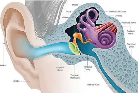
The ear is made up of three parts: the outer, middle, and inner ear3 (Figure 1.1).
The outer ear is the external part of the ear and acts as a funnel to conduct air vibrations through to the eardrum, which collects sound waves and includes the auricle, auditory canal and tympanic membrane1,4.
The middle ear is located between the external and inner ear and transmits sound from the outer ear to the inner ear. The middle ear consists in the tympanic membrane, tympanic cavity and ossicles (malleus, incus and stapes)1,4.
The inner ear is the sensory organ that is the deepest part of the whole ear and is located in the bony labyrinth and includes the cochlea, semicircular canals and vestibular system5,6. The
membranous labyrinth is a continuous system of ducts filled with endolymph and it lies within the bony labyrinth surrounded by perilymph. The perilymph is a fluid that presents an ion concentration similar to the concentration of all other extracellular fluids, contrary to the endolymph that presents an ionic content similar to the contents of the intracellular fluids, with high [K+] and low [Na+]7,8.
The vestibular system, plays a major role in the sense of balance and it is the sensory system that provides the leading contribution about movement and sense of balance7. Together with the
cochlea it constitutes the labyrinth of the inner ear. The vestibule contains the utricle and the saccule, two components of the membranous labyrinth, which contain a small patch of hair cells and their supporting cells, which collectively are known as macula. The macula which is located in the utricle and saccule, is a specialised area of the sensory receptor cells7,8.
The semicircular canals are part of our balance and equilibrium system and contain sensory receptors5.
Figure 1.1-Diagram of the ear. Adapted image by https://www.earq.com/hearing-loss/ear-anatomy.
2
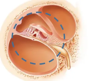
Inside the inner ear exists the cochlea, a structure with the form of a snail-like spiral in the temporal bone and it is divided into three fluid-filled parts (Figure 1.2). The scala vestibuli (upper compartment), scala tympani (lower compartment), which contains perilymph and the scala
media (middle compartment) that contains endolymph9,10
.
The organ of Corti (Figure 1.3) is the sensitive element in the inner ear and it is situated on the basilar membrane, one of the compartments of the cochlea. It contains hair cells which protrude from its surface. Individual hair cells have multiple strands called stereocilia. They act as a transducer, converting vibration into nerve impulses causing displacement of cochlear fluid and movement hair cells at the organ the Corti to produce electrochemical signals11,12
.
Figure 1.2- Schematic view of the cochlea. (1) the scala media; (2) the scala vestibule and (3) the scala tympani. Adapted image by http://www.cochlea.eu/en/cochlea.
Figure 1.3- Representation of the organ of Corti (circle). Adapted image by http://www.gettyimages.pt/fotos/organ-of-corti.
3
1.2 Epidemiology of deafness
Deafness or hearing loss refers to the inability to hear sounds, totally or partially. It is the most common sensory impairment in humans and affects more than 5% of the population (360 million people, including 32 million of children)13.
Approximately 1/1000 children are born deaf and a third of the population over 65 is also affected14,15.
Clinically, hearing loss may be classified according to the time of the manifestation of the first symptoms, degree of severity, origin and type16. Hearing loss can occur at any age and may
be congenital, with symptoms appearing at birth or later on the life15. When hearing loss occurs
after the acquisition of speech it is known as post-lingual hearing loss; hearing loss appearing before speech is called pre-lingual16. To establish the degree of hearing loss, sound is measured
by its intensity (dB) and frequency (Hz) in an audiogram. A person with a hearing loss between 21-40dB is classified as having mild hearing loss; between 41-70 dB as having moderate loss; severe hearing loss is between 71-95 dB; over 95dB is defined as profound hearing loss16,17.
Factors causing hearing loss may be environmental, genetic or a combination of both4,18. An
example of an environmental factor is a pre- or post-natal infection such as cytomegalovirus19–21.
Genetic factors cause various types of hearing loss, but the majority of genetic cases of hearing loss are associated to non-syndromic autosomal recessive heredity, where loss of hearing is the only pathology22,23.
Hearing loss can also be caused by a mix of environmental and genetic factors such as the A1555G mutation in the mitochondrial DNA and exposure to ototoxic antibiotics22,24,25.
Hearing loss can be classified in different types: conductive hearing loss, sensorineural hearing loss, and mixed hearing loss, depending on the part of the ear that is affected24,26.
Conductive hearing loss is due to alterations in the outer and/or middle ear that affects the conduction of sound along the ear canal, from thepinna to the eardrum19,27.
Sensorineural hearing loss affects the inner ear, including the cochlea, the auditory nerve or both. In these cases the sound reaches the cochlea correctly but is not converted into nerve impulses, so the conduction of these nerve impulses through the auditory nerves is inhibited28.
Mixed hearing loss is a mixture of conductive hearing loss and sensorineural hearing loss. To clarify the type of hearing loss it is important to assess the hearing threshold through an audiogram as well as to distinguish between the different forms of hearing loss (genetic or acquired) by using a precise diagnosis that must also include the family history, the otological and clinical analysis and genetic analysis16,27
.
1.3 Age Related Hearing loss
Age-related hearing loss (ARHL), also known as presbycusis is an important problem in society and refers to sensorineural hearing impairment in elderly individuals29. Presbycusis is
characterized by progressive, bilateral, symmetrical hearing decline in clinical and audiological terms30,31. The highest frequencies of the auditory spectrum are the first to be affected, with the
lowest frequencies being the last to be damaged. Consequently, individuals with presbycusis can’t rely on their hearing to overcome limitations of impaired vision and slowed reaction time. In addition, age-associated decline in concentration and memory contribute to difficulty understanding speech, especially in noisy situations. Thus, there is a decrease in communicative capacity, which causes harmful effects on the quality of life of individuals, resulting in emotional problems and social isolation32,33.
ARHL is considered to be a multifactorial progressive disease caused by many factors, such as ototoxic drugs, noise, otologic disease history, metabolic changes, hormones, diet, and immune
4
system that are superimposed upon an intrinsic, genetically controlled, aging process34. Several
intrinsic (genetic predisposition) and extrinsic (exposure to intense noise) factors are thought to affected the inner ear throughout life and which cumulatively lead to a decrease in cochlear transduction of acoustic signals35.
According to the World Health Organization (WHO), by 2025 there will be approximately 1.2 billion people in the world over 60 years old and, consequently, ARHL will continue to be a problem in the coming years and might be a huge public health problem for societies36,37.
1.4 Genetic studies
Research using genetic and molecular studies has meant we have taken great strides in the study of hearing loss. It is estimated that over 50% of cases are caused by genetic factors and that about 60% of people with hearing loss show monogenic transmission36,38
.
Hereditary hearing loss is known as syndromic when in addition to hearing loss, a person has other characteristics or alterations specific to a syndrome. However, around 70% of the cases are associated with non-syndromic hearing loss39.
Approximately 80% of the non-syndromic hearing loss show autosomal recessive heredity (DFNB loci), 17% show an autosomal dominant inheritance pattern (DFNA loci), and in a minority of cases (1-2%) the cause of pathology is in chromosome X (DFN loci) or in the mitochondrial genomes27.
Studies carried out in molecular genetics have identified around 170 loci associated with non-syndromic hearing loss, and over 60 genes have been identified so far40,41.
Locus DFNB1, located on chromosome 13q11-12, is associated with autosomal recessive non-syndromic sensorineural hearing loss, pre-lingual onset and mostly profound42,43.
This locus contains two genes - GJB2 and GJB6 – which belong to the same cluster and codify two transmembrane proteins - connexin 26 (Cx26) and connexin 30 (Cx30) – expressed in the Organ of Corti inside the cochlea and both are essential in the for the hearing process42,44
.
1.5 Connexins- Structure and function
Connexins (Cx) are a family of transmembrane proteins found in most vertebrate tissues. There are at least 21 different connexins in the human species and at least one type of connexin can be found in almost all cells of the body, at some stage of development45,46.
In the human genome there are more than 20 genes codifying for connexins, thus mutations in these genes can lead to profound or congenital diseases, namely hearing loss. Mutations in connexins are in fact associated with various illnesses, such as neurological diseases, skin diseases and cataracts47,48.
The two connexins, Cx26 and Cx30, are both found inside the cochlea at hair cells of the Corti Organ, and play a key role in the hearing process allowing the flow of potassium ions49. All
the members of the connexin family share the same make up of nine domains: the amino terminal domain (-NH2), the carboxyl terminal domain (-COOH), two extracellular loops, one cytoplasmic loop and four transmembrane domains50–52. The most conserved regions in this family of proteins
are the extracellular loops, involved in the docking of proteins the process necessary for the formation of intercellular channels53. The most different regions between the various connexins
are the cytoplasmic loop and the C-terminal region, which may vary either in sequence or in length and these regions are the most susceptible to post-translational modification, for example, by phosphorylation47. However, it is believed these regions play an important regulatory role since
as observed by crystallography the N-terminal region of the protein is part of the pore structure of the intercellular canal54.
5
The connexins join together in hexameric channels, forming connections or hemichannels, with two sets of 6 connexins from each of the adjacent cell55. The oligomerization of the six
connexins happens progressively at the endoplasmic reticulum and/or the Golgi47. After their
synthesis, the connexins are modified post-translationally and packed in vesicles of the Golgi membrane to be transported correctly to the membrane56. Finally, docking occurs on the cell
membrane from each of the adjacent cell, allowing contact between them and creating an intercellular pore57,58.
The connexons, formed by 6 units of identical connexins, are classified as homomeric, being heteromeric when are composed by different connexins as subunits, namely of connexin 26 and 3059. The heteromeric channels have different characteristics, both in terms of shape and charge,
thus regulatory properties can be different60.
Connexons are thus organized as potassium channels also acting as gap junctions, a type of intercellular communication located in specialized regions of the membrane, thus essential for the normal physiological processes of adjacent cells. Their distribution along the inner ear ensures the cochlear homeostasis being its distribution fundamental for the recycling of potassium ions at the organ of Corti61–63.
1.5.1 Connexin 26
Connexin 26 (Cx26), also known as the Beta-2 gap junction protein, is the most frequently connexin associated with hearing loss. The protein is codified by gene GJB2, presenting 226 amino acids and a molecular mass of 26 kDA14,64.
Cx26 is found in the cochlea, at the organ of Corti, at hair cells and support cells and contributes to the recycling of K+ ions and glucose homeostasis65,66.
The GJB2 gene is a small gene with 5500 bp long, just two exons being the coding region almost entirely within the exon 267,68. This gene is considered to be the most frequent target for
mutations associated with autosomal recessive neurosensorial, bilateral and congenital hearing loss. There are around 110 recessive mutations described leading to the production of truncated proteins or non-functional proteins. However, the majority of modifications associated with the
GJB2 gene are alterations caused by nucleotide substitutions and deletions69,70.
The type and frequency of mutations are heavily influenced by the geographical distribution of populations. In Caucasian populations, modification c.35delG is the most common, characterized by the deletion of a guanine at nucleotide 3557,71. This modification causes a
frameshift at the beginning of the protein and consequently leads to a truncated protein with only 12 amino acids, where the 13th is a STOP codon72.
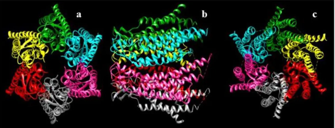
Figure 1.4- Connexon image obtained by CHIMERA software. (a) Schematic three dimensional view of the possible structure of connexions formed by connexin 26 (each colour represents a different connexin). View of the channel from the extracellular side. (b) Side view. (c) View of the channel from the cytoplasmic side.
6
There are other mutations which are more common in other populations, such as the c.167delT, which is predominant among Ashkenazi Jews, the c.235delC found in Asians, the V37I modification in Taiwan and the 235delC modification in Japanese populations73.
Also W24X is very common among Romanian as well as Portuguese populations14.
1.5.2 Connexin 30
Connexin 30 (Cx30), also known as the Beta-6 gap junction protein, is codified by the GJB6 gene presenting 10430 pb65. This gene is a neighbor of GJB2 gene at chromosome 13 and both
account for the DFNB1 locus. Cx30 is a slightly larger protein than the Cx26, containing 261 amino acids74,75.
In the GJB6 gene, there are four mutations associated with hearing loss (T5M, 63delG, G11R and A88V) and two big deletions: one of 309kb, del(GJB6-D13S1830) and the other of 232kb, del(GJB6-D13S1854)76,77. These two deletions delete the CRYL1 gene and the exon 1 and 2 of
the GJB6 gene, as well as the sequence between the two genes78,79.
Del(GJB6-D13S1830) and del(GJB6-D13S1854) can be found in deaf people, both in homozygotes or in heterozygote compounds if associated to other mutations in GJB2 gene79,80.
2. Functional studies
Proteins are not only the most abundant biological macromolecules, they are also the most complex biomolecules in terms of conformational stability and biological versatility81. After
being synthesised, proteins need to acquire a specific three-dimensional structure, through a process known as folding81–83.
Proteins can become misfolded due to alterations in the polypeptide chain, which causes aberrant structures associated to negative effects and consequently damage the normal biological functioning of the human body84,85. Challenges of the folding process include a specific protein
quality control system in charge of degrading misfolding proteins. This system is made up of molecular chaperones, proteases and factors that regulate activity or allow communication between the various components86–88.
The molecular chaperones recognise structural signs presented by misfolded proteins. Proteins are ubiquitinated and when detected are sent to the proteolytic pathway to be degraded by proteasomes in the cytoplasm86,89,90
.
Functional studies are thus the group of studies undergone to identify alterations in protein conformation and expression. Since this type of studies were developed in the present work considering Cx26 protein, further explanations will be particularized with GJB2 gene and Cx26. The first step in the in vitro functional study is to reconstruct the wild-type sequence and the mutations of the Cx26 in a heterologous system, through the transfection of cell lines that do not express this protein endogenously. Consequently it will be possible to obtain cells that start to express this foreign DNA previously inserted. This insertion is confirmed by using a specific antibody and immunofluorescence techniques also crucial for localization of the protein inside the cell, and so to evaluate cell traffic.
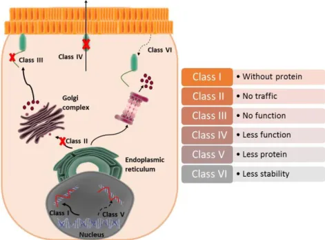
Mutations are grouped in classes according to their functional effect representing an advantage for future clinical treatments, since it is expected that some therapeutic strategies could be applied for mutations of different proteins members of the some class91,92. Possible classes are:
class I, class II, class III, class IV, class V and class VI (Figure 2.1).
Class I (absence of protein) – mutations that inhibit protein synthesis. The mutations belonging to class I inhibit the translation and production of proteins due to nonsense mutations
7
which cause STOP codons prematurely or due to alterations that affect splicing locations, for example. The mutations in this class cause the total absence of proteins91,93.
Class II (absence of cell traffic) - mutations inhibit cell traffic. Mutations can lead to misfolding avoiding protein migration inside the cell. Consequently it is possible to observe the retention of the protein in the endoplasmic reticulum and later its rapid degradation into proteasomes94.
Class III (absence of function) - mutations that inhibit the function of the protein to for channels. This mutations cause a reduction in the activity of the channel, which remains closed and can cause a loss of permeability due to reduction in the size of the pore95. Mutations can also
be associated to a negative effect in relation to the wild-type protein. This dominant-negative effect on the Cx26 can reduce or even inhibit the normal activity of the potassium channel94,96.
Class IV (reduced function) - mutations do not affect the translation of the protein nor the cell traffic. The connexins migrate correctly to the membrane and create ionic channels. However, their functional activity is reduced97,98.
Class V (reduced protein expression) - includes mutations that cause low levels of expression. These mutations are associated with low levels of mRNA91,99.
Class VI (low stability) - mutations that cause a reduction in the stability of the protein when in the surface of the cell94,100.
8
2.1 Mutations p.Leu213X, p.Gly160Cys and p.Gly160Ser
Besides the mutations already mentioned there are many others involved in hearing loss. Frameshift mutations and others leading to partial production of Cx26 represent about 28% of all
GJB2 mutations. The vast majority of Cx26 mutations (79%) are point mutations, caused by
substitutions or nucleotide deletion in the connexin sequence78.
Mutation p.Leu213X (c.638T>A), is a recessive mutation located at the C-terminus, the most variable region within the connexins family. It is thought that this mutation may have a role in regulatory processes. This mutation is characterised by the alteration of the 213 cordon (TTG), which codifies the leucine with a STOP codon (TAG)101.
Mutation p.Gly160Ser (c.478G>A) is located on the second extracellular domain of the Cx26. It is characterised by the alteration of a guanine for an adenine, replacing one glycine (GGC) for a serine (AGC)72.
Mutation p.Gly160Cys (c.478G>T), is similar to the previous one since it occurs in the same nucleotide, however, the guanine is replaced by a thymine, and the amino acid generates a cysteine (TGC)102.
The pathogenicity of these mutations is still not fully clarified since they were recently identified in the Portuguese populations, thus further studies are necessary to assess its pathogenicity. Knowing the structure of one protein allows to use computer analysis to predict the pathogenicity of the mutated protein by comparing 3D structures.
There are several procedures for determining a protein’s 3D structure, being X-ray crystallography or nuclear MRI the most commonly used103. However, although these methods
are quite precise, they are very expensive and time consuming. Therefore, computational methods, using modelling prevision software are quicker and cheaper and still very accurate contributing for in silico modelling, based on the biological principal that the sequence determines the structure, which determines the function104. Tools used for modelling analysis are quite
accurate, leading to their use in drug design, virtual screening, protein engineering and site-specific mutagenesis105. These methods are also very complex since they must consider a large
amount of data, although a short approach is possible to have a preliminary prediction.
3. Objectives
Throughout this study two main objectives have been established:
1. To genetically characterise the population of elderly individuals with presbycusis in regards to genes GJB2 and GJB6 and so contribute to the study of the role of the DFNB1 locus in this type of hearing loss. To achieve this goal we screened for mutation in the coding region of
GJB2 gene and the two major deletions described in GJB6 gene.
2. Study new mutations (p.Leu213X, p.Gly160Ser, p.Gly160Cys, p.Ala40Gly) in GJB2 gene identified in the Portuguese population, to better understand their pathogenicity. To achieve this goal we selected three mutations for functional studies with transfected HeLa cells and we have developed some conformational models to the four mutation based in simple hypothesis of conexon structures.
9
4. Materials and methods
4.1 Description of the study sample
In the present work it was considered a sample of individuals (n= 200) aged between 50 and 90 years old). Individuals were selected from different regions of Portugal and most present age related hearing loss (ARHL). Blood samples, clinical and audiological studies were realized by ENT clinicians and by audiologists. A survey was taken to all individuals including a clinical history to help avoid confounding factors, such as smoking habits or high blood pressure, and thus the genetic diagnosis. Samples were blind coded to ensure anonymity using the designation “PRE” followed by a number for each sample. All participants signed an informed consent and were volunteers.
4.2 Extraction of DNA
Blood samples were collected in FTA cards. The cards were labelled and stored at room temperature. The DNA extraction was performed with the NZY Tissue gDNA Isolation Kit (NZYTech, Lisboa), following the manufacturer’s instructions (Annexes A).
4.3 Amplification by Polymerase Chain Reaction (PCR)
4.3.1 GJB2 gene amplification by Polymerase Chain Reaction (PCR)
Exon 2 of GJB2 gene was amplified by standard PCR. Table 4.1 shows the primers used to amplify the whole coding exon and the accepted splicing location. The PCR reaction was optimized in the thermal cycler Biometra T Professional.
The PCR reaction was prepared for a total volume of 25μL. Both primers were used at a concentration of 10μM. The reaction mixture also included Xpert TaqPlus Mastermix (2x) (Grisp, Porto), containing dNTPs and MgCl2 , sterile ultra-pure water. Finally we added DNA and started
the run with an optimized program for GJB2 gene (Annexes B).
For each PCR reaction, a negative control without DNA was considered to confirm the presence/absence of contaminants in the PCR reagents.
Table 4. 1- PCR Primers used to amplify the exon 2 in GJB2. Primers with the respective sequence, melting temperature and size of the fragments they amplify.
4.3.2 GJB6 gene amplification by Polymerase Chain Reaction (PCR)
All the samples were screen for the two great deletions in the GJB6 gene, del(GJB6-D13S1830) and del(GJB6-D13S1854). This analysis was performed with a PCR Multiplex, thus both deletions could be detected simultaneously.
Table 4.2 presents the primers used, respectively to amplify exon 1, to detect deletion del(GJB6-D13S1854) and primers to detect deletion del(GJB6-D13S1830).
The PCR mix included each primer at 10μM, reaction mixture 2X KAPA2G Robust HotStart (Grisp, Porto) containing dNTPs and MgCl2, sterile ultra-pure water to a final volume of 25 μL.
For each PCR (Annexes C) a negative control was realized, without DNA, to confirm the presence/absence of contaminants in the reagents in the PCR mixture.
Primer’s name Sequence Tm Amplicon size
2AF 5’-AAGTCTCCCTGTTCTGTCCT-3’
60.7ºC 928pb
10
Table 4.2-PCR Primers used to amplify GJB6. Primers with the respective sequence, melting temperature and size of the fragments they amplify.
4.4 Electrophoresis of the PCR products
All the products of PCR were observed in electrophoresis, using 2% agarose gel (SeaKem® LE Agarose, Lonza), in a buffer solution TBE 0.5X. Midori Green Advanced DNA Stain (Grisp, Porto), was used as the DNA intercalling agent, seen under a blue light. The gel was photographed using the camera system FastGene® FAZ Digi (Grisp, Porto).
The GRS Ladder 100 bp (Grisp, Porto) was used to compare the size of the DNA fragments. The loading buffer was a solution of 0,25% bromophenol blue, 0,25% xylene cyanol, 10mM of sodium hydroxide and 95% formamide.
4.5 Sequences analysis
Prior to sequencing the PCR products obtained through the PCR reaction products must be purified. Purification eliminates primers, enzyme, buffer solution or DNA molecules not consumed during the PCR. Consequently, purification is necessary to prevent potentially hybridisation to non-specific and so DNA during sequencing. The samples were sent to a commercial company, STAB Vida, where purification and sequencing was carried out. The sequences were analyzed using Chromas Lite 2.1.1 software and compared to the standard sequence using the NCBI BLAST programme.
4.6 Cell culture
HeLa cells were used in the present project. HeLa cells are derived from a human cell line of cervical cancer. This cell line was chosen for the functional studies since the endogenous expression of connexins is absent.
For cell culture, a CO2 independent medium was used (CO2 (1x), Gibco Products, Life
Technologies), supplemented with 10% foetal bovine serum (FBS) (Gibco Products, Life Technologies), 2mM of L-glutamine (Gibco Products, Life Technologies) and PSN (100X) (Gibco Products, Life Technologies). Cells were maintained in an incubator at 37ºC without CO2,
since this media contains a buffer solution with β-glycerophosphate and sodium bicarbonate, which increases the production and use of CO2 by the cells.
Cells grew on the inner surface of the 25cm² flasks, adhering in a monolayer. After reaching a confluence of approximately 80%, the cell culture was transferred to new T25cm² flasks using Trypsin EDTA (0.05%) in DPBS (1X) (Grisp, Porto). Procedure include removal of the growth
Primer’s name
Sequence Tm Amplicon size
Cx30 Ex1A 5’-CGTCTTTGGGGGTGTTGCTT-3’ 60ºC 333pb Cx30 Ex1 B 5’-CATGAAGAGGGCGTACAAGTTAGAA-3’ Del BK1 5’-TCATAGTGAAGAACTCGATGCTGTTT-3’ 60ºC 564pb Del BK2 5’-CAGCGGCTACCCTAGTTGTGGT-3’ GJB6-1R 5’-TTTAGGGCATGATTGGGGTGATTT-3’ 60ºC 460pb BKR-1 5’-CACCATGCGTAGCCTTAACCATTTT-3’
11
medium followed by two washes with PBS and Trypsin EDTA (0.05%) in DPBS (1X) (Grisp, Porto). After adding the trypsin to the culture, flasks were incubated at 37ºC for about 5 minutes until the dissociation of the cells occurred. Finally, the cells were centrifuged at 2000xg for 5 minutes and after the removal of the Trypsin EDTA (0.05%) in DPBS (1X) (Grisp, Porto), they were resuspended in 1mL of growth medium. Cells were placed in a new 25cm² flask to obtain a culture with a confluence of approximately 15%.
4.6.1 Freezing the cells
The freezing process began by washing the cultures with PBS (10X) (Gibco Products, Life Technologies) buffering solution to remove residual growth medium in the flask. Cells were detached from the flask by using Trypsin EDTA (0.05%) in DPBS (1X) (Grisp, Porto). The suspended cells were put in a 1.5mL microtube, centrifuged at 2000g for 5 minutes to create a pellet and to eliminate the trypsin in the supernatant which was discarded. The pellet was then resuspended in freezing medium with 70% CO2 independent culture medium (1x) (Gibco
Products, Life Technologies), 20% FBS (Gibco Products, Life Technologies) and 10% DMSO. Cells were initially frozen at -20ºC for 1 hour and then kept at -80ºC to maintain cell viability.
4.6.2 Thawing cells
A 1.5mL microtube of cells kept at -80ºC was thawed in a water-bath at 37ºC for 1 minute. It is essential that the thawing is done quickly to maintain cell integrity. After thawing, each microtube was centrifuged for 5 minutes at 1000g. The supernatant was carefully discarded to eliminate most of the DMSO and the pellet was resuspended with approximately 1mL of growth medium supplemented with CO2 independent medium (1x) (Gibco Products, Life Technologies),
10% FBS (Gibco Products, Life Technologies), 2mM of L-glutamine (Gibco Products, Life Technologies) and PSN (100X) (Gibco Products, Life Technologies). The final volume was transferred to a 25cm² culture flask, where another 4mL of supplemented growth medium was added to make a final volume of 5mL.
4.7 Cell transfection
HeLa cells cultured in 25cm² flasks at 37ºC, with confluence between 80-90% were used for transfection.
After washing the cells with PBS (10X) (Gibco Products, Life Technologies), they were trypsinized with 1mL of trypsin (0.05%, Grisp Porto) and followed the procedure already described.
Cells were gently resuspended in 1 mL of the growth medium.
In a 6-well microplate, 2 mL of growth medium was added to each well, plus 40 μL of cell suspension and incubated at 37ºC for 24 hours until the cells adhered to the bottom.
Two different mixes were prepared in micro-tubes (volumes described correspond to the quantity necessary for one well):
Mix A: 95 μL of Opti-MEM® Reduced Serum Medium (Gibco Products, Life Technologies) and 5 μL of Lipofectamina® 2000 (Invitrogen™) and rested for 5 minutes.
Mix B: 100 μL of Opti-MEM® Reduced Serum Medium and 1μg of mutated DNA (plasmid) and 2μg of control DNA (plasmid).
Both mixes (A and B) were joined and incubated for 20 minutes at room temperature. Meanwhile, cells were washed 3 times with PBS 1x buffering solution to remove the growth medium from the wells. After the 20 minutes of incubation, 300 μL of Opti-MEM® and 200 μL of the mixed solution (A and B) were added to each well and incubated at 37ºC for 6 hours.
12
After this period, the medium was removed and 2mL of growth culture medium supplemented with of Opti-MEM® was added to each well. In this step a coverslip was placed in the bottom of each well allowing cells to grow on this glass surface that will be used for staining the cells. The plates were incubated for 24 hours at 37ºC to ensure enough time for cells to adhere and express Cx26.
At the end, the transfected cells were used for an immunofluorescence assay and protein Cx26 was extracted for Western Blot using microwell plates where cells grew without coverslips, as described below.
4.8 Subcellular expression and localization of Cx26
4.8.1 Western Blot
This technique106 was realized in order to measure the concentration of protein Cx26
expressed by the transfected HeLa cells. The procedure includes different steps as described in the sections below.
4.8.1.1 Sodium dodecyl sulphate polyacrylamide gel electrophoresis (SDS-PAGE)
The pellets obtained from scraping the cells out of the wells were centrifuged at 2000g for 5 minutes after resuspension in lysis buffer 2% Igepal (Annexes D) and incubated 1 hour in ice and shaken for 30 seconds every 15 minutes. The samples were centrifuged at 8000rpm for 10 minutes and the supernatant was removed to another tube.
Protein quantification was carried out using the Pierce™ BCA Protein Assay Kit (Life Technologies, Rockford, USA), following the manufacturer’s recommendations.
Two gels were prepared at 12.5% acrylamide separation gel and a 4% acrylamide concentration gel, as described (Annexes E). These separation gels were polymerized and covered with distilled water to accelerate the polymerization process. After, the ethanol (ethanol absolute, Fisher Scientific) was removed and the concentration gel was polymerized with the respective comb to avoid the formation of air bubbles at the surface of the gel.
Before putting the samples in the wells of the gel, samples were denatured at 95ºC for 5 minutes. After this, 40μL of each sample and 5 μL of marker (Protein Marker II®, NZYTech) was added to the gel. Electrophoresis ran at 60 V during the concentration phase and at 100 V during the separation phase for a total of 2h.
4.8.2 Protein transfer
Having been separated electrophoretically, the proteins must be transferred to a PVDF membrane. This was done by submerging the PVDF membrane in methanol for 2 mins and then in H2Od for 5 min. The transfer was carried out for 1 hour at 200mA, under refrigeration. The gel
from the first electrophoresis is placed in contact with the PVDF membrane and both were placed between two sheets of filter paper and two sponges soaked in transfer buffer forming a sheath (figure 4.1). The sheath must be placed in order to allow the current flow from the negative pole (cathode) to the positive pole (anode), so the proteins migrate properly from the gel to the membrane.
After the transfer, the membrane was washed 3 times in PBST (0.1% (v/v) Tween 20 in PBS) for 10 minutes each wash and then blocked with 10mL of a solution of milk in PBST 5% (p/v) for 1 hour.
After one hour, the primary antibody for connexin 26 was added (connexin 26/GJB2 antibody, Invitrogen), and left overnight at 4ºC.
13
The following day, the primary antibody (Connexin 26 N-19 policlonal, goal, IgG, Santa Cruz Biotechnology, INC) was removed by washing the membrane 3 times with PBST for 5 minutes. After washing, the secondary antibody was incorporated (Goat, Anti Rabbit, IgG, TRITC, Novex) for 1 hour at room temperature and then washed 3 times with PBST, each wash lasting for 5
minutes.
4.8.3 Revelation and quantification of band intensities
During the membrane revelation procedure, 1mL of WesternBright™ ECL Luminol/enhancer solution and 1 mL of WesternBright™ Peroxide Chemiluminescent Detection Reagent (WesternBrightTM ECL, GrISP, Porto) were mixed in one eppendorf tube and then placed on the membrane. Resulting image was obtained with the ChemiDoc™ XRS+ System (BioRad).
4.9 Immunofluorescence
For the immunofluorescence, transfected cells were cultivated on coverslips placed in the bottom of the wells of the microwell plates as described above.
After checking at the microscope that the culture reached up to 80% of confluence, medium was removed and cells were washed with PBS three times for 5 minutes each wash. After this, 2% formaldehyde was added during 15-20 minutes, to fix the cells. The cells were then washed 3 times with PBS 1x, during a total of 15 minutes.
The cell membrane was permeabilized with 0.1% Triton in PBS for 10 minutes and then blocked with 2% BSA in PBST during 30 minutes. Cells were then incubated with Phalloidin (CF™ dye phalloidin conjugates, Biotium) during 30 minutes and then washed with PBST 5 times, each wash lasting for 5 minutes.
Cells were then incubated with the primary antibody (Connexin 26 N-19 policlonal, goal, IgG, Santa Cruz Biotechnology, INC) in a dilution of 1:10 in BSA 2% and incubated overnight, at 4ºC. After, cells were washed with PBST 5 times, each wash lasting 5 minutes.
The secondary antibody was diluted at a proportion of 1:500 (Goat, Anti Rabbit, IgG, TRITC, Novex) in BSA 2% and incubated for 30 minutes with the cells, being then washed with PBS 1x. The coverslips were now ready to be placed in a glass slide, but first coverslips were covered with glycerol + DAPI (1:100000), placed in the slides keeping the cells between both glasses. The Figure 4.1-Scheme of gel protein transfer to the PVDF membrane. Adapted image by http://www.abcam.com/
14
preparation is then sealed with nail polish and was ready to be observed under a fluorescence microscope (Olympus bmx 60 microscope).
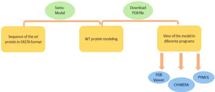
4.10 Methodology models of protein Cx26
The wild-type sequence obtain at the NCBI database was used as control or reference sequence for the Cx26 without mutations (wt).
The reference sequence or wt sequence (FASTA sequence) was used to compare possible alterations in the conformation of the Cx26 mutated. Four different mutated Cx26 were studied separately always considering that the connexons are homotypic, or in other words, formed only by identical Cx26 proteins. The Cx26 channels obtained when using the selected software’s were automatically obtained as a 3D model built in the models was formed automatically.
Three different programs were chosen to visualize the model of the wt proteins and of the mutated ones, PDB Viewer, CHIMERA and PYMOL all obtained from public domains. The pdb format is the a.a. sequence obtained at the PDB viewer and recognized by all the other softwares was obtained from the protein FASTA sequence. Since the four mutations under study are new ones, it does not exist any FASTA sequence for them, consequently, the corresponding FASTA mutated sequences were written manually in Word using the wt sequence as the basis where the mutations were introduced in the respective changed nucleotide (Annex F).
The models obtained were developed following the procedure referred in figure 4.2. Using cartoon view in PYMOL, only the locations of the mutations in the study were chosen. The visualizations obtained were made by superimposing the mutated protein on the wt protein. A summary of the methodology followed in the present project is presented in figure 4.3.
15
16
5. Results and discussion
A total of 200 samples were analyzed in the present study, obtained from individuals selected all over Portugal. Individuals were men (31.5%, n=63) and women (68.5%, n=137) with ages between 50-90 years old (Table 5.1). The majority of individuals (n=159) present presbyacusis and only 4.5% (n=9) of the individuals present normal hearing level (<20dB), being noted that 32 individuals do not have information about the hearing status.
Table 5.1-Distribution of the sample according the sex.
From the ones presenting presbyacusis, the moderate hearing loss was the degree most common (35.5%, n=72) and only 1% (n=2) of the individuals present profound hearing loss (Table 5.2).
Table 5.2-Characterization of the sample considering the degree of hearing loss.
5.1 DFNB1 Analysis
5.1.1 GJB2 gene
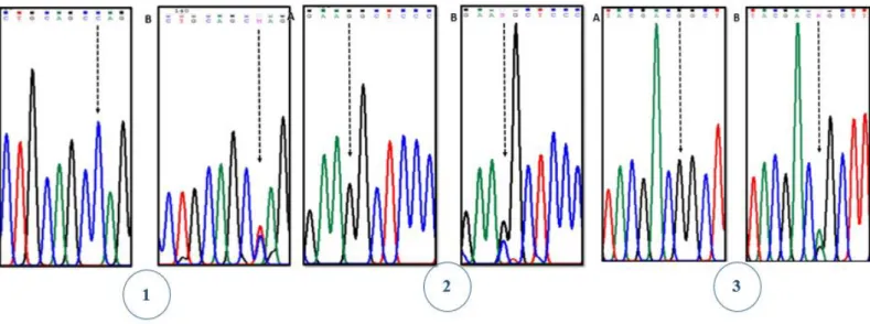
The analysis of the exon 2 of the GJB2 gene was carried out on 80 random samples from our total sample. Of the 80 samples studied, 41 (51.25%) were men and 39 (48.75%) were women.
Three mutations (Figure 5.1) were identified in heterozygousity, p.Arg143Gln, p.Ala40Gly e p.Met93Ile in four individuals (5%) and curiously, mutation A40G was observed in two different individuals. Thus, 76 (95%) of the individuals didn’t present any mutation in the coding region of GJB2 gene. The frequency of the variants found in the total of 160 chromosomes was 2.50% (Table 5.3). N % Men 63 31.5 Women 137 68.5 Total 200 100 Hearing status n % Normal (<20dB) 9 4.5 26-40dB 66 33.5 41-60dB 72 35.5 61-80dB 19 9 >81dB 2 1 Without hearing information 32 16.5 Total 200 100
17
Figure 5.1-Electrophoretograms of GJB2 mutations identified for the first time in our sample. (1) wild type (A) and (B) mutation p.Arg143Gln (c.425G>A); (2) wild type (+/+) (A) and (B) mutation p.Ala40Gly (c.119G>C) and (3) wild type (A) and (B) mutation p.Met93Ile (c.279G>A).
Table 5.3-Frequency of the 3 mutations.
The alteration p.Arg143Gln was found in one individual, (coded as PRE165) and occurs in nucleotide 428 (c.G428A). This mutation may cause mild to profound bilateral sensorineural hearing loss. Several studies suggest that this mutation must alter the protein structure and probably partially avoid the role of Cx26 protein107.
The variant p.Ala40Gly was found in two different individuals (coded as PRE138 and PRE564) and result from the replacement of a C nucleotide by an A at position 119. Studies of the pathogenicity of this variation are still not conclusive but some authors shown its association with severe non-syndromic hearing loss51.
The variant p.Met93Ile, found in one individual (coded as PRE 198), result from a G replaced by and A at nucleotide 279. Studies of this mutation suggest that it is associated with moderate to severe hearing loss108,109.
Carriers of this variants were men and women and are included in the group of moderate HL. Thus, no association with severity of presbyacusis neither with sex of elderly individuals are observed especially considering the small number of carriers identified.
n %
R143Q 1 0.63
A40G 2 1.25
M93I 1 0.63