UNIVERSIDADE DA BEIRA INTERIOR
Faculdade de Ciências da Saúde
Development of intelligent vehicles for delivery
of anti-tumoral drugs
Helena Carina Canhoto de Andrade Pissarra
Dissertação para obtenção do Grau de Mestre em
Ciências Biomédicas
(2º ciclo de estudos)
Orientador:
Prof. Doutor Ilídio Joaquim Sobreira Correia
UNIVERSIDADE DA BEIRA INTERIOR
Faculdade de Ciências da Saúde
Desenvolvimento de veículos inteligentes para
entrega de fármacos anti-tumorais
Helena Carina Canhoto de Andrade Pissarra
Dissertação para obtenção do Grau de Mestre em
Ciências Biomédicas
(2º ciclo de estudos)
Orientador:
Prof. Doutor Ilídio Joaquim Sobreira Correia
v
“Nobody said it was easy It's such a shame for us to part Nobody said it was easy No one ever said it would be this hard Oh, take me back to the start”vii
Acknowledgments
At the end of this master thesis, there are a lot of people that I would like thank, by the support and courage. One way or another, they all contributed for the realization of this work.
First, I would like to thank to my supervisor Professor Ilídio Correia for the unconditional support, guidance, patience, and help during my master’s degree. His wise advices help me to grow as an investigator and person. I also feel the need to acknowledge him for having believed in my capabilities and for making me believe in myself. It has been a privilege to work with him.
I also would like to thank to Sónia Miguel, MSc for the help in acquiring scanning electron microscopy and Confocal Laser Scanning Microscopy images of the produced nanoparticles. I would like to thank to all my group colleagues for their support, knowledge sharing, support and courage that they gave me in the bad moments, but also for all the moments of fun and happiness that we had together, without a doubt, I have been blessed with a friendly and cheerful group.
To my closest friends that have always accompanied me. They always believed in me and they helped me to always have a smile in my face. Thank you all from the bottom of my heart.
Lastly, and most importantly, I would also like to express my sincere thanks to my family, particularly to parents and my brothers. Thanks for the unconditional support and for always believing in me when I doubted about myself. To them I dedicate this thesis.
ix
Abstract
The female reproductive system is essential for the maintenance of the human species. However, it can be affected by several diseases such as ovarian cysts or cancer, pelvic infection, cervical cancer, cervical cysts and polyps, endometrial cancer, endometriosis, endometrial polyps, hyperplasia and fibroids, tipped uterus, prolapsed uterus and breast cancer, which can lead to infertility problems or patient death. Cervical cancer remains as one of the leading causes of women’s death worldwide, and it does not present any signs or symptoms. It has been intimately related whit Human Papilloma Virus duo to the genetic mutations that this virus can cause, when it enters in the cervical cells. For the treatment of cervical cancer surgery, radiotherapy or chemotherapy, alone or combined can be used. Chemotherapy use different drugs as therapeutic agents and the most used in cervical cancer are cisplatin, ifosfamide, paclitaxel, irinotecan and gemcitabine. These drugs can be administered either orally or intravenously. The lack of specificity of these chemotherapeutics agents avoids their accumulation at the target tissue and allows its spreading along the body causing side effects in the healthy tissues. In an attempt to minimize side effects and improve the efficacy of the currently available treatments, new therapeutic approaches are currently being developed. New drug delivery systems are currently being developed to allow the transport of a therapeutic substance through the body and a sustained drug release at the target tissue. Among the different drug delivery systems, nanoparticles are the most used due to their higher capacity to be internalizing by cells and release the drug that they transport inside them. There are several types of nanoparticles that belong to two main groups: organic and inorganic. The magnetic nanoparticles constitute the larger group of inorganic nanoparticles, whereas liposomes, dendrimers and polymeric nanoparticles belong to the organic group.
The objective of the present work was to produce polymeric poly-ε-caprolactone/poly-methyl methacrylate nanoparticles with a sustained drug release to be used for cancer treatment. The produced nanoparticles were characterized by Scanning Electron Microscopy, Transmission Electron Microscopy, Dynamic Light Scattering and Fourier Transform Infrared spectroscopy. Moreover, Cisplatin was used as model drug for the characterization of the loading and release profiles of these nanocarriers, as well as to evaluate the therapeutic potential of the produced system. In addition the in vitro studies were performed to evaluate the cellular internalization and biocompatibility of the produced nanocarriers, and also characterize the release profile and therapeutic efficacy of the loaded drug to cancer cells. The results obtained, suggest that the produce nanosystem has suitable properties to be used as a drug delivery system for cancer therapy.
x
Keywords:
Cervical cancer, Drug delivery systems, Nanoparticles, Poly-ε-caprolactone, Poly-methyl methacrylate.
xii
Resumo
O sistema reprodutor feminino é essencial para garantir a continuidade da espécie humana. No entanto este está sujeito a várias doenças tais como quistos ou cancro nos ovários, infeções pélvicas, cancro, quistos e pólipos no colo do útero, cancro do endométrio, endometriose, pólipos no endométrio, hiperplasia, miomas e cancro da mama, que podem levar a problemas de infertilidade ou mesmo à morte das doentes. O cancro do colo do útero continua a ser uma das principais causas de morte entre mulheres em todo o mundo, e não apresenta sinais ou sintomas. Esta doença está intimamente relacionada com o Papiloma Vírus Humano, devido ao poder mutagénico que este vírus tem após infetar as células do colo do útero. No tratamento do cancro do colo do útero pode ser usada cirurgia, radioterapia e quimioterapia, sozinhas ou combinadas. A quimioterapia usa fármacos como agentes terapêuticos, sendo os mais usados a cisplatina, ifosfamida, paclitaxel, irinotecano e gencitabina, que podem ser administrados quer por via oral ou intravenosa. A falta de especificidade dos fármacos usados faz com que eles não se acumulem apenas no tecido alvo mas também se espalhem pelo corpo afetando os em tecidos saudáveis e destruindo órgãos vitais. Na tentativa de minimizar os efeitos colaterais e melhorar os resultados obtidos com as terapias disponíveis, tem sido desenvolvidas novas abordagens que assentam no desenvolvimento de sistemas de entrega de fármacos. Estas formulações/dispositivos podem transportar uma substância terapêutica através do corpo e promover a sua libertação durante o período de tempo necessário e no órgão alvo. As nanopartículas são os sistemas mais utilizados devido à grande capacidade para entrar nas células e libertar o fármaco no seu interior. Na atualidade existem vários tipos de nanopartículas, sendo divididas em dois grandes grupos: orgânicas e inorgânicas. Dentro das nanopartículas inorgânicas podemos encontrar as nanopartículas magnéticas. No que se refere as nanopartículas orgânicas as mais estudadas são os lipossomas, dendrímeros, e as nanopartículas poliméricas.
O objetivo do presente trabalho foi produzir nanopartículas poliméricas de poli-ε-caprolactona/poli-metil metacrilato que permitissem o transporte de fármacos e a sua libertação em células cancerígenas para serem utilizadas no tratamento do cancro. As nanopartículas produzidas foram caracterizadas por microscopia eletrónica de varrimento, microscopia eletrónica de transmissão, dispersão dinâmica de luz e espectroscopia de infravermelho. Estudos in vitro foram realizados para avaliar a biocompatibilidade e a capacidade das nanopartículas serem internalizadas pelas células cancerígenas, assim como avaliar a eficiência de encapsulação do fármaco no interior da nanopartículas e a sua libertação, usando Cisplatina como fármaco modelo, de forma a permitir caracterizar a eficiência terapêutica do sistema apresentado. Os resultados obtidos sugerem que as nanopartículas produzidas constituem bons candidatos para futuras aplicações de entrega de fármacos à nano-escala para terapia do cancro.
xiii
Palavras-chave
Cancro do colo do útero, Nanopartículas, Poli-ε-caprolactona, Poli-metil metacrilato, Sistema de entrega de fármacos.
xv
Resumo Alargado
O sistema reprodutor feminino é essencial para garantir a continuidade da espécie humana. Ele é responsável pela receção do óvulo para possível fertilização, suportar o desenvolvimento do feto e permitir o seu nascimento. Este sistema tem ainda a capacidade de produzir certas hormonas, como o estrogénio e progesterona, que influenciam não só o sistema reprodutor feminino mas também outros órgãos. Da sua composição fazem parte os ovários e ovidutos, o útero, a vagina, a genitália externa e as glândulas mamárias. Tal como todos os sistemas do corpo, este está sujeito a diferentes desequilíbrios dos quais podem resultar doenças como quistos ou cancro nos ovários, infeções pélvicas, cancro, quistos e pólipos no colo do útero, cancro do endométrio, endometriose, pólipos no endométrio, hiperplasia, miomas e cancro da mama. O cancro do colo do útero continua a ser uma das principais causas de morte de mulheres em todo o mundo. Este tipo de cancro não apresenta sinais ou sintomas e tem sido intimamente relacionada com o Papiloma Vírus Humano (HPV). No entanto, existem outros fatores de risco como sejam o tabagismo, outras doenças sexualmente transmissíveis, o uso prolongado de contracetivos orais e alterações genéticas. A prevenção desta doença pode ser feita através de vacinação contra o vírus HPV e pela redução da exposição aos outros fatores de risco. A quimioterapia é uma das terapias mais usadas para tratar esta doença. A cisplatina, ifosfamida, paclitaxel, irinotecano e gencitabina são exemplos dos fármacos mais usados no tratamento deste tipo de cancro individualmente ou de uma forma combinada. No entanto, sendo uma terapia sistémica envolve todo o corpo e tem uma elevada toxicidade associada. Esta toxicidade traduz-se em náuseas, vómitos, diminuição da produção de glóbulos brancos e vermelhos, diminuição da resposta a infeções, danos nos rins, entre outros. Na tentativa de minimizar estes efeitos colaterais e melhorar a eficácia terapêutica das terapias disponíveis, têm sido desenvolvidas novas abordagens terapêuticas que incluem o desenvolvimento de sistemas direcionados de entrega de fármacos. Estas formulações/dispositivos podem transportar substâncias terapêuticas através do corpo promovendo uma libertação prolongada do fármaco, durante o período necessário e no órgão alvo. Na atualidade existem vários tipos de sistemas entrega de fármacos, como sejam microesferas, nanopartículas, ou implantes biodegradáveis para libertação controlada e sustentada de fármacos (géis, filmes, andaimes, etc). No entanto, as nanopartículas são as mais utilizadas devido à possibilidade de lhes ser conferida seletividade através da funcionalização da sua superfície com ligandos específicos, e ainda devido à maior capacidade entrar destes entrarem nas células e libertar o fármaco dentro destas. Os fármacos podem encontrar-se ligados à superfície do nanotransportador, encapsulados no seu interior ou dissolvidos na sua matriz. As nanopartículas têm também a vantagem de conferir proteção aos fármacos, aumentar do tempo de meia-vida do fármaco na circulação sanguínea, e ainda permitir a entrega de mais do que um composto ao mesmo tempo. Atualmente existem vários tipos de nanopartículas, sendo divididas em dois grandes grupos: orgânicas e inorgânicas. Dentro das nanopartículas inorgânicas podemos encontrar as nanopartículas magnéticos que
xvi
podem ser usadas não só na entrega de fármacos, mas também em técnicas de diagnóstico e noutros tipos de terapias. As nanopartículas poliméricas mais estudadas são os lipossomas, dendrímeros e nanopartículas poliméricas. As nanopartículas poliméricas podem ser produzidas com polímeros naturais ou sintéticos que podem ou não ser biodegradáveis. Existem vários métodos para a produção de nanopartículas poliméricas que podem determinar o tamanho e a carga a superfície que as partículas possuem.O objetivo do presente trabalho foi produzir nanopartículas de poli-ε-caprolactona/ poli-metil metacrilato que permitissem efetuar uma libertação controlada do fármaco ao longo do tempo, a fim de serem utilizadas no tratamento do cancro. As partículas foram produzidas através de uma adaptação do método de nanoprecipitação anteriormente descrito na literatura. A Microscopia Eletrónica de Varrimento e a Microscopia Eletrónica de Transmissão foram usados para analisar a morfologia das partículas. A dispersão dinâmica de luz foi usada para avaliar o tamanho e o zeta-potencial da nanopartículas produzidas. A Espectroscopia Infravermelha permitiu também a análise da composição química das nanopartículas produzidas. A quantidade de fármaco incorporado nas nanopartículas e o seu perfil de libertação foi também caracterizado para avaliar a aplicabilidade do sistema na entrega de fármacos, usando a Cisplatina como fármaco modelo. Este fármaco permitiu também verificar a eficácia terapêutica do sistema apresentado. Para avaliar a internalização das nanopartículas pelas células cancerígenas procedeu-se à obtenção de imagens de Microscopia Confocal. A citotoxicidade dos nanoveículos produzidos foi avaliada através de estudos in
vitro usando uma linha de células cancerígena (Hela) e uma linha celular normal (fibroblastos
humanos). As nanopartículas com o fármaco incorporado provocaram uma diminuição significativa da proliferação das células cancerígenas para 30% ao fim de 7 dias. Os resultados obtidos revelam que as nanopartículas produzidas possuem as propriedades necessárias para serem usados na entrega de fármacos na terapia do cancro.
xviii
Table of Contents
Chapter I - Introduction
1. Introduction ... 2
1.1. Female Reproductive System ... 2
1.2. Female Reproductive System Disorders ... 4
1.2.1. Cervical cancer ... 4
1.2.1.1. Diagnosis of cervical cancer ... 5
1.2.1.2. Prevention and treatment of cervical cancer ... 5
1.3. Drug Delivery Systems ... 8
1.3.1. Nanotechnology ... 9
1.3.2. Types of nanoparticles ... 9
1.3.2.1. Inorganic Nanoparticles ... 10
1.3.2.1.1. Magnetic nanoparticles ... 10
1.3.2.2. Organic Nanoparticles ... 11
1.3.2.2.1. Liposomes ... 11
1.3.2.2.2. Dendrimers nanocarriers ... 12
1.3.2.2.3. Polymeric nanoparticles ... 13
1.3.3. Poly-ε-caprolactone nanoparticles ... 17
1.3.4. Poly (methyl methacrylate) nanoparticles ... 20
1.3.5. Characterization of nanoparticles properties ... 22
1.3.5.1. Particle size and shape ... 22
1.3.5.2. Nanoparticles Surface properties ... 23
1.3.5.3. Drug loading and release profile ... 26
1.3.6. Mechanisms of cellular uptake ... 26
1.4. Objectives ... 30
Chapter II - Materials and Methods
2. Materials and Methods ... 32
2.1. Materials ... 32
2.2. Methods ... 32
2.2.1. Synthesis of PCL/PMMA nanoparticles ... 32
xix
2.2.3. Transmission electron microscopy analysis of nanoparticles ... 33
2.2.4. Characterization of Size and Zeta Potential of nanoparticles ... 33
2.2.5. Fourier Transform Infrared spectroscopy analyses of nanoparticles ..
... 33
2.2.6. Characterization of the loading profile of the nanoparticles ... 33
2.2.7. Characterization of the release profile of the nanoparticles ... 34
2.2.8. Characterization of the cell proliferation in the presence of
PCL/PMMA nanoparticles ... 34
2.2.9. Characterization of the biocompatibility of PCL/PMMA nanoparticles .
... 34
2.2.10.
Characterization of the cellular uptake of PCL/PMMA nanoparticles
... 35
2.2.11.
IC50 determination of cisplatin ... 35
2.2.12.
Evaluation of the cytotoxic profile of the produced loaded
nanoparticles ... 36
2.2.13.
Statistical analysis ... 36
Chapter III - Results and Discussion
3. Results and Discussion ... 38
3.1. Particles morphology, size and zeta potential characterization ... 38
3.2. Fourier Transform Infrared Spectroscopy Analysis of the produced
nanoparticles ... 40
3.3. Characterization of the loading profile of the produced nanoparticles .. 43
3.4. Characterization of the release profile of the vehicles ... 44
3.5. Evaluation of PCL/PMMA nanoparticles biocompatibility ... 45
3.6. Qualitative evaluation of the in vitro transfection ... 50
3.7. Determination of the inhibitory concentration of free Cisplatin in
Hela-Cells ... 51
3.8. Characterization of the cytotoxic profile of the produced cisplatin load
nanoparticles ... 52
Chapter IV - Conclusions and Future Perspectives
4. Conclusions and Future Perspectives ... 55
Chapter V - Bibliography
xxi
List of Figures
Figure 1: Illustration of the human female reproductive system ... 2
Figure 2: Mechanism of action of cisplatin ... 7
Figure 3: Biomedical applications of magnetic nanoparticles ... 10
Figure 4: Representation of the traditional phospholipids liposomes ... 11
Figure 5: Representation of a dendrimers ... 12
Figure 6: Schematic representation of polymeric nanoparticles ... 13
Figure 7: Schematic representation of the emulsification/solvent evaporation
technique ... 14
Figure 8: Representation of the nanoprecipitation method ... 15
Figure 9: Schematic illustration of the emulsification/solvent diffusion technique .. 16
Figure 10: Schematic illustration of the salting-out technique ... 17
Figure 11: Representation of ε-caprolactone and poly-ε-caprolactone structure ... 18
Figure 12: Representation of methyl methacrylate and poly (methyl methacrylate)
structure ... 20
Figure 13: Biodistribution and clearance of nanoparticles ... 23
Figure 14: Representation of targeted nanoparticle delivery to cancer cells ... 26
Figure 15: Mechanisms of nanoparticles internalization in cells ... 28
Figure 16: SEM and TEM images of PCL/PMMA NPs. ... 38
Figure 17: SEM and TEM images of cis-PCL/PMMA NPs. ... 38
Figure 18: Size, PDI and zeta potential of PCL/PMMA NPs and cis-PCL/PMMA NPs .... 39
Figure 19: FTIR spectra of the produced nanoparticles and of the used compounds
used for their production ... 41
Figure 20: FTIR spectra of the produced nanoparticles and cisplatin ... 42
Figure 21: Calibration curve and its respective equation used to calculate the
encapsulation efficiency of cisplatin in PCL/PMMA NP. ... 43
Figure 22: Release profile of cisplatin from cis-PCL/PMMA NP formulations at ph=7.4.
... 45
Figure 23: Inverted Microscope Images of HFib cells 24, 48 and 72h after being
seeded with PCL/PMMA nanoparticles ... 46
Figure 24: Inverted Microscope Images of Hela cells 24, 48 and 72h after being
seeded with PCL/PMMA nanoparticles ... 47
Figure 25: Evaluation of the cellular viability after cells being exposed to the
produced nanoparticles . ... 49
Figure 26: CLSM images of Hela cells ... 51
Figure 27: IC 50 determination of Cisplatin anti-tumoral activity in Hela cancer cells.
... 52
Figure 28: Evalution of the cytotoxic effect of Cisplatin when delivered by PCL/PMMA
NP ... 53
xxiii
List of Tables
Table 1: Examples of PCL nanoparticles reported in literature ... 19
Table 2: Examples of PMMA nanoparticles produced so far ... 21
Table 3: Ligands that have been used for functionalized the NPs surface ... 25
Table 4: Stirring time and encapsulation efficiency of different formulations ... 44
xxv
List of Abbreviations
BSA Bovine serum albumin
CLSM Confocal Laser Scanning Microscopy
CEA Carcinoembryonic antigen
Cis-PCL/PMMA NP Cisplatin loaded Poly-ε-caprolactone/Poly-methyl metacrylate nanoparticles
CTR1 Copper transporter 1
DDS Drug delivery system
DMEM-F12 Dulbecco’s Modified Eagle’s Medium - F12
DMEM-HG Dulbecco’s Modified Eagle’s Medium – High Glucose
DMSO Dimethyl sulfoxide
DNA Deoxyribonucleic Acid
EDTA Ethylenediaminetetraacetic Acid
EE Encapsulation Efficiency
EtOH Ethanol
FBS Fetal Bovine Serum
FTIR Fourier Transform Infrared Spectroscopy
GM-CSF Granulocyte–macrophage colony-stimulating factor
Hela Human cervical cancer cells
HFib Human Fibroblast cells
HPV Human Papilloma Virus
k- Negative control
K+ Positive control
LTTs Ligand-targeted therapeutics
MPS Mononuclear phagocytic system
MTS 3-(4,5-dimethylthiazol-2-yl)-5-(3-carboxymethoxyphenyl)-2-(4-sulfophenyl)-2H tetrazolium reagent NGR Asn–Gly–Arg tripeptide PAA PCL Poly-acrylic acid Poly-ε-caprolactone PAMAM Polyamidoamine
PCL/PMMA NP Poly-ε-caprolactone/Poly-methyl metacrylate nanoparticles
PCR Polymerase chain reaction
PEG Poly(ethylene-glycol)
PFA Paraformaldehyde
PLA Poly-lactic acid
PLGA Poly-lactic-co-glycolic acid
PMMA Poly-methyl methacrylate
xxvi
Pt Platinum
PVA Poly-vinyl alcohol
PVP Poly-vinyl pyrrolidone
RGD Arg–Gly–Asp tripeptide
ROS Reactive oxygen species
SEM Scanning electron microscopy
SiO2 Silicon dioxide
SnCl2 Tin(II) chloride
TAG72 Oncofetal antigen tumour-associated glycoprotein-72
TCA Tricarboxylic acid
TiO2 Titanium dioxide
Chapter I:
Introdution
Chapter I - Introduction
2
1. Introduction
The female reproductive system is essential for the maintenance of the human species. It is responsible for receiving the oocytes for fertilization, supports the development of the fetus and allows child birth (Kobayashi and Behringer, 2003). Furthermore, it is also involved in different functions such as the production of oocytes and sexual hormones, such as estrogen and progesterone, which have influence in the reproductive system and other organs such as brain, skin, bone and vascular system (Junqueira and Carneiro, 2004).
According with GLOBOCAN 2012, an estimate of the incidence of mortality and prevalence from the world health organization, there were 14.1 million new cancer cases and 8.2 million cancer deaths, in 2012 worldwide (WHO, 2014). Among all types of cancer, the cervical cancer is the fourth most abundant, accounting for 8% of all cancers in women, and it is responsible for 7% of deaths caused by cancer in 2012. (WHO, 2014). Despite the great efforts done so far, the survival rate of patients suffering this disease is still not satisfactory. Due to that, it is essential to develop new therapies that reduce side effects for patients, mortality rate, and also decrease costs associated with treatment in clinical practice (Jin et al., 2014, Lohcharoenkal et al., 2014).
1.1. Female Reproductive System
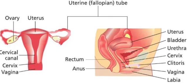
Female reproductive system is comprised by a pair of ovaries and oviducts, uterus, vagina, external genitalia (see Figure 1) and mammary glands (Spencer, 2009).
Chapter I - Introduction
3
As we can see in figure 1, the ovaries are two small oval glands located in the pelvic portion of the abdomen (Markovic and Markovic, 2008). They are responsible for the production of the female gametes (oocytes) and for the secretion of hormones namely estrogen and progesterone. The external surface of each ovary is covered by a epithelium (Jones and Lopez, 2006), also known as germinal epithelium (Van De Graaff, 2002). This layer is composed by cuboidal epithelial cells. The tunica albuginea, a collagenous connective tissue, is located immediately bellow (Van De Graaff, 2002). The interior of the ovary has two regions: the cortex and medulla (Saladin and Miller, 1998). The outer cortex is responsible for the development of the germ cells while the central medulla contain arteries, veins, lymphatic vessels and nerves (Saladin and Miller, 1998, Jones and Lopez, 2006).The oviducts (or fallopian tubes) are funnel-shaped long muscular tubes that connect the ovary to the upper part of the uterus (Markovic and Markovic, 2008, Van De Graaff, 2002, Saladin and Miller, 1998). It can be divided in infundibulum (at ovarian end), the ampulla (the middle part of the tube) and the isthmus (at uterus end) (Saladin and Miller, 1998). This is a very important site because it is where the ovum meets sperms and the fertilization takes place (Markovic and Markovic, 2008). They are composed by three layers: the internal mucosa composed by ciliated columnar epithelium, the muscularis (the middle layer) composed by a circular and a longitudinal layer of smooth muscle, and the serous layer (the external layer) that is part of the visceral peritoneum (Van De Graaff, 2002, Junqueira and Carneiro, 2004). The mucosa epithelium has secretory cells that secret mucus which covers the internal surface of the fallopian tubes. This mucus is responsible for nutrition and protection of the oocyte. This mucus is also important for the sperm activation (Junqueira and Carneiro, 2004). The muscular layer is responsible for the movement of the oocyte through the lumen of the uterine tube (Van De Graaff, 2002). The main function of serous layer is the structural support (Tate et al., 2003).
Connected with the oviducts we can find the uterus. Uterus is a pear-shaped muscular organ located in the pelvic cavity behind the bladder and in front of the bowel. It is responsible for receiving the blastocyst and providing a site for his implantation, for the gestation takes place, and also contracts during the bird of the baby (Van De Graaff, 2002). It can be divided in corpus uteri (the wider upper portion of the uterus) and the cervix uteri (the lower, narrower part of the uterus) (Markovic and Markovic, 2008). The wall of the corpus uteri is composed by three layers: the perimetrium, myometrium and endometrium (Jones and Lopez, 2006, Van De Graaff, 2002, Junqueira and Carneiro, 2004, Saladin and Miller, 1998, Tate et al., 2003). The perimetrium is the most external layer of the uterus and is composed by a thin visceral peritoneum (Van De Graaff, 2002, Tate et al., 2003). The myometrium, the higher layer of the uterus, is composed of smooth muscle running in all directions (Saladin and Miller, 1998, Van De Graaff, 2002). The endometrium is the most internal layer and is composed by a stratum functionalis and stratum basalis (Jones and Lopez, 2006). The stratum functionalis consists in a lining epithelium, uterine glands and is shed during every
Chapter I - Introduction
4
menstruation (Jones and Lopez, 2006, Saladin and Miller, 1998). The stratum basalis is a highly vascularized structure and serves to regenerate the stratum functionalis after every menstruation (Van De Graaff, 2002).Underneath the uterus is located the vagina that connects the uterine cavity with external genitals (Markovic and Markovic, 2008). It is a muscular tube that receives the sperm during the coitus, acts as a birth canal during parturition and provides an exist for the menstruation to get out of the body (Van De Graaff, 2002, Saladin and Miller, 1998). The vagina wall is composed by an external fibrous layer, a middle muscle layer and an internal mucosal layer (Van De Graaff, 2002). The fibrous layer is an elastic connective tissue that supports nerve bundles that control blood flow and the contraction of smooth muscle of the vaginal tissue. Moreover, it has some free sensory nerve endings, mainly near the vaginal opening (Jones and Lopez, 2006, Van De Graaff, 2002). The muscle layer is composed by smooth muscle that allows the vagina dilatation (Tate et al., 2003). The mucosal layer consists of nonkeratinized stratified squamous epithelium that forms a series of transverse folds called vagina rugae (Van De Graaff, 2002). Vagina has no glands, but it is lubricated by the transudation of serous fluid through its walls and by mucus from the cervical glands above it (Saladin and Miller, 1998). The epithelial cells are rich in glycogen. This glycogen is metabolised to produce lactic acid, which is responsible for decrease of pH value, for about 3.5-4, and consequently protect vagina against some microorganisms (Junqueira and Carneiro, 2004, Saladin and Miller, 1998). The vulva (pudendum) is the external part of the female reproductive system and consists of several female organs such as labia major, labia minor, clitoris, opening of the vagina and the urethra (Markovic and Markovic, 2008). It is a highly vascularized region with a sympathetic and parasympathetic innervation as well as extensive somatic neurons that respond to sensory stimulation (Van De Graaff, 2002).
1.2. Female Reproductive System Disorders
The female reproductive system can be affected by various diseases/disorders such as ovarian cysts or cancer, pelvic infection, cervical cancer, cervical cysts and polyps, endometrial cancer, endometriosis, endometrial polyps, hyperplasia and fibroids, tipped uterus, prolapsed uterus and breast cancer. These diseases/disorders can be induced by infections caused by virus and bacteria or genetic mutations, which lead to uncontrolled growth of cells. Also other diseases may potentiate the appearance of these diseases, such as obesity or diabetes (Jones and Lopez, 2006).
1.2.1. Cervical cancer
As already mentioned, cervical cancer remains as one of the leading causes of death in women across the world (Markovic and Markovic, 2008). This type of cancer doesn’t present
Chapter I - Introduction
5
any signs or symptoms. It has been intimately related whit Human Papilloma Virus (HPV) (Mota, 2012). HPV was identified as viral particles in 1949 and nowadays are known almost 100 different types (from these 100 types about 18 have been identified as being implicated in the majority of cervical cancers) (Jones and Lopez, 2006). The prevalence of HPV DNA in women withcervical cancer
has been shown to range from 63% to 98% (Chin’ombe et al., 2014). The HPV can integrate the deoxyribonucleic acid (DNA) of the host cell and promote the synthesis of some viral proteins such as E6 and E7. These proteins are oncogenic proteins that bind to p53 and retinoblastoma protein (pRB), which are two tumor suppressors’ proteins and cause their inactivation. Thereby, with the inactivation of these two proteins, cell cycle arrest is prevented without causing significant transformation (Mota, 2012, Tornesello et al., 2013). However, there are other factors that promote this type of cancer such as smoking, sexually transmitted diseases, prolonged use of oral contraceptives and genetic alterations (Mota, 2012). Women with a greater number of sexual partners have an increased risk of be infected with the virus (HPV) or other pathogenic agents (Jones and Lopez, 2006).1.2.1.1. Diagnosis of cervical cancer
The diagnosis of this disease is extremely important so that the treatment can be administered as soon as possible. There are some methods to investigate the presence of cervical cancer, such as, cervical cytology (pap smear), DNA identification of oncogenic HPV and colposcopy with directed biopsy (Mota, 2012).
Pap smear is one of the most commonly used tests for screening and diagnosis cervical cancer. It consists of 3 basic steps: sample collection with a medical device to scrap cervical epithelium, sample processing using Papanicolaou stains and interpretation of the results using cytopathology (Markovic and Markovic, 2008).
Other possible method is the identification of oncogenic HPV DNA in the host (namely the high-risk HPV types include types 16, 18, 31, 33, 35, 39, 45, 51, 52, 55, 56, 58, 59, 66, 68, 73, 82 and 83) (Al Moustafa et al., 2014) that can be performed by polymerase chain reaction (PCR) . This technique consists in a chemical reaction that results in the synthesis of a large number of target DNA strands, in this case HPV DNA (Olesen et al., 2014).
At last, colposcopy arises as a diagnostic medical procedure where a speculum is used to see female organs such as vulva, vagina and cervix. If a sick-looking tissue is observed, a biopsy can be collected and then histologic analysis be performed (Markovic and Markovic, 2008).
1.2.1.2. Prevention and treatment of cervical cancer
The prevention of cervical cancer is a very important step and may depend not only on the reduction of risk factors (mentioned above), but also by vaccination against HPV (Markovic and Markovic, 2008). Moreover, a deep knowledge of the disease will contribute to decrease the number of infected women in the near future. However these measures will not allow
Chapter I - Introduction
6
eradicates the disease, so it is important understand and improve the currently available therapies.There are three conventional therapies that can be used for cervical cancer treatment (depending on the stage): surgery, radiotherapy and chemotherapy. These therapies can be used alone or combined.
The surgery can be done in 3 ways: the removal of the tumor mass without invasion of the surrounding tissues; to remove the tumor mass and part of surrounding tissue; remove involves the removal of part of the tumor mass for the restitution of function of other organs affected by the tumor. The application of this therapeutic approach is mainly dependent on the stage of development of the cancer (Markovic and Markovic, 2008).
Radiotherapy uses ionizing radiation (beta particles, alpha particles or X-rays) for cancer treatment, causing DNA damage of cells (Markovic and Markovic, 2008). The main disadvantage of this technique is the lack of selectivity; therefore it damages not only the cancer cells but also the healthy ones. However, healthy cells have DNA repair systems available and allow their survival (Khan and Khan, 2003). Radiotherapy is classified according to the type of energy used. There are two radiation therapeutic techniques used in the treatment of cervical cancer: teletherapy and brachytherapy (Khan and Khan, 2003, Rosenberg, 2008). Teletherapy uses X-ray applied from the outside of the body, while brachytherapy involves the intake of a radioactive agent that will release alpha or beta particles, depending on the radioactive agent (Khan and Khan, 2003).
An alternative to surgery and radiology is chemotherapy, which uses drugs as therapeutic agents. Inversely to the surgery and radiotherapy, chemotherapy is a systemic therapy that involves all body and causes toxicity (Markovic and Markovic, 2008). The drugs are frequently administered orally or intravenously. The observed lack of selectivity of drugs leads to unspecific uptake of drugs by all type of cells, leading to various side effects (Jain, 2008). This toxicity and side effects are caused by the accumulation of drugs within non-target tissues. This can damage vital organs such as liver and heart and trigger future complications. The drugs that are commonly used for cervix cancer treatment are cisplatin, ifosfamide, paclitaxel, irinotecan, and gemcitabine in combination with cisplatin (Markovic and Markovic, 2008).
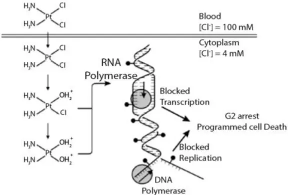
Cisplatin is part of a unique and important class of platinum (Pt) antitumor agents (Cepeda et al., 2007). The mechanism of action of cisplatin involve the binding of the drug to DNA, causing the subsequent cell death by apoptosis, necrosis or both (Fuertes et al., 2003a, Galluzzi et al., 2013). Cisplatin has been used in the treatment of other types of cancer such as testicular, ovarian, bladder, head and neck, oesophageal and small cell lung cancer (Giaccone, 2000). It has been reported that this drug is able to enter into the cell either by passive diffusion or through a gated channel (Ciarimboli, 2012). The input of cisplatin through
Chapter I - Introduction
7
the cell via passive diffusion is unspecific and only depends of the concentration of cisplatin in the medium (Ciarimboli, 2012). On the other hand cisplatin can enter into the cell through some specific channels. A channel that has been most closely associated with the input of cisplatin to the cell is a high-affinity copper transporter 1 (CTR1) (Florea and Büsselberg, 2011). Some studies suggest that CTR1 is predominantly in perinuclear vesicles of some cell lines, whereas in others it is at the plasma membrane (Liang et al., 2014, Safaei and Howell, 2005, Zisowsky et al., 2007). The mechanism used for the entrance of cisplatin into the cell trough CTR1 is not yet fully known, however there are some hypotheses to explain this transport: platinum (Pt) drugs may be transported across the plasma membrane through a pore formed by the three transmembrane domains of CTR1 (Klomp et al., 2003, Safaei and Howell, 2005, Petris et al., 2003).However, there are other factors that influence the uptake of the Pt drugs by CTR1 channel, such as temperature, pH, K+ionsand reducing agents (Safaei and Howell, 2005). The uptake
of cisplatin leads to an accumulation of the drug inside the cell, where the Cl- concentration
lower than that found in blood. In such conditions, cisplatin Cl- group is released, conferring a
positive charge to cisplatin that is now able to interact with nucleophilic sites on intracellular macromolecules like proteins, RNAs, and DNA adducts. Adduct formation results in the inhibition of DNA replication, RNA transcription, arrest at the G2 phase of the cell cycle, and/or programmed cell death (Kartalou and Essigmann, 2001, Galluzzi et al., 2013, Ciarimboli, 2012)(see figure 2).
Chapter I - Introduction
8
Due to its high affinity for nucleophilic groups, cisplatin reacts with different cellular components such as membrane phospholipids, cytoskeletal microfilaments, and thiol-containing molecules which leads to only approximately 1% of the intracellular cisplatin will be able to interact with nuclear DNA (Galluzzi et al., 2013). These nuclear and protein damages leads to the activation of apoptotic pathways, through the activation of caspase-3, caspase-6, and caspase-7 with the subsequent cleavage of key substrates (Fuertes et al., 2003b).However, in cisplatin resistant cell lines a decreased uptake or increased efflux of the drug has been observed. Moreover, the increased inactivation of cisplatin by intracellular proteins such as glutathione, the increased repair of cisplatin-DNA adducts and defects in the apoptotic response pathway also promote the resistance of cancer cells (Galluzzi et al., 2013, Galluzzi et al., 2011). The decrease in the uptake can be explained by the loss of efficiency of CTR1 (due to the factors affecting the uptake), low concentration of cisplatin outside the cell and decreased expression of CTR1 (Ciarimboli, 2012). On the other hand, it has been previously reported in literature that the two copper efflux transporters ATP7A and ATP7B regulate the efflux of cisplatin. These two copper efflux transporters are located in trans-Golgi network and export cisplatin by exocytosis (Prohaska and Gybina, 2004, Ciarimboli, 2012, Kuo et al., 2007). There are other studies that described the multidrug resistance is associated with protein 2 (ABC-transporter 2) (Liedert et al., 2003). ABC-transported 2 works as an ATP-dependent conjugate export pump that require the formation of Pt-glutathione conjugates for the cisplatin efflux from the cell (Kuo et al., 2007).
Therapies based on this PT drug family have many side effects such as nausea and vomiting, decreased production of blood cells and platelets in bone marrow, decreased response to infection, induce damage in the kidneys, neurons and hearing loss (Florea and Büsselberg, 2011).
In an attempt to minimize the side effects and also improve the efficacy of the currently available therapeutics, new approaches are currently being explored. New drug delivery systems (DDS) are being designed and developed to encapsulate and deliver therapeutic agents (Jain, 2008, Haley and Frenkel, 2008).
1.3. Drug Delivery Systems
DDS are formulations/devices that can be loaded with a therapeutic substance through the body. Furthermore, these carriers can promote a sustained release of the therapeutic substance along time, at a specific target organ (Jain, 2008).
Conventional therapies to cancer, like chemotherapy, involve the administration of free therapeutic agents to the patient. However, they have a limited effectiveness, lack of
Chapter I - Introduction
9
selectivity and poor biodistribution (Wilczewska et al., 2012). Such therapies can be improved by encapsulating the drug in a DDS. These systems upgrade the efficacy and safety of the treatments (Jain, 2008), since the drug is carried till the place of action, minimizing the interaction with others tissues, and protecting the drug from rapid degradation and clearance from the body. Furthermore, a lower concentration of drug is required to achieve the aimed therapeutic effect in the organism (Wilczewska et al., 2012) .Several types of DDS such as microspheres, nanoparticles (NPs), gels, films and scaffolds have been developed so far (Jain, 2008). Between them NPs are the most used ones, since they have several advantages including small size, which allows their transport along the entire body. Furthermore, the surface of these carriers can also be functionalized with specific ligands that drive the NPs to the target cell and have a high loading capacity (Jain, 2008, Davis, 2008).
1.3.1. Nanotechnology
Nanotechnology is a field of research that involves the development of new materials at the nanoscale level (Bhushan, 2010), ranging from 10nm to 1000nm (Mohanraj and Chen, 2007). Such materials can be used in different fields such as energy, environment and medicine (Gu et al., 2013). In energy field it allows the production of photovoltaic solar cells with higher efficiency, while reducing their manufacturing and electricity production costs at an unprecedented rate (Serrano et al., 2009). In the environment field, sensor system based in nanocrystalline materials with high spatial resolution and sensitivity, can complement and improve the effectiveness of conventional analytical instruments for environmental monitoring (Rickerby and Morrison, 2007). In medicine these materials (NPs) can be used in imaging and diagnostic proposes, and also as vehicles for therapeutic agent/drug delivery (Gu et al., 2013).
Nanomedicine focused on cancer therapeutics is a particularly interdisciplinary field, which gathers knowledge from Biology, Chemistry, Engineering, Physics and Medicine. The developed of DDS, that can increased drug bioavailability in diseased tissues and also minimize side effects in healthy cells is of crucial importance for improve patients’ quality of life and also increase life expectancy.
1.3.2. Types of nanoparticles
The different types of NPs produced are classified according to their composition into two groups: inorganic and organic NPs (Yezhelyev et al., 2006).
Inorganic NPs are characterized by their stability, good loading capacity and controlled release of drug. However, inorganic NPs often present cytotoxic effects to the human body.
Chapter I - Introduction
10
Magnetic NPs constitute the larger group of this type of NPs (Xu et al., 2006, Sahoo and Labhasetwar, 2003).Organic DDS have suitable properties for their application as anti-cancer therapeutics. They have high biocompatibility, biodegradability, high loading capacity and versatile chemical composition that allows their modification with bioactive macromolecules. Liposomes, dendrimers and polymeric NPs are members of this group (López-Dávila et al., 2012, Zhang et al., 2008a).
1.3.2.1. Inorganic Nanoparticles 1.3.2.1.1. Magnetic nanoparticles
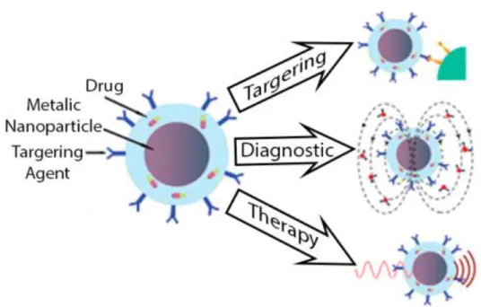
Magnetic NPs are generally composed by a magnetic core and can be coated with several materials (silica, gold, polymers, etc.). Their applications involve the delivery of a drug to a specific tissue, diagnostic (Magnetic Resonance imaging) or therapy (hyperthermia/thermal ablation) (Arruebo et al., 2007) (see Figure 3). A very interesting property of magnetic NPs is their ability to obey to the Coulomb’s law. Such feature enables an external magnetic gradient to guide them to a specific site, maintaining them at the site of action until needed and then allow their removal (Pankhurst et al., 2003).
The most investigated magnetic NPs are those produced with iron, their alloys and oxides (Wilczewska et al., 2012). These NPs are the only magnetic NPs approved for clinical use by Food and Drug Administration (Wilczewska et al., 2012). When they were used as a DDS the drug can be linked to the NP by a covalent bound, electrostatic interaction, absorbed or
Chapter I - Introduction
11
adsorbed (Figuerola et al., 2010, Yallapu et al., 2011, Wu et al., 2010). Magnetic NPs can cause oxidative stress by promoting the production of Reactive oxygen species (ROS) and the activation of phagocytosis and cytokine-release function of macrophages. However, these NPs have several advantages such as simple synthesis, chemical stability in physiological conditions and the possibility of chemical modification through their coating (Figuerola et al., 2010, Asmatulu et al., 2005, Karimi et al., 2013). The coating of these NPs with polymers such as gelatine, chitosan, poly(ethylene-co-vinyl acetate), poly(vinyl pyrrolidone) (PVP), poly(lactic-co-glycolic acid) (PLGA), poly(ethylene glycol) (PEG), and poly(vinyl alcohol) (PVA), oleic acid and dextran or other materials like proteins (like albumin) can improve their biocompatibility (Wilczewska et al., 2012, Jain et al., 2005, Berry et al., 2003, Peng et al., 2008, Karimi et al., 2013).1.3.2.2. Organic Nanoparticles 1.3.2.2.1. Liposomes
Liposomes are spherical, self-closed structures, that have an aqueous core, composed of phospholipids and steroids (Wilczewska et al., 2012, Torchilin, 2005). Liposomes usually present a concentric lipid bilayer but in some cases can have several bilayers. Their size can vary between 100 and 5000 nm, depending on the number of bilayers that they have on their composition. The number of bilayers and size allow the classification of the liposomes into four categories: small unilamellar, oligolamellar, large unilamellar and multilamellar vesicles (Mallick and Choi, 2014). They are able to load hydrophobic and hydrophilic drugs as presented in Figure 4. The water-soluble drugs are carried inside the liposomes, in the aqueous phase, whereas the water-insoluble drugs can be incorporated into the liposomal membrane.
These carrying systems are biocompatible, biodegradable and can carry a variety of different molecules like neurotransmitters, antibiotics, anti-inflammatories, genes and drugs. Figure 4: Representation of the traditional phospholipids liposomes. a- water-soluble drug, b- water-insoluble drug (adapted from (Torchilin, 2005)).
Chapter I - Introduction
12
Liposomes are formed through the hydrophilic/hydrophobic interactions between lipid/lipid and lipid/water. In the production process the surface of the liposomes can be modified with specific proteins, antigens or other biological substances, which will improve their selectivity to the target tissue. Liposomes present some limitations such as low encapsulation efficiency, fast burst release of drugs and poor storage stability (Bamrungsap et al., 2012). In the market there are some liposome based formulations for cancer therapy, being Doxil® the most used in the clinical (Barenholz, 2012).1.3.2.2.2. Dendrimers nanocarriers
Dendrimers have well-defined size and structure and they have been produced with different molecules such as glycogen, amylopectin and proteoglycans (Wilczewska et al., 2012).
These structures are composed by a core, dendrons and surface active groups (Figure. 5). Dendrimers can be produced by two methods: the divergent approach and the convergent approach (Svenson and Tomalia, 2012). The main difference between these two methods is the direction of dendrimer growth. In divergent method, dendrimer growth starts from a polyfunctional core and proceeds radially (Hierold et al., 2010). In the convergent method the dendrimer growth begins from the exterior of the molecule and continues inward by coupling end groups to each branch of the monomer (Ledin et al., 2011). The modification of the surface amine groups of dendrimers with neutral or anionic groups can be easily produced in order to reduce its toxicity. This functionalization consists in a reaction between the amine groups in the dendrimer surface and the functionalization agent. The drug can be covalent or noncovalent bonded to the dendrimer. In the noncovalent approach the drug, generally hydrophobic, is incorporated into dendrimer core and it is difficult to control their release from the dendrimer core, which limits their use. when the drug is covalently bond to the dendrimer periphery, its release can be controlled by incorporation of degradable linkages between the drug and the dendrimer (Gillies and Frechet, 2005).
Polyamidoamine (PAMAM) is one of dendrimers already in commercialization and one of the most used for biomedical applications (Gillies and Frechet, 2005). Polypropyleneimine Figure 5: Representation of a dendrimers. a- core, b- dendrons, c- active groups (adapted from (Svenson and Tomalia, 2012)).
Chapter I - Introduction
13
dendrimers have been commercialized and investigated for their biological application, but the presence of multiple cationic amine groups leads to a significant toxicity (Malik et al., 2000).Furthermore, polyaryl ether dendrimers, have been tested for drug delivery applications, but their poor water solubility demands the extensive use of solubilizing groups at their periphery (Liu et al., 2000).1.3.2.2.3. Polymeric nanoparticles
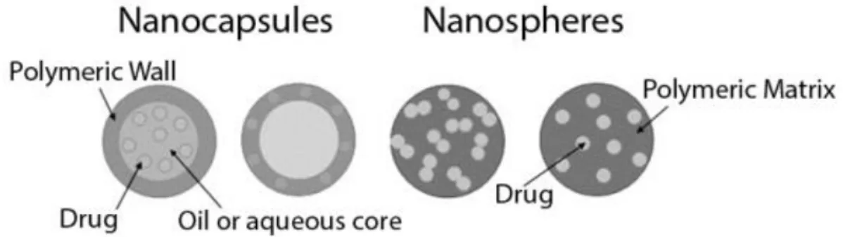
Polymeric NPs are core-shell spherical structures that can be produced with natural (Serum albumin, Gelatin, Chitosan, etc.) and/or synthetic (Poly-ε-caprolactone (PCL), Poly lactic acid (PLA), Poly methyl methacrylate (PMMA), Poly glycolic acid, etc.) polymers (Pinto Reis et al., 2006a). They can be designated as nanocapsules, if the drug is confined in the core of nanoparticle, or nanospheres, if the drug is physically and uniformly dispersed in the polymeric matrix (see Figure 6).
Polymeric nanocarriers provide several advantages, such as better overall drug/carrier stability (Sutton et al., 2007) and sustained drug controlled release (Soppimath et al., 2001). The synthetic polymers have some advantages over the natural, since they can provide a temporally controlled drug delivery of bioactive pharmaceutics for longer periods than those presented by some natural polymers, such as chitosan or alginate, which suffer from extensive swelling and also some biodegradation (Panyam and Labhasetwar, 2012).
Different methods, like nanoprecipitation, emulsification/solvent evaporation, emulsification/solvent diffusion, salting out, dialysis, superficial fluid technology, spray-drying and crystallization have been used for polymeric NPs synthesis (Mohanraj and Chen, 2007, Pinto Reis et al., 2006a, Vauthier and Bouchemal, 2009, Rao and Geckeler, 2011, Miladi et al., 2013, Mora-Huertas et al., 2010).
The emulsification/solvent evaporation (see Figure 7) was the first method developed to prepare polymeric NPs from a preformed polymer (Rao and Geckeler, 2011, Vanderhoff et al., 1979). This technique consists on the formation of a simple or double emulsion (using
high-Figure 6: Schematic representation of polymeric nanoparticles (adapted from (Pinto Reis et al., 2006b))
Chapter I - Introduction
14
speed homogenization or ultrasonication) and the evaporation of the organic solvent, by continuous magnetic stirring or under reduced pressure, which leads to the precipitation of the polymer and the subsequent obtention of the particles (Miladi et al., 2013). The size of the obtained particles can be controlled by changing the stirring rate, type and amount of dispersing agent, viscosity of organic and aqueous phase and temperature (Pinto Reis et al., 2006a). Generally, this method of simple emulsion is used for the encapsulation of hydrophobic drugs. On the other hand, when hydrophilic drugs are aimed to be encapsulated, the double emulsion is more appropriate. This double emulsion consists in the dispersion of the primary emulsion in a second aqueous phase, before organic solvent evaporation (Miladi et al., 2013).Another method described in the literature is nanoprecipitation or solvent displacement method (see Figure 8). This method was developed by Fessi et al. (Fessi et al., 1989) and consist in a spontaneous emulsification of the organic internal phase containing the dissolved polymer into the aqueous external phase (Pinto Reis et al., 2006a). The organic phase must be miscible in the aqueous phase and easy to remove by evaporation. It can be an organic solvent or a mixture of organic solvents. The solvents frequently used are acetone, dimethyl sulfoxide, isopropyl alcohol, ethanol or ethyl lactate, ethyl acetate, acetonitrile, etc. (Anton et al., 2008).
Figure 7: Schematic representation of the emulsification/solvent evaporation technique (adapted from (Raemdonck et al., 2014)).
Chapter I - Introduction
15
The decrease of interfacial tension between the two phases results in the rapid diffusion of the organic phase (organic solvent and polymer) into the aqueous phase (non-organic solvent supplemented with one or more naturally or synthetic surfactants). This increase the surface area and leads to the formation of small droplets of organic solvent (Rao and Geckeler, 2011). The main variables that induce the formation of the spontaneous emulsification are the conditions used for adding the organic phase into the aqueous phase such as injection and agitation rate, the method of addition and the proportions between the two phases. One of the limitations of this technique is that only water-miscible solvents can be used, in which the diffusion rate is enough to produce spontaneous emulsification (Pinto Reis et al., 2006a). This technique is more adequate to encapsulate lipophilic drugs than hydrophilic drugs. Such can be explained by the week interaction between the drug and the polymer. In the hydrophilic drugs, the drug will tend to diffuse from the organic phase to the external aqueous medium during the spontaneous emulsification process of the polymer, reducing the amount of encapsulated drug (Khan and Schneider, 2013).The emulsification/solvent diffusion (see Figure 9) is other technique used to produce polymeric NPs and was developed by Leroux and collaborators (Leroux et al., 1995). In this method it is required an organic phase, that contains the partial water-soluble organic solvent, the polymer, the hydrophobic drug, and two aqueous phases. The first aqueous phase contains the stabilizer agent solution and the second that is called the dilution phase, is mainly composed for a large volume of water (Miladi et al., 2013). In the encapsulation of a hydrophilic drug it is needed the use of an aqueous inner phase to dissolve the drug. To Figure 8: Representation of the nanoprecipitation method (adapted from (Khan and Schneider, 2013)).
Chapter I - Introduction
16
obtain the emulsification both the organic phase and the aqueous phase must be saturated to ensure the initial thermodynamic equilibrium of both liquids (Pinto Reis et al., 2006a, Miladi et al., 2013).The emulsification can be obtained under vigorous agitation. The next step is the dilution of the previous emulsion with a large amount of dilution phase (usually pure water). This dilution results in the diffusion of the organic solvent contained in the dispersed droplets leading to the precipitation of the polymer (Vauthier and Bouchemal, 2009), which results in the formation of the NPs. The organic solvent can be then eliminated by distillation or cross-flow filtration (Mora-Huertas et al., 2010). The main conditions that affect the size of the particles are the organic/aqueous phase ratio, emulsification stirring rate, volume of water for the dilution and the temperature (Mora-Huertas et al., 2011).
The salting out method (see Figure 10) is currently used for the preparation of polymeric NPs from the preformed polymer, and it was first developed by Bindschaedler et al. (Bindschaedler et al., 1990). It is very similar to the emulsion/solvent diffusion method (Pinto Reis et al., 2006a). This method requires a water-miscible solvent and involves a salting-out process, that, by turn, requires the dissolution of a high concentration of salt in the aqueous phase which will result in the loss of miscibility between the two phases. The polymer and the drug (lipophilic) are dissolved in a water-miscible solvent and then the mixture is emulsified into an aqueous phase containing the salting-out agent and the stabilizer.
Figure 9: Schematic illustration of the emulsification/solvent diffusion technique (adapted from (Pinto Reis et al., 2006a)).
Chapter I - Introduction
17
Figure 10: Schematic illustration of the salting-out technique (adapted from (Pinto Reis et al., 2006a))After the emulsion, like in the emulsion/solvent diffusion, a large amount of water is added to this solution, in a way that the concentration of salting out agent will decrease and the miscibility of the two phases will increase (Rao and Geckeler, 2011). Then the water-miscible solvent diffuse in the aqueous phase, the polymer precipitation is induced and forms the NPs. So, the polymer solvent and the salting-out agent are eliminated (Vauthier and Bouchemal, 2009). In this technique the increase of temperature is not necessary, allowing the encapsulation of heat-sensitive substances. However, this technique present the disadvantage of requiring extensive nanoparticle wash steps (Pinto Reis et al., 2006a).
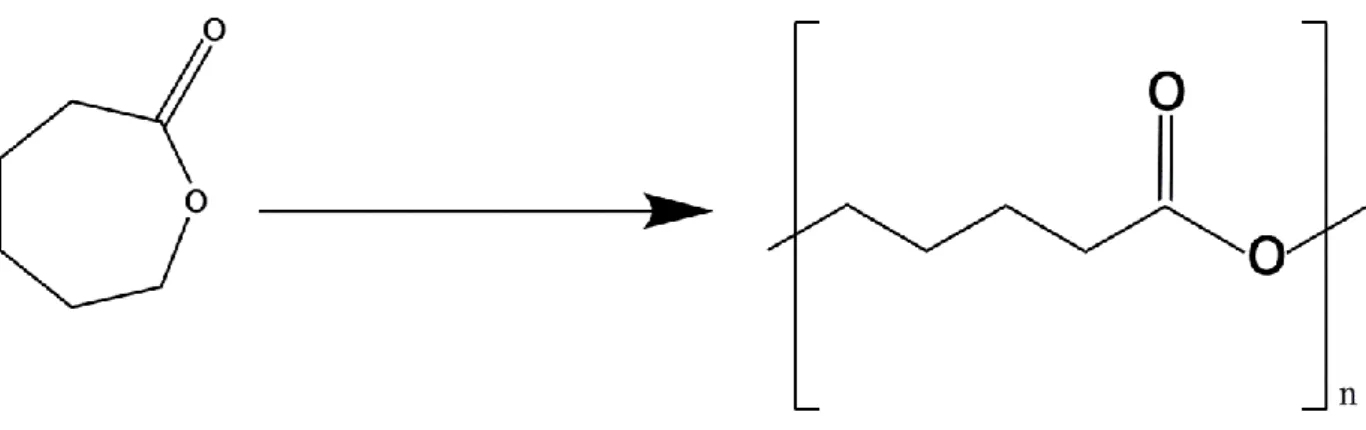
1.3.3. Poly-ε-caprolactone nanoparticles
PCL is a synthetic, hydrophobic, semi-crystalline and biodegradable polymer obtained by polymerization of ε-caprolactone (see Figure 11). The monomer (ε-caprolactone) can be obtained from the oxidation cyclohexanone by peracitic acid (Rocca et al., 2003). One of the most used methods for PCL preparation is the ring opening polymerization. There are four main mechanisms of ring opening polymerization: anionic, cationic, monomer-activation and coordination insertion. All of them result in a polymeric chain composed of several replications of the extended monomer (Labet and Thielemans, 2009).
Chapter I - Introduction
18
PCL is insoluble in water and alcohols, has a low solubility in polar solvents (acetone, acetonitrile, dimethylformamide, etc) and is soluble in aromatic and chlorine solvents (dichloromethane, chloroform, carbon tetrachloride, benzene, etc) (Sinha et al., 2004, Pohlmann et al., 2013). PCL suffers slow degradation by enzymatic or non-enzymatic processes. In the non-enzymatic cleavage of PCL (hydrolytic degradation) the water permeability is the limiting step because it starts in the amorphous regions. This region is auto-catalyzed by carbonyl end groups of fragmented polymeric chain (Jenkins and Harrison, 2006, Sinha et al., 2004). This process of PCL degradation is too slow and can take several months or even years (Woodruff and Hutmacher, 2010). Alternatively, the enzymatic fragmentation is faster and involves the activity of lipase. This enzyme is responsible for the cleavage of esters groups’ with the release of carboxyl groups. This group can undergo phagocytosis and used in tricarboxylic acid (TCA) cycle (Woodruff and Hutmacher, 2010). PCL can be combined with different polymers to modify its mechanical, physical and ionic properties and the biodegradability profile. This polymer, alone or conjugated, have been used for the production of implants, nanofibres, scaffolds, composites, films, hydrogels, micelles, microspheres and NPs (Dash and Konkimalla, 2012).
In the literature there are several examples of NPs produced with PCL are described (Table 1).
Figure 11: Representation of ε-caprolactone (left) and poly-ε-caprolactone (right) structure (adapted from (Labet and Thielemans, 2009)).
Chapter I - Introduction
19
Table 1: Examples of PCL nanoparticles reported in literature. BSA - Bovine serum albumin; MPEG - Methoxy poly(ethylene glycol); PEG - Poly(ethylene glycol); EE – Encapsulation Efficiency.Polymer Drug Method of
Production Mean size EE% Reference
PCL BSA Double emulsion/solvent evaporation 276 -308 nm 55-80% (Lamprecht et al., 1999) PCL Exemestane Interfacial deposition method 115 – 350 nm 23 – 84%
(Kumar and Sawant, 2013)
PCL Uncaria
tomentosa
Emulsion solvent
evaporation 223 – 408 nm 33 – 88% (Ribero et al., 2013) PCL/
Dextran Doxorubicin
Nanoprecipitation
method 95 – 123 nm 42-52% (Li et al., 2013)
PCL tamoxifen Solvent
displacement 100 – 300 nm 64 %
(Chawla and Amiji, 2002) PCL -
MPEG Ibuprofen
Emulsion/solvent
diffusion 85 – 97 nm 48 – 64 % (Baimark, 2009)
PCL - PEG Honokiol Solvent diffusion
method ~132 nm 20% (Gou et al., 2009)
PCL –
l-Lactide nimodipine
Precipitation
method 81 – 132 nm 19 – 91 % (Ge et al., 2000)
PCL/ magnetic Cisplatin Single emulsion/solvent evaporation ~160 nm 7% (Yang et al., 2006) Gemcitabine 25%
As we can see in table 1, several PCL based NPs have been produced and used over the years (Lamprecht et al., 1999, Kumar and Sawant, 2013, Ribero et al., 2013, Li et al., 2013, Chawla and Amiji, 2002, Baimark, 2009, Gou et al., 2009, Ge et al., 2000, Yang et al., 2006). They can be produced with several sizes, depending mainly on the molecular weight of the polymer used. This system based on PCL are characterised by an initial burst followed by a sustained release during several hours or days. PCL NPs can be loaded with several drugs either hydrophilic or hydrophobic. Their good biocompatibility makes them good candidates to be
Chapter I - Introduction
20
used in the treatment of several diseases such as neuroblastoma, breast and ovarian cancer (Li et al., 2013, Chawla and Amiji, 2002, Gou et al., 2009).Shenoy and collaborators loaded PCL NPs with tamoxifen and evaluated their biodistribution
in vivo using female athymic mice (4/6 weeks old) (Shenoy and Amiji, 2005). Their results
suggested that the produced NPs were transferred from the circulatory compartment to the tumor site. They concluded that after 1 hour of injection, 90% of free drug administered reached the liver, while only 70% attained this organ, when loaded NPs were used. 6 hours post injection the concentration in the liver of both formulations (free drug and inside NPs) was 2 and 7%, indicating possible degradation/metabolism of the drug. These results suggest that PCL NPs delivery the drug at the tumour tissue, minimizing the amount of drug that reaches the liver and subsequently degraded.
1.3.4. Poly (methyl methacrylate) nanoparticles
PMMA is a synthetic, biocompatible and non-degradable polymer obtained through the polymerization of the methyl methacrylate monomer (see Figure 12) (Bettencourt and Almeida, 2012). PMMA is soluble in several organic solvents like trichloromethane and trichloroethylene, dimethylformamide and chloroform (Evchuk et al., 2005). Despite its low solubility in water, this polymer has been reported as being soluble in a mixture of water and alcohol (Hoogenboom et al., 2010).
PMMA has been reported to be used in several biomedical applications such as a bone cement (in total hip replacement), vertebral stabilization agent in patients with osteoporosis, a prosthetic material in dental and mandibular corrections and as a permanent implant in the form of intraocular lens used in cataract surgery (Carvalho Costa et al., 2009, Schade and Roukis, 2010). Furthermore, PMMA particles have been the first ones to be developed for vaccination purposes (Kreuter and Speiser, 1976, Kreuter et al., 1976). Since then several Figure 12: Representation of methyl methacrylate (left) and poly (methyl methacrylate) (right) structure (adapted from (Bettencourt and Almeida, 2012)).
Chapter I - Introduction
21
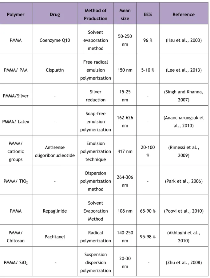
micro and NPs have been produced using this polymer alone or combined with others materials (Table 2).Table 2: Examples of PMMA nanoparticles produced so far. PAA – Poly acrylic acid; TiO2 -
Titanium dioxide; SiO2 - Silicon dioxide; EE - Encapsulation Efficiency.
Polymer Drug Method of Production
Mean
size EE% Reference
PMMA Coenzyme Q10 Solvent evaporation method 50-250 nm 96 % (Hsu et al., 2003)
PMMA/ PAA Cisplatin
Free radical emulsion polymerization 150 nm 5-10 % (Lee et al., 2013) PMMA/Silver - Silver reduction 15-25 nm -
(Singh and Khanna, 2007) PMMA/ Latex - Soap-free emulsion polymerization 162–626 nm - (Anancharungsuk et al., 2010) PMMA/ cationic groups Antisense oligoribonucleotide Emulsion polymerization technique 417 nm 20-100 % (Rimessi et al., 2009) PMMA/ TiO2 - Dispersion polymerization method 264-306 nm - (Park et al., 2006) PMMA Repaglinide Solvent Evaporation Method 108 nm 65-90 % (Poovi et al., 2010) PMMA/ Chitosan Paclitaxel Radical polymerization 140-250 nm 95-98 % (Akhlaghi et al., 2010) PMMA/ SiO2 - Suspension dispersion polymerization 20-30 nm - (Zhu et al., 2008)