Axonal Hyperpolarization in Amyotrophic Lateral Sclerosis
Marisa Tavares Brum
Orientador: Prof. Dr. Mamede de Carvalho, Faculdade de Medicina da Universidade de Lisboa
Curso de Mestrado em Neurociências Dissertação
Todas as afirmações efectuadas no presente documento são da exclusiva responsabilidade do seu
autor, não cabendo qualquer responsabilidade à Faculdade de Medicina de Lisboa pelos conteúdos nele apresentados.
A impressão desta dissertação foi aprovada pelo Conselho
Científico da Faculdade de Medicina de Lisboa em reunião de
22 de Setembro de 2015.
‘‘Let us keep looking, in spite of everything. Let us keep searching. It is indeed the best method of finding, and perhaps thanks to our efforts, the verdict we will give such a patient tomorrow will not be the same we must give this man today”
INDEX
1. Abstract 6
2. Resumo 8
3. Background 13
3.1 Amyotrophic lateral sclerosis 13
3.1.1 Axonal excitability 16
3.1.2 Excitability Studies 18
3.1.3 Threshold tracking in ALS patients 24
4 . Objectives 26 5 . Methods 26 5.1 Population of study 26 5.2 Procedures 27 5.2.1 Study Procedures 27 5.2.2 Statistical analysis 30
5.2.3 Ethical considerations and regulatory 31
6 . Results 31
6.1 Population 31
6.2 Axonal excitability 32
6.2.1 Baseline 32
6.2.2 Before versus after exercise 34
6.2.2.1 Controls 34
6.2.2.2 ALS Group 1 37
6.2.2.3 ALS Group 2 39
7 . Discussion 42
8 . Conclusion 47
9 . Acknowledgments 48
10. Bibliography 48
11. Annex - Measures of axonal excitability for each
Abstract
Background: Amyotrophic lateral sclerosis (ALS) is a progressive fatal
neurodegenerative disorder of uncertain pathogenesis. Nerve excitability examination (Threshold Tracking) is a novel method that complements conventional nerve conduction studies. This technique provides insight into peripheral nerve function and the physiological mechanisms underlying axonal membrane properties. Our aim is to study the effect of contraction on ion channels function of peripheral axons in ALS patients, in muscles with different degree of motor unit loss.
Methods: We examined the median nerve of 10 healthy controls and 22
ALS patients divided in 2 groups according to the M-wave peak-to-baseline amplitude of the ABP response (ALS group 1 > median value, ALS group 2 < median value) using the automated threshold-tacking program, QTRAC. Differences in baseline values were tested through the One-way analysis of variance (ANOVA), ALS group 1 vs ALS group 2 vs controls. Paired-t test was applied to compare results before and after intervention (contraction) in each group.
Results: No significant differences in demographic characteristics were
found among groups: age, disease duration, gender (p>0.05). At baseline, values were not significantly different among the three groups for all the measurements investigated. In the group of healthy controls, comparing measurements before and after exercise did show a significantly smaller resting I/V slope (p<0.001), greater threshold changes in TEd peak and TEd20 (peak) – (p<0.000 and p=0.001, respectively) and reduced refractoriness at 2 ms (p=0.001), after
exercise. In ALS group 1, we observed a statistically significant reduction in the relative refractory period, as well in the refractoriness at 2 and 2.5 ms (p=0.001 for all). Moreover, the superexcitability at 5 ms was significantly increased (p=0.001), following contraction. In ALS patients group 2, the only change observed was a significant threshold reduction during the early phases of hyperpolarization, TEh (10-20 ms) - (p<0.001).
Conclusion: Changes in agreement with hyperpolarization was observed
in controls after exercise, as expected. In ALS patients, our observations
suggest a dysfunctional Na+/K+ pump, in particular in weak muscles. This
might imply that in weak muscles, exercise in can be detrimental to axons, which are unable to compensate for repeated depolarization.
Key words: Amyotrophic Lateral Sclerosis; Axonal Excitability; Axonal
Resumo
Introdução: A esclerose lateral amiotrófica (ELA) é uma doença
neurodegenerativa progressiva e fatal. Na sua forma clássica a ELA afeta os neurônios motores em duas ou mais regiões do corpo. Atinge o segundo neurónio motor que reside no corno anterior da medula espinhal e tronco cerebral, assim como o primeiro neurónio motor presente no córtex. A idade de início é variável, tipicamente ocorre pelos 58-63 anos. É uma doença com relativa baixa incidência, mas com prognóstico reservado, dado que 50% dos doentes morrem nos 30 meses após o início dos sintomas. Na ausência de um marcador biológico estabelecido, o diagnóstico é essencialmente clínico, dependente da demonstração de sinais do 1º e do 2º neurónio. No entanto, a electromiografia é especialmente importante neste contexto, dado poder revelar envolvimento do 2º neurônio motor em músculos clinicamente normais. O principal fator que leva ao defeito funcional na ELA é a fraqueza muscular devida à perda de neurônios motores. Assim, intervenções que possam aumentar a força em músculos preservados, como o exercício, poderia preservar a função e, possivelmente, ter impacto na sobrevida do doente. No entanto, por outro lado, o exercício poderá condicionar o esgotamento de unidades motoras disfuncionais e, assim, ter um efeito lesivo. No entanto, não existem dados robustos na literatura que esclareçam esta controversia.
O estudo de condução nervosa convencional, apesar de ser o gold standard para estudo do nervo periférico, fornece pouca informação sobre a excitabilidade da membrana axonal. O threshold tracking é uma
técnica complexa, recente, que permite o estudo da excitabilidade dos axónios, desta forma complementando os estudos convencionais da condução nervosa convencionais. Em particular, esta técnica esclarece os mecanismos fisiológicos subjacentes às propriedades da membrana axonal, como por exemplo o estudo dos diferentes canais iônicos, e da eficácia da bomba-sódio-potássio, ativada por despolarizações repetidas. Já foram realizados alguns estudos sobre a excitabilidade axonal em doentes com ELA. O objectivo do nosso trabalho é estudar o efeito do exercício (contração muscular) na função dos canais iônicos dos axónios periféricos, em músculos com diferentes graus de perda de unidades motoras, desta forma esclarecendo a função da bomba-sódio-potássio subjacente ao processo de hiperpolarização.
Métodos: Foram examinados 10 controlos saudáveis e 22 doentes com
o diagnóstico de ELA, sendo estes divididos em 2 grupos de acordo com a amplitude da resposta motora por estimulação do nervo mediano com captação no músculo abductor curto do polegar (ELA grupo 1 superior à mediana, ELA grupo 2 inferior à mediana, definida a amplitude como o pico-à-linha-de-base) ou de acordo com a força muscular. Neste protocolo foi estudado o segmento do nervo mediano no punho, antes e depois da contração máxima do músculo abductor curto do polegar (1 minuto, antes de cada teste), com o uso do programa automático QTRACT. As variáveis estudadas foram: strength-duration time constant; reobase; threshold electrotonus; no recovery-cycle; current threshold. Para determinar diferenças nas avaliações iniciais entre os três grupos (ELA grupo 1, ELA grupo 2 e controlos) foi utilizado o teste
one-way (ANOVA). O teste t-student emparelhado foi aplicado comparar em cada grupo os resultados antes e depois da intervenção (contração muscular). O estudo foi aprovado pela comissão de ética da Faculdade de Medicina da Universidade de Lisboa-CHLN.
Resultados: No grupo de doentes com ELA 13 eram do sexo feminino, a
mediana da idade de início foi de 58,5 anos (variando de 38 a 76), a mediana da duração da doença foi de 11,5 meses (variando de 4 a 36), a forma de início foi bulbar (disartria) em nove doentes, em cinco a doença teve início nos membros superiores e em oito doentes nos membros inferiores. Todos os doentes estavam sob tratamento com riluzol.
A população de controle de 10 indivíduos tinha uma idade mediana de 51,9 (variando de 30 a 70 anos). Não se observaram diferenças significativas relativamente às características demográficas entre os dois grupos de ELA, em particular quanto à idade, duração da doença e sexo (p>0,05). Na avaliação inicial não foram encontradas diferenças significativas entre os três grupos para todos os parâmetros investigados. Comparando nos diferentes grupos as alterações promovidas pela contração muscular, no grupo de controles saudáveis o estudo evidenciou uma redução do resting I/V slope após o exercício (p<0,001), assim como um aumento no limiar na TEd pico e TEd20 (pico) - p<0,000 e p = 0,001, respectivamente. Relativamente à refratoriedade aos 2 ms, esta foi significativamente menor após o exercício (p = 0,001). No grupo ELA 1 observou-se uma redução estatisticamente significativa no período refractário relativo e na refratoriedade aos 2 e 2,5 ms (p=0,001), para além de um aumento na superexcitabilidade aos 5 ms (p = 0,001). No
grupo 2 dos doentes com ELA a única alteração significativa observada foi uma redução (p <0,001) do limiar nas fases iniciais de hiperpolarização, Teh (10-20 ms).
Discussão: A esclerose lateral amiotrófica é caracterizada por
progressiva fraqueza e atrofia muscular, fadiga, espasticidade, disfunção bulbar e insuficiência respiratória, levando a severo defeito funcional e, em fases mais tardias, à ventilação mecânica e morte. Apesar dos recentes avanços no entendimento da fisiopatologia da ELA, a causa ainda permanece desconhecida. O efeito do exercício físico na ELA tem sido considerado um tema controverso. Em sujeitos saudáveis, a atividade muscular pode produzir hiperpolarização axonal substancial. Muitos estudos têm descrito que a contração voluntária ativa a atividade
da bomba Na+/K+ da membrana axonal, a qual tenta estabilizar o
potencial de membrana após a ter cessado a contração, resulta em hiperpolarização.
No nosso estudo, no grupo dos controlos saudáveis verificou-se hiperpolarização nos axónios motores após a ativação voluntária isométrica. Tal ficou demonstrado pela significativa redução da resting I/V slope e da refratoriedade aos 2 ms, bem como pelo aumento do limiar no TEd pico e TEd20 (pico) após a contração. Nos doentes com ELA grupo 1 os parâmetros de refratoriedade foram reduzidos, como fisiologicamente antecipado. No entanto, a resting I/V slope ficou inalterado após o exercício, sugerindo uma disfunção minor da bomba Na+/K+. No grupo ELA 2 podemos afirmar que as características da hiperpolarização encontradas nos grupos de controlos saudáveis e grupo
de ELA 1 estavam ausentes, sugerindo uma disfunção major da bomba Na+/K+.
Em conclusão, os nossos resultados indicam uma disfunção da bomba
Na+/K+ nos doentes com ELA, em particular em músculos com maior
perda de unidades motoras. Isto pode suportar um efeito prejudicial do exercício, pois em grupos musculares muito afectados, os axónios são incapazes de compensar a despolarização repetida. Um novo estudo incluindo uma maior população de doentes com ELA e com um protocolo mais elaborado, por exemplo com diferentes tempos e graus de contração, deve ser programado.
Palavras-chave: Esclerose Lateral Amiotrófica; Excitabilidade axonal;
Background
Amyotrophic lateral sclerosis
Amyotrophic lateral sclerosis (ALS) is a progressive fatal neurodegenerative disorder of uncertain pathogenesis. The disorder is named for its underlying pathophysiology, with “amyotrophy” referring to the atrophy of muscle fibers, a consequence of anterior horn cells degeneration. “Lateral sclerosis” refers to the changes seen in the lateral columns of the spinal cord as upper motor neuron (UMN) axons are replaced by fibrous astrocytes (gliosis) following Wallerian degeneration (Charcot and Joffroy, 1869). In its classic form, ALS affects motor neurons at two or more regions of the body. It affects lower motor neurons (LMNs) that reside in the anterior horn of the spinal cord and brainstem, and corticospinal UMNs that are located in the precentral gyrus (Swash and Desai, 2000).
In sporadic cases of ALS the mean age onset is 58-63 years and the lifetime risk of developing ALS is 1 in 350–500, for male sex (Miller et al. 2013); indeed, the incidence is slightly higher for male sex. Only 5% of cases have an onset before the age of 30 years (Haverkamp et al. 1995), but young-onset sporadic cases are increasingly recognized (Gouveia & de Carvalho 2007). ALS is a progressive disorder, with 50% of patients dying within 30 months after symptoms onset, although 20% can survive between 5 years and 10 years after presentation (Talbot, 2009).
The clinical diagnosis of ALS is dependent on the demonstration of LMN and UMN signs, which progress within the same region and spread to attain other body regions. Babinski sign, sustained clonus, pathologically
hyperactive deep tendon reflexes and spasticity are universally accepted manifestations of UMN involvement. However, LMN signs are usually identified by muscle weakness, atrophy out of proportion to disuse, attenuation or loss of deep tendon reflexes and fasciculations (Traynor et al., 2000; Kiernan et al., 2011). Fasciculations are a prominent early feature of ALS and are derived from ectopic activity of motor axons, but a motor neuron cell body origin cannot be discarded (de Carvalho & Swash 2012).
Most ALS cases (approximately two thirds of patients with typical ALS) have a spinal form of the disease: symptoms may start either distally or proximally in the upper or lower limbs. Nearly 25% of the patients have bulbar-onset ALS: dysarthria as initial symptoms. In this group of patients, limbs symptoms can develop almost simultaneously with bulbar manifestations or, for most of the cases, will occur later within 1–2 years. All patients with bulbar symptoms will develop dysphagia and sialorrhea due to difficulty in swallowing saliva, as well as mild UMN type bilateral facial weakness, which affects the lower part of the face. 'Pseudobulbar' symptoms such as emotional lability are seen in a significant number of cases (Wijesekera & Leigh 2009).
In the absence of an established biological marker, ALS is primarily a clinical diagnosis. However, clinical neurophysiological investigation is especially important in this context (de Carvalho et al,, 2008), because it can extend the clinical findings by revealing lower motor neuron involvement in muscles otherwise regarded as unaffected.
The management of ALS is supportive, palliative and multidisciplinar (Wijesekera & Leigh 2009). Riluzole is the only approved drug that has been shown to have a modest effect in prolonging life (Bensimon et al. 2002).
Respiratory failure and other respiratory complications account for the majority of deaths in ALS. Although respiratory function is a frequent late complication, it may be a presenting feature (de Carvalho et al. 1996). Several different factors have been shown to predict survival in ALS. The most consistent negative predictive factors for survival are: elderly age, low body-mass index, short diagnostic delay, bulbar and respiratory presentation, rapid clinical and respiratory decline (Chiò et al.).
Non-invasive ventilation (NIV) is widely used to improve alveolar hypoventilation in ALS. Several studies indicate a longer survival when NIV is used. Health-related QoL is improved in patients under NIV, in particular in those with none to moderate bulbar dysfunction (Vrijsen et al. 2013). Forced vital capacity, vital capacity, nasal inspiratory pressure obtained during a sniff, the size of the phrenic nerve motor response and mean nocturnal oxygen saturation have been considered as respiratory markers related to prognosis in ALS (Pinto & Carvalho 2014).
The major factor leading to disability in ALS is muscle weakness due to motor neuron loss. So, increasing the strength of muscles whose innervation is not significantly affected could preserve function and possibly could as well impact on the survival. Exercise is one potential
mode to increase muscle strength (de Almeida et al. 2012). Despite, improper and unsupervised exercise may lead to severe injuries in ALS patients, such as falls; supervised strength training seems to be beneficial in improving absolute muscle strength and delaying muscle loss. Moderate exercise appears to not be harmful in patients, but no studies have been yet determined whether it is beneficial (Bello-Haas et al. 2007; Pinto et al. 1999; McCrate & Kaspar 2008). Possibly, the training should be implemented as early as possible in the natural course of disease to maximize the strength of affected muscles (Pinto et al. 1999; Pinto et al. 2012).
Axonal excitability
The original formulation of a model for axonal sodium (Na+) channel
function and nerve impulse generation by Hodgkin and Huxley remains a landmark in human physiology (Hodkin & Huxley, 1952). In a myelinated nerve (figure 1), multiple layers of myelin sheaths wrap tightly around the axon along most of its length except at small gaps between each myelin sheath. The insulated areas are known as the internodes and the gaps, where voltage-gated sodium channels predominate is called the nodes of Ranvier.
Figure 1: Axolema channel distribution.
Different channels are distributed unevenly along the axonal membrane: sodium (Naþ) channels are found in high concentrations at the node, as are slow Kþ channels. Fast potassium channels (K+) are almost exclusively paranodal. Inward rectifier channels (Ih), permeable to both K+ and Na+ ions, act to limit axonal hyperpolarization, whereas the Na+/K+ pump serves to reverse ionic fluxes that may be generated through activity. Adapted from Krishnan, et al, Journal of the Peripheral Nervous System 13:7–26 (2008).
Axonal excitability depends primarily on the presence of high concentrations of Na+ channels in the nodal membrane, but it is also controlled by a variety of other types of ion channel and ion pumps, that affect the excitability through their effects on membrane potential. The
classical transient Na+ current is activated rapidly by membrane
depolarization and then inactivates, so that further Na+ ions cannot pass
no matter how much the membrane is depolarized. The remaining
persistent Na+ current (INap) is activated equally rapidly, but at
membrane potentials that are ~10–20 mV more negative (i.e., less depolarized), so that it contributes disproportionately to subthreshold excitability behaviour. At the most negative potentials over which the current activates, inactivation is minimal, giving rise to a persistent inward leak of sodium ions at the resting potential (Krishnan et al. 2008).
Potassium channels are divided into 3 subgroups: fast channels, slow
channels, and inward rectifier channels. There are few fast K+ channels at
the human node of Ranvier. Fast K+ channels are located in a tight band
in the justaparanodal region where they contribute to the resistance of the internodal membrane and limit the depolarizing after-potential. Slow K+
channels are, like Na+ channels, more concentrated at the node than in
the internode, but their kinetics are too slow to allow them to affect the action potential directly. They help determine the resting membrane potential and contribute to accommodation to long lasting depolarizing
stimuli. The inward rectification K+ channels are activated by
hyperpolarization, in this way limiting hyperpolarization that develops due to the increased activity of the Na+/K+ pump (Krishnan et al. 2008).
The repolarization is achieved by inactivation of Na+ channels and current
leak to the internode. Resting membrane potential is determined by those channels that are open at or near rest (both voltage-dependent and voltage-independent) and by the activity of the Na+/K+ pump, and this implies that it is largely determined by the properties of the internodal membrane. The major contributors to resting membrane potential are probably slow and fast K+ channels, persistent Na+ channels, and the Na+/K+ pump (Lin et al. 2006)
Excitability Studies
Although conventional nerve conduction studies (NCS) remain the gold standard neurophysiologic assessment of peripheral nerve, it has been
known for many years that the insight into peripheral nerve pathophysiology provided by conventional NCS is limited (Krarup & Moldovan 2009). Conventional electrophysiological tests of nerve function focus on the number of conducting fibers and their conduction velocity. These tests are sensitive to the integrity of the myelin sheath, but provide little information about the axonal membrane (Bostock et al. 1998). On the other hand, nerve excitability examination is a relatively novel method that complements conventional nerve conduction studies. Assessment of nerve excitability in human peripheral nerves provides insight into peripheral nerve function and the physiological mechanisms underlying axonal membrane properties (Bostock et al., 1998; Krishnan et al., 2008). Measurement of nerve excitability by threshold tracking may be used to test nerve excitability, which depends on the membrane properties of the axons at the site of stimulation. This technique, studding changes in membrane potential, infers the activity of a variety of ion channels, energy-dependent pumps and ion exchange processes activated during the process of impulse conduction.
In the context of nerve excitability studies, “threshold” usually means “threshold current”, the minimal current required to excite a single axon or group of axons (as distinct from the “threshold potential,” which is the membrane potential at which an all-or-none response is initiated). For single axons, the threshold current is defined as the current that excites the unit on 50% of occasions.
Threshold determination is a trial-and-error process, so the use of a computer to control the output of a current source, depending on the response of the axon or group of axons stimulated, is the preferable method to track the threshold (Burke et al. 2001).
The absolute value of the threshold current usually provides little information. More information is provided by comparing the difference in threshold current following different patterns of stimulation according to different stimulation protocols. The development of a semi-automated computer controlled protocol to perform threshold tracking, known as TROND (originally developed for a course on nerve excitability in Trondheim, Norway), has enabled the use of the technique in many neuromuscular and nerve disorders (Kiernan et al. 2001; Kiernan et al. 2000).
The ‘Trond’ protocol enables multiple excitability properties of motor axons to be measured within 10 minutes (Kiernan et al. 2000). The protocol comprises the following sequence of recordings (Kiernan et al., 2000; Burke et al., 2001; Lin et al., 2006):
(i) Stimulus-response and strength-duration relationships. Stimulus-response curves are recorded separately for test stimuli of durations 0.2 ms and 1 ms. The ratio between the 0.2 ms and 1 ms stimuli required to evoke the same responses are used to estimate the strength-duration time constant and rheobase of axons with different threshold.
(ii) Threshold electrotonus. Prolonged subthreshold currents are used to alter the potential difference across the internodal axonal membrane, a process referred to as electrotonus. The changes in threshold associated with electrotonus normally have a similar time course to the changes in membrane potential and are known as threshold electrotonus. Test stimuli of 1-ms duration are used to produce the target CMAP (40% of maximal), and threshold tracking is used to record the changes in threshold induced by subthreshold polarizing currents, 100 ms in duration, set to be +40% (depolarizing) and -40% (hyperpolarizing) of the control threshold current.
(iii) Current-threshold relationship. The current-threshold
relationship is a threshold analogue of the current-voltage ('I/V') relationship, and depends on the rectifying properties of the internodal axolemma. Threshold is tested with 1 ms pulses at the end of subthreshold polarizing currents lasting 200 ms. The polarizing current is altered in a ramp fashion from +50% (depolarizing) to -100% (hyperpolarizing) of the control threshold in 10% steps.
(iv) Recovery cycle. The final part of the protocol records the recovery of excitability at intervals from 2 to 200 ms following a supramaximal conditioning stimulus. Immediately after an impulse the axons are refractory, but this is normally followed by phases of superexcitability and late subexcitability.
As indicated above, threshold (current) refers to the stimulus current required to excite a single unit or to evoke a compound potential that was
a defined fraction of the maximum (40%) and rheobase is the threshold for stimuli of infinitely long duration. Strength-duration time constant (SDTC) is an apparent membrane time constant inferred from the relationship between threshold current and stimulus duration, indirectly
reflecting nodal persistent Na+ conductance. Changes in membrane
potential affect strength duration properties, so the SDTC increases with
membrane depolarization and decreases with membrane
hyperpolarization (table 1).
Following conduction of a single nerve impulse, an axon undergoes a well characterized and reproducible sequence of excitability changes before returning to its resting state. This predictable sequence of excitability changes constitutes the recovery cycle. The mechanisms responsible for both the absolute and the relative refractory period (RRP) are fairly well known: absolute refractoriness is due to inactivation of transient Na+ channels, and the RRP is due to the gradual recovery of Na+ channels
from inactivation and the increased of membrane permeability to K+ ions
(slow channel). The RRP is dependent on membrane
depolarization/hyperpolarization condition (Burke et al. 2001). Superexcitability is due to a depolarizing after-potential and is, in part, determined by paranodal fast K+ channels, it is strongly dependent on membrane potential, with depolarization leading to a reduction in superexcitability and hyperpolarization causing an increase (Barrett & Barrett 1982). Late subexcitability reflects hyperpolarization of the
membrane potential due to current through slow K+ channels at the nodes
also changes according to the electrochemical gradient for K+ ions, which limits its use as an indicator of membrane potential.
Although insufficient to produce an action potential, subthreshold currents exert significant effects on axonal excitability and resting membrane potential. These excitability changes are often mediated via capacitative charging of the internode following impulse generation, resulting in activation or deactivation of voltage-dependent ion channels in the internode (Bostock et al. 1998). So, the threshold electrotonus is the only method with which to examine the behaviour of internodal conductances in human axons. In TE, the threshold changes produced by prolonged subthreshold depolarizing or hyperpolarizing currents are measured. The threshold changes generally parallel the electrotonic potentials responsible for them and therefore reflect membrane potential. The threshold changes in relation to the onset of the polarizing currents reflect different accommodation properties. Accommodation was defined as the tendency of the membrane potential to return toward the resting level despite a sustained depolarizing or hyperpolarizing stimulus. Accommodation after depolarization reflects the accommodation to the
depolarizing current activation of the slow K+ conductance, probably
reflecting the major factor producing accommodation to prolonged depolarizing currents. Inward rectification is activated by hyperpolarizing currents and acts to limit the degree of hyperpolarization. Membrane depolarization causes a ‘‘fanning-in’’ appearance of TE, while hyperpolarization leads to a ‘‘fanning out’’ (Krishnan et al., 2008; Lin et al. 2011).
Table 1: Major excitability parameters.
Determinants of each parameter and the response of that parameter to alteration in membrane potential. Adapted from Krishnan, et al, Journal of the Peripheral Nervous System 13:7–26 (2008).
Legend: RRP: relative refractory period.
Threshold tracking in ALS patients
When axons are diseased or damaged, disturbed axonal excitability may result in the inability to maintain conduction of a significant impulse train (Kaji et al. 2000). Alternatively ectopic activity may develop at a focus of hyperexcitability leading to symptoms of paresthesia, pain or fasciculation
Excitability parameter Major determinant Effect of depolarization
Effect of hyperpolarization
Unconditioned threshold Structural changes, geometric factors Strength-duration time constant Nodal persistent Na+ conductance Rheobase Nodal persistent Na+ conductance Threshold electrotonus
Nodal and internodal conductance
Fanning-in appearance
Fanning- out appearance
Refractoriness and RRP Nodal transient Na+ channel
Superexcitability Paranodal fast K+ channel
(Mogyoros et al. 2000).
In ALS patients a number of studies have revealed increased sodium conductance and reduced inward potassium current across the axon membrane in peripheral nerve, leading to an increased of spontaneous depolarization (Nakata et al. 2006).
Recently a study of nerve excitability testing in SOD1G127X mouse model
of ALS showed that axons recovering from repetitive stimulation had changes in excitability suggestive of membrane hyperpolarization, which was smaller in the SOD1G127X than in wild-type mice, consistent with a reduced Na+/K+ pump activity (Alvarez et al. 2013).
Most studies with threshold tracking in patients with ALS were directed to understand the mechanism of ectopic activity. To our knowledge no study has ever addressed the effect of exercise on the axonal excitability in ALS patients. The causes of exercise-related neurotoxicity observed in ALS remain to be dissected in detail. A plausible explanation is based
upon the dysfunction of the energy-dependent, axonal Na+/K+
electrogenic pump, which affects restoration of Na+ and K+ gradients after
high-frequency impulse activity such as physical exertion (de Almeida et al. 2012).
Objectives
Our aim is to study the effect of exercise in ion channels function of peripheral axons in ALS patients, in muscles with different degree of motor units loss.
Methods
Population of study
We investigated 22 patients followed in the Neuromuscular Unit of the Department of Neurosciences - Hospital Santa Maria (Centro Hospitalar Lisboa-Norte), with the diagnosis of ALS.
The inclusion criteria were: patients classified with probable or definite ALS according to the revised El Escorial criteria; with a supportive EMG investigation as defined by the Awaji criteria (de Carvalho et al. 2008); aged between 18-76 years; with no clinical signs of respiratory impairment; disease duration shorter than 24 months; ALS-FRS (functional scale) > 25; normal conventional nerve conduction of the investigated median nerve. The exclusion criteria were: patients with other neuromuscular disorders (such as polyneuropathy); patients with other neurological disorders (such as fronto-temporal dementia); ALS patients with abductor pollicis brevis (APB) muscle strength less than 4
on the MRC scale or with a M-wave < 1 mV of amplitude (peak-to-baseline), in both hands; patient with medication that could modify nerve excitability; patient with marked upper limb spasticity (>2 on the modified Asworth scale) (Pandyan et al., 1999).
A control population of 10 healthy controls matched for age and gender was investigated to compare with ALS patients. This population was formed by workers and their relatives of the Institution. None of these control subjects developed ALS or any other neuromuscular condition in the subsequent year.
The clinical history, focused neurological examination and medication frequently administered by the healthy controls were reviewed to exclude peripheral neuropathy, and any predisposing conditions such as neurotoxic medications, carpal tunnel syndrome or diabetes.
Procedures
Study Procedures
Median nerve excitability at wrist was tested before and after exercise, following the protocol detailed above. The right hand was selected, unless APB strength was below 4 on the MRC scale on the right side. The exercise corresponded to repeated maximal contraction of the target APB for 1 minute, against resistance provided by the investigator, in ALS patients and controls. The force of contraction was monitored by the examiner and by visual inspection of the electrical signs produced by muscle activation. The maximal contraction of the APB was performed
before the first electrophysiological test (SDTC), and before every other test integrated in this protocol.
Excitability studies are performed by stimulating the selected median nerve at wrist in all subjects, while seated in a relaxed position, after full muscle contraction (see above). The arm is resting on a pillow kept warm by a heater device. Before starting the study, the hand of the subject is immersed in warm water, or heated on the pillow, as necessary to reach a minimum skin temperature of 32ºC. A thermistor is used to measure skin temperature at the beginning of the study, after each test over the course of the investigation protocol, and at the end of the study. The study is interrupted and the hand warmed every time the measured skin temperature fell below 32ºC. Excitability measurements were performed using the TRONDE protocol of the QTRAC program (Professor Hugh Bostock, Institute of Neurology, Queen Square, London, UK), which compares threshold, defined as the stimulus current required to achieve a target response under a variety of conditions. The EMG signal was recorded through a NL844 - Four Channel AC Preamplifier (Digitimer, Welwyn Garden City, UK) connected to a NeuroLog System (Digitimer, Welwyn Garden City, UK) and filtered between 2Hz and 10kHz. The active electrode is placed overlying the motor point of the APB and the reference on the proximal phalanx (20 mm diameter disk, E.K50430-001, Digitimer, Welwyn Garden City, UK). Stimulus waveforms are generated by the test computer and converted to current by a DS-5 isolated linear bipolar constant-current source (Digitimer, Welwyn Garden City, UK) with a maximal output ±50 mA. The stimulus currents are applied via
non-polarizable electrodes (20 mm diameter disk, E.K50430-001, Digitimer, Welwyn Garden City, UK) with the active electrode over the nerve at the wrist and the reference electrode ~10 cm proximal at the lateral region of the forearm. Test current pulses of 0.2 or 1 ms are applied regularly at 0.8 s intervals and are combined with suprathreshold conditioning stimuli or subthreshold polarizing currents as required. The amplitude of the compound muscle action potential (CMAP) is measured from baseline to negative peak. For all tracking studies, the target CMAP was set to 40% of the peak response.
The following excitability variables were derived from the tests performed:
• Strength-duration time constant (SDTC) is inferred from the relationship between threshold current and stimulus duration and reflects resting nodal sodium current.
• Rheobase is the threshold for a current of infinitely long duration and is related to SDTC by Weiss’s law. SDTC and rheobase are calculated by measuring threshold for stimuli from 0.2 to 1 ms and plotting stimulus charge versus duration.
• Threshold electrotonus (TE) is the selected method to examine the internodal conductances in human axons. In TE, the threshold changes produced by prolonged subthreshold depolarizing or hyperpolarizing currents are measured. With the use of subthreshold polarizing currents of 100millisecond duration and 20% and 40% (depolarizing [TEd]) and -20% and -40% (hyperpolarizing [TEh]) of the control threshold current,
the threshold is tested at different time points during and after the 100-millisecond polarizing currents.
• Recovery-cycle is investigated by double stimulation technique in which a supramaximal conditioning stimulus is given followed by the submaximal test stimulus, separated by variable interval (2 to 200 ms), to evaluate the refractory, supernormal and late subnormal periods.
• Current threshold (IV) relationship describes the maximal extent of threshold changes, resulting from long duration (200 ms) polarizing currents, with a strength from + 50 to – 100% of the resting threshold current.
Statistical analysis
The population of ALS was split in 2 groups as defined by the M-wave amplitude of the APB muscle, respectively above (ALS group 1) and below (ALS group 2) the median value in the total group of 22 ALS patients. One-way analysis of variance (ANOVA) was used to determine whether there were significant differences between the means of the three independent groups (ALS group 1, ALS group 2, and controls). Paired-t test was applied to compare results before and after intervention (exercise) in each group. The p value was corrected for multiple comparisons, thus p≤0.001 was accepted as a statistically significant value.
Ethical considerations and regulatory
The Ethics Committee of the Institution (Faculty of Medicine, University of Lisbon, Centro Hospitalar Lisboa-Norte) approved the study. This study was conducted according to the rules and laws, including the Good Clinical Practice and Ethical Principles that have their origins in the Declaration of Helsinki.
Results
Population
We investigated 22 ALS patients and 10 healthy control subjects.
All ALS patients were under treatment with riluzole. We subdivided the group of patients in two groups, according to the M-wave peak-to-baseline amplitude of the ABP response: 11 patients (Group 1) with values above the median (2.84 mV) and another 11 (Group 2) with an amplitude below the median value. No significant differences in demographic characteristics were between groups: age, disease duration, gender (p>0.05).
For the whole ALS group, 13 patients were women, the median age at onset was 58.5 years (ranging from 38 to 76), median disease duration was 11.5 months (ranging from 4 to 36), the presentation was bulbar (dysarthria) in nine patients, five had a upper limbs onset and in the remaining eight patients the disease commenced in the lower limbs.
A control population of 10 healthy subjects, seven women, matched for age (median age 51.9, ranging from 30 to 70 years) underwent the same protocol for comparison. No subject had diabetes or clinical symptoms of neurological disorders, in particular suggestive of carpal tunnel syndrome.
Axonal excitability
A - Baseline
Baseline values were not significantly different among the three groups for all the measurements investigated. See Table 2 and Figure 2:
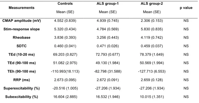
Table 2 - Baseline results of the three studied groups.
Results of all comparisons among the three groups at baseline. ALS Group-1 amplitude > median value, 2.84 mV); ALS Group-2 (M-amplitude < median value, 2.84 mV); NS – non-significant p value; SDTC - Strength duration time constant
Measurements Controls Mean (SE) ALS group-1 Mean (SE) ALS group-2 Mean (SE) p value CMAP amplitude (mV) 4.552 (0.839) 4.939 (0.745) 2.306 (0.153) NS Stim-response slope 5.320 (0.434) 4.784 (0.569) 5.830 (0.835) NS Rheobase 3.836 (0.393) 3.256 (0.443) 4.119 (0.742) NS SDTC 0.460 (0.041) 0.471 (0.026) 0.459 (0.037) NS TEd (10-20 ms) 69.203 (0.827) 72.793 (0.677) 78.379 (1.649) NS TEd (90-100 ms) 51.082 (2.975) 49.130 (1.984) 50.569 (1.994) NS TEh (90-100 ms) -110.993(18.113) -82.798 (31.588) -127.713 (6.553) NS RRP (ms) 2.673 (0.095) 2.672 (0.091) 2.659 (0.128) NS Superexcitability (%) -20.516 (1.005) -27.206 (1.934) -27.206 (1.934) NS Subexcitability (%) 16.604 (2.885) 16.532 (1.946) 10.015 (1.351) NS
Figure 2: Baseline results of the three studied groups.
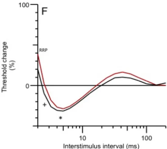
Figure comparing the three groups at baseline. Red represents healthy controls; green represents ALS Group-1; Blue represents ALS-Group-2. It is represented the absolute stimulus-response relationship (top left), the normalized stimulus-response relationship (top right), the current-threshold relationship (middle-left), current-threshold change-stimulus width ratio (middle right), threshold-tracking (bottom left) and recovery-cycle (bottom right). .1 1 10 Peak r esp on se (m V) 10 5 10
Stimulus current (mA)
A 0 100 Peak r esp on se (% ma x) 100 150
Stimulus (% mean threshold)
B -500 0 Threshold reduction (%) -100 0 Curr en t (% th re sho ld) C 0 10 Th re sho ld ch ar ge (m A.ms) -1 0 1 Stimulus width (ms) D -200 -100 0 100 Th re sho ld r ed uctio n (% ) 0 100 200 Delay (ms) E 0 100 Th re sho ld ch an ge (% ) 10 100 Interstimulus interval (ms) F
B - Before versus after exercise
Controls
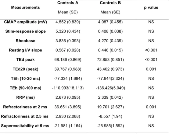
In the group of healthy controls comparing measurements before and after exercise did show significantly smaller resting I/V slope after exercise (p<0.001). In addition, we observed greater threshold changes during depolarizing threshold electrotonus, TEd peak and TEd20 (peak) – (p<0.000 and p=0.001, respectively). Finally, the refractoriness at 2 ms was significant reduced after the exercise (p=0.001). The results are summarized in Table 3 and Figure 3.
Table – 3: Control groups, comparison before vs after muscle
contraction. Measurements Controls A Mean (SE) Controls B Mean (SE) p value CMAP amplitude (mV) 4.552 (0.839) 4.087 (0.455) NS Stim-response slope 5.320 (0.434) 0.408 (0.038) NS Rheobase 3.836 (0.393) 4.270 (0.439) NS Resting I/V slope 0.567 (0.028) 0.446 (0.015) <0.001
TEd peak 68.186 (0.869) 72.853 (0.851) <0.001 TEd20 (peak) 39.767 (0.988) 43.402 (0.973) 0.001 TEh (10-20 ms) -77.334 (1.694) -77.944(2.324) NS TEh (90-100 ms) -110.993(18.113) -136.426(5.049) NS RRP (ms) 2.673 (0.095) 2.339 (0.042) NS Refractoriness at 2 ms 36.651 (3.895) 19.701 (2.627) 0.001 Refractoriness at 2.5 ms 2.930 (2.088) -8.557 (1.94) NS Superexcitability at 5 ms -21.981 (1.164) -26.985(1.592) NS
Control group: Comparing different parameters before (controls A) and after muscle contraction (controls B). NS – non-significant p value.
Figure – 3: Control groups, comparison before vs after muscle
contraction.
Figure demonstrating the control group before and after muscle contraction. Red represents the control group at rest and black after contraction. We observe the threshold-tracking (left), the recovery-cycle (middle) and IV current voltage relationship (right). * shows significant differences for TEd peak and TEd 20 peak, + for refractoriness at 2 ms and # for I/V current voltage relationship.
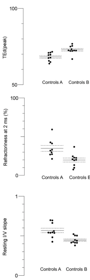
Figure 4: Control groups, mean values before vs after muscle
contraction.
Illustration of the control group mean values and standard error before (controls A) and after (controls B) contraction. Top, TEd (peak) significantly increase after contraction; middle, refractoriness at 2 ms significantly decrease; bottom, IV slope is reduced after exercise.
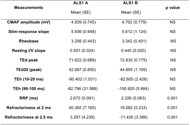
ALS Group 1
In ALS group 1 (with M-wave amplitude > median value) we observed a statistically significant reduction in the RRP, as well in the refractoriness at 2 and 2.5 ms (p=0.001 for all). Moreover, the superexcitability at 5 ms was significantly increased (p=0.001). The results are summarized in Table 4 and Figure 5 and 6.
Table – 4: ALS groups 1, comparison before vs after muscle contraction.
Measurements ALS1 A Mean (SE) ALS1 B Mean (SE) p value CMAP amplitude (mV) 4.939 (0.745) 4.702 (0.779) NS Stim-response slope 5.936 (0.648) 5.612 (1.124) NS Rheobase 3.256 (0.443) 3.342 (0.401) NS Resting I/V slope 0.551 (0.024) 0.445 (0.020) NS TEd peak 71.622 (0.689) 72.830 (0.775) NS TEd20 (peak) 42.087 (0.850) 44.655 (1.159) NS TEh (10-20 ms) -80.402 (1.531) -82.605 (2.426) NS TEh (90-100 ms) -82.798 (31.588) -150.920 (5.984) NS RRP (ms) 2.672 (0.091) 2.326 (0.063) 0.001 Refractoriness at 2 ms 40.392 (7.160) 18.082 (5.233) 0.001 Refractoriness at 2.5 ms 3.297 (4.239) -11.426 (3.386) 0.001
Superexcitability at 5 ms -28.052 (1.968) -31.562 (2.008) 0.001
ALS group 1: Comparing different parameters before (ALS1 A) and after muscle contraction (ALS1 B). NS – non-significant p value.
Figure 5: Recovery cycle in ALS group 1.
Recovery cycle in ALS group 1, before (red) and after exercise (black). Significant changes were observed in the RRP (relative refractory period), refractoriness at 2 and 2,5 ms (+); and in the superexcitability period at 5 ms (*).
Figura 6: ALS group 1, mean values before (A) vs after (B) muscle
contraction.
Figure comparing the ALS group 1, before (ALS5 A) and after (ALS5 B) contraction. Mean values and standard errors. At the top the refractoriness at 2 ms (reduced), at bottom the superexcitability (increased).
amplitude < median value) demonstrated steep changes during the early phases of hyperpolarization, TEh (10-20 ms), with significant reduction from - 66.840 (SE=1.855) at rest to -78.253 (SE=1.788) following contraction (p<0.001). These changes have been described as ‘fanning-out’. The results are summarized in Table 5 and Figure 7 and 8.
Table – 5: ALS 2 groups, comparison before vs after muscle contraction.
Measurements ALS2 A Mean (SE) ALS2 B Mean (SE) p value CMAP amplitude (mV) 2.306 (0.153) 2.332 (0.325) NS Stim-response slope 4.860 (0.355) 4.353 (0.369) NS Rheobase 4.119 (0.742) 4.791 (.425) NS Resting I/V slope 0.537 (0.023) 0.485 (0.032) NS TEd peak 66.807 (1.441) 71.245 (1.618) NS TEd20 (peak) 38.446 (1.484) 42.914 (1.590) NS TEh (10-20 ms) - 66.840 (1.855) -78.253 (1.788) 0.000 TEh (90-100 ms) -127.713 (6.553) -135.318 (6.841) NS RRP (ms) 2.659 (0.128) 2.355 (.052) NS Refractoriness at 2 ms 39.057 (9.515) 18.475 (4.227) NS Refractoriness at 2.5 ms 1.997 (4.952) -9.884 (3.041) NS Superexcitability at 5 ms -27.713 (1.793) -30.708 (2.580) NS
ALS group 2: Comparing different parameters before (ALS2 A) and after muscle contraction (ALS2 B). NS – non-significant p value.
Figure 7: Threshold-tracking in ALS group 2.
Threshold-tracking in ALS group 2, before (red) and after contraction (black). The change is consistent with the early ‘fanning out’ in threshold electrotonus.
Figura 8: ALS group 2, mean values before (ALS4 A) vs after (ALS4 B)
Figure comparing the ALS group 2, before (ALS4 A) and after (ALS4 B) contraction. Mean values and standard errors. Threshold electrotonus hyperpolarization is reduced after exercise.
Discussion
ALS is characterized by progressive muscle weakness and atrophy, fatigue, spasticity, bulbar dysfunction and respiratory insufficiency, leading to disability, and in later stages, to mechanical ventilation and death (Jackson and Bryan, 1998). Despite recent advances in understanding the pathophysiology of ALS, the cause and pathogenesis of ALS remain elusive.
The effect of physical exercise in ALS has been considered controversial. Most studies only documented a slight trend towards to less deterioration on most scales (Drory et al. 2001; Sanjak et al. 1987; Pinto et al. 2012). On the other hand, there are epidemiological studies supporting that regular strenuous exercise can increase the risk of developing ALS later
in life (Mitchell, 2000). The controversy generated by exercise in ALS
patients started with Sinaki and Mulder (1978) who showed a potential negative effect in rapidly progressive cases. However, another study (DeLisa et al. 1979) showed that exercise was beneficial in the initial phase of the disease by preventing fatigue, muscular weakness and disuse weakness. Bohannon and colleagues (Bohannon et al.,1983)
demonstrated that a gradually resisted isometric exercise increased muscular power in ALS patients. As a whole, physical exercise encompasses motor neuron activation, which thereby triggers the oxidative stress, free radical production and glutamate stimulation. These exercise-induced effects are strongly regulated under physiological conditions, which do not occur in ALS subjects where there is a higher risk of becoming neurotoxic (de Almeida et al. 2012).
In healthy people, muscular activity can produce substantial axonal hyperpolarization. A study developed by Vagg and their colleagues describe that maximal voluntary contraction for as little as 15 seconds can lead to a reduction in excitability lasting for 5–10 minutes, and that longer periods of contraction can cause changes that last up to 15–20 min. In their study, a short maximal voluntary contraction of the ABP increased the threshold of median motor axons (measured using 1-ms test pulses) by 15±20% and contraction for 1 min increased threshold by 40% (Vagg et al., 1998). The author also considered that the extent of the activity-dependent hyperpolarization in motor axons following their voluntary activation is quite prominent and could be a significant limiting factor for impulse conduction if the safety margin was impaired, as anticipated in Guiilian Barré Syndrome and Multifocal Motor Neuropathy. Many other studies have described that voluntary contraction activates
the axonal membrane Na+/K+ pump, which attempts to return the resting
membrane potential to baseline after contraction has ceased, resulting in activity dependent hyperpolarization (Burke et al. 2001; Bostock & Grafe 1985; Bostock & Bergmans 1994; Morita et al. 1993).
The threshold electrotonus and strength-duration studies in ALS suggest that potassium conductances may be reduced (increased threshold changes in TEd 10–30 ms and TEd 90–100 ms and greater
supernormality) and persistent Na+ conductance increased (higher
strength–duration time constant), both of which would increase axonal excitability (Kanai et al. 2006a; Nakata et al. 2006; Boërio et al. 2010; Vucic & Kiernan 2006; Mogyoros et al. 1998). However, results depends
on the size of the motor response, in particular K+ conductances (Kanai et
al. 2006b). The above mentioned changes in threshold electrotonus of in ALS patients are described as fanning-out, related to their resemblance to a Japanese fan (Kaji R. 1997; Horn et al., 1996). The early fanning-out tend to be normalized during hyperpolarization, due to activation of inward rectification by the hyperpolarization activated current Ih (hyperpolarizing threshold electrotonus 90–100). The observed “pseudo-normalization” of excitability changes as the disease progresses may indicate that the most abnormal axons have already died (Winhammar et al., 2005).
In our study we did not find significant differences between ALS group 1, ALS group 2 and controls at baseline. This probably derives from the small number of patients in each group, the recruitment criteria of APB M-wave response > 1 mv and the corrected p value. Indeed, other studies have included patients with very low motor response, < 1mV (Kanai et al. 2006b).
The group of controls showed hyperpolarization in motor axons after isometric voluntary activation. These feature was demonstrated in
significantly smaller resting I/V slope after exercise. When the slope of the current-threshold relationship decrease reflects inward rectification accommodating to hyperpolarization and activation of the inwardly rectifying cation conductance. They also exhibited significantly greater threshold changes in depolarizing threshold electrotonus, both in TEd and TEd20 peaks, during a conditioning current after contraction. So, in depolarizing threshold electrotonus immediately after the initiation of the conditioning pulse, excitability of the membrane changes as a consequence of its cable properties (F phase). When the membrane is hyperpolarized, as after contraction, an increase in threshold is required. Finally, the refractoriness, which corresponds to the gradual recovery of Na+ channels from inactivation, is significant reduced after the exercise, as expected in membrane hyperpolarization.
In ALS group 1, refractory parameters are reduced and superexcitability increased as physiologically presumed. However, resting I/V slope after
exercise as not altered by exercise in this group, suggesting minor Na+/K+
pump dysfunction.
In ALS group 2, the specific change observed can understandable as a component of the ‘fanning-out’, but without other variation this alteration is quite weak to represent a meaningful modification. We can state that features of hyperpolarization after exercise were not recorded, strongly
suggesting a major dysfunction of the Na+/K+ pump dysfunction.
Vucic, et al (Vucic et al. 2007) observed a greater increase in threshold following voluntary contraction in patients with ALS compared with
controls. These alterations were associated by changes in other excitability parameters indicating membrane hyperpolarisation. However,
Na+/K+ pump overactivity was not the cause, as derived from the curves
of threshold variation following exercise. It was accepted the role of the higher firing rates of surviving motor units in ALS patients with muscular
weakness. Our results, following a different methodology, shows a Na+/K+
pump dysfunction when the number of functional axons is small.
Limitations of the study
Our exploratory study included a small population of patients and controls. The patients were investigated only once; as such consistency of the results was not supported by repeated investigations. In addition, the same patients were not evaluated longitudinally to observe changes over the course of the disease in the same group.
Much additional information can be obtained about the properties of disturbed axons from studies of their excitability properties with threshold tracking techniques, so this technique is concerned to find the pathophysiological mechanisms of the diseases more than disease diagnosis. However, as other techniques, this has as well some limitations. Threshold measurements only test the nerve at the point of stimulation, so unlike conduction studies they are not useful for focal neuropathies, unless it is possible to stimulate at or close to the lesion site. Threshold tracking tests only the axons with thresholds close to the chosen target response, so that conditions affecting the excitability of only
a minority of axons, whether the least or the most excitable, may be undetected. Axons that have degenerated or which are blocked between stimulation and recording site cannot be tested. Interpretation may be challenging as changes in parameters may have different explanations; modeling has been helpful in the use of the methods in clinical neurophysiology, but we had no opportunity to model our results.
Conclusion
In conclusion, our observations suggest a dysfunctional Na+/K+ pump in ALS, in particular in weak muscles. This implies that exercise can be detrimental to axons, which are unable to compensate repeated depolarization. Further studies, in a larger population of ALS patients is advisable.
Acknowledgments
I want to thank my thesis supervisor Prof. Dr. Mamede de Carvalho for all the effort and encouragement in the elaboration of this thesis.
I also want to thank to Eng. Isabel Casanova for all the support and help in the technique application.
Bibliography
De Almeida, J.P.L. et al., 2012. Exercise and amyotrophic lateral sclerosis. Neurological sciences : official journal of the Italian Neurological Society and of the Italian Society of Clinical Neurophysiology, 33(1), 9–15. Alvarez, S. et al., 2013. Peripheral motor axons of SOD1(G127X) mutant mice are susceptible to activity-dependent degeneration. Neuroscience, 241, 239–49.
Anon, Charcot JM, Alexis Joffroy A. Deux cas d’atrophie musculaire progressive avec lésions de la substance grise et des faiseaux antérolatéraux de la moelle épinière. Archives de physiologie normale et pathologique, Paris, 1869, 2: 354-367, 629-649, 744-760.
Anon, Kaji R. Physiological and technical basis of peripheral nerve and motoneurons testing. In: Kimura J, Kaji R, editors. Physiology of ALS and related diseases. Amsterdam: Elsevier; 1997. p. 15–41.
Barrett, E.F. & Barrett, J.N., 1982. Intracellular recording from vertebrate myelinated axons: mechanism of the depolarizing afterpotential. The Journal of physiology, 323, 117–44.
Bello-Haas, V.D. et al., 2007. A randomized controlled trial of resistance exercise in individuals with ALS. Neurology, 68(23), 2003–7.
Bensimon, G. et al., 2002. A study of riluzole in the treatment of advanced stage or elderly patients with amyotrophic lateral sclerosis. Journal of neurology, 249(5), 609–15.
Boërio, D. et al., 2010. Excitability properties of mouse motor axons in the mutant SOD1(G93A) model of amyotrophic lateral sclerosis. Muscle & nerve, 41(June), 774–784.
Bohannon, R.W., 1983. Results of resistance exercise on a patient with amyotrophic lateral sclerosis. A case report. Physical therapy, 63(6), 965– 8.
Bostock, H. et al., 1998. Threshold tracking techniques in the study of human peripheral nerve. Muscle & Nerve, 21(February), 137–158.
Bostock, H. & Bergmans, J., 1994. Post-tetanic excitability changes and ectopic discharges in a human motor axon. Brain : a journal of neurology, 117 (5), 913–28.
Bostock, H. & Grafe, P., 1985. Activity-dependent excitability changes in normal and demyelinated rat spinal root axons. The Journal of physiology,
Burke, D., Kiernan, M.C. & Bostock, H., 2001. Excitability of human axons. Clinical Neurophysiology, 112, 1575–1585.
De Carvalho, M. et al., 2008. Electrodiagnostic criteria for diagnosis of ALS. Clinical neurophysiology : official journal of the International Federation of Clinical Neurophysiology, 119(3), 497–503.
De Carvalho, M. et al., 1996. Motor neuron disease presenting with respiratory failure. Journal of the neurological sciences, 139, 117–22.
De Carvalho, M., Costa, J. & Swash, M., 2005. Clinical trials in ALS: a review of the role of clinical and neurophysiological measurements. Amyotrophic lateral sclerosis and other motor neuron disorders : official publication of the World Federation of Neurology, Research Group on Motor Neuron Diseases, 6(4), 202–12.
De Carvalho, M. & Swash, M., 2012. Fasciculation potentials: still mysterious. Clinical neurophysiology : official journal of the International Federation of Clinical Neurophysiology, 123(2), 227–8.
Chiò, A. et al., 2009. Prognostic factors in ALS: A critical review. Amyotrophic lateral sclerosis : official publication of the World Federation of Neurology Research Group on Motor Neuron Diseases, 10(5-6), 310– 23.
DeLisa, J.A. et al., 1979. Amyotrophic lateral sclerosis: comprehensive management. American family physician, 19(3), 137–42.
Drory, V.E. et al., 2001. The value of muscle exercise in patients with amyotrophic lateral sclerosis. Journal of the neurological sciences, 191(1-2), 133–7.
Gouveia, L.O. & de Carvalho, M., 2007. Young-onset sporadic amyotrophic lateral sclerosis: a distinct nosological entity? Amyotrophic lateral sclerosis : official publication of the World Federation of Neurology Research Group on Motor Neuron Diseases, 8(6), 323–7.
Haverkamp, L.J., Appel, V. & Appel, S.H., 1995. Natural history of amyotrophic lateral sclerosis in a database population. Validation of a scoring system and a model for survival prediction. Brain : a journal of neurology, 118 (3), 707–19.
Hodgkin, A.L. & Huxley, A.F., 1952. Propagation of electrical signals along giant nerve fibers. Proceedings of the Royal Society of London. Series B, Containing papers of a Biological character. Royal Society (Great Britain), 140, 177–83.
Jackson, C.E. & Bryan, W.W., 1998. Amyotrophic lateral sclerosis. Seminars in neurology, 18(1), 27–39.
Kaji, R. et al., 2000. Activity-dependent conduction block in multifocal motor neuropathy. Brain : a journal of neurology, 123(8), 1602–11.
Kanai, K. et al., 2006a. Altered axonal excitability properties in amyotrophic lateral sclerosis: Impaired potassium channel function related to disease stage. Brain, 129, 953–962.
Kanai, K. et al., 2006b. Altered axonal excitability properties in amyotrophic lateral sclerosis: impaired potassium channel function related to disease stage. Brain : a journal of neurology, 129(4), 953–62.
Kiernan, M.C. et al., 2011. Amyotrophic lateral sclerosis. Lancet, 377, 942–55.
Kiernan, M.C. et al., 2000. Multiple measures of axonal excitability: a new approach in clinical testing. Muscle & nerve, 23, 399–409.
Krarup, C. & Moldovan, M., 2009. Nerve conduction and excitability studies in peripheral nerve disorders. Current opinion in neurology, 22, 460–466.
Krishnan, A. V. et al., 2008. Assessment of nerve excitability in toxic and metabolic neuropathies. Journal of the Peripheral Nervous System, 13, 7– 26.
Lin, C.S.-Y. et al., 2011. Modulatory effects on axonal function after
intravenous immunoglobulin therapy in chronic inflammatory
demyelinating polyneuropathy. Archives of neurology, 68(7), 862–9. Lin, C.S.Y. et al., 2006. Assessment of nerve excitability properties in peripheral nerve disease. Handbook of Clinical Neurophysiology, 7, 381– 403.
McCrate, M.E. & Kaspar, B.K., 2008. Physical activity and neuroprotection in amyotrophic lateral sclerosis. Neuromolecular medicine, 10(2), 108–17.
Miller, R.G. et al., 2013. Quality improvement in neurology: amyotrophic lateral sclerosis quality measures: report of the quality measurement and reporting subcommittee of the American Academy of Neurology. Neurology, 81(24), 2136–40.
Mitchell, J.D., 2000. Guidelines in motor neurone disease (MND)/amyotrophic lateral sclerosis (ALS) - from diagnosis to patient care. Journal of neurology, 247, 7–12.
Mogyoros, I. et al., 1998. Strength-duration properties of sensory and motor axons in amyotrophic lateral sclerosis. Brain : a journal of neurology, 121(5), 851–859.
Mogyoros, I., Bostock, H. & Burke, D., 2000. Mechanisms of paresthesias arising from healthy axons. Muscle & nerve, 23(3), 310–20.
Morita, K. et al., 1993. Posttetanic hyperpolarization produced by electrogenic Na(+)-K+ pump in lizard axons impaled near their motor terminals. Journal of neurophysiology, 70(5), 1874–84.
Nakata, M. et al., 2006. Distal excitability changes in motor axons in amyotrophic lateral sclerosis. Clinical Neurophysiology, 117, 1444–1448.
Pandyan, A.D. et al., 1999. A review of the properties and limitations of the Ashworth and modified Ashworth Scales as measures of spasticity. Clinical rehabilitation, 13(5), 373–83.
Pinto, A.C. et al., 1999. Can amyotrophic lateral sclerosis patients with respiratory insufficiency exercise? Journal of the neurological sciences, 169(1-2), 69–75.
Pinto, S. & Carvalho, M. de, 2014. Breathing new life into treatment advances for respiratory failure in amyotrophic lateral sclerosis patients. Neurodegenerative disease management, 4(1), 83–102.
Pinto, S., Swash, M. & de Carvalho, M., 2012. Respiratory exercise in amyotrophic lateral sclerosis. Amyotrophic lateral sclerosis : official publication of the World Federation of Neurology Research Group on Motor Neuron Diseases, 13(1), 33–43.
Sanjak, M., Reddan, W. & Brooks, B.R., 1987. Role of muscular exercise in amyotrophic lateral sclerosis. Neurologic clinics, 5(2), 251–68.
Sinaki, M. & Mulder, D.W., 1978. Rehabilitation techniques for patients with amyotrophic lateral sclerosis. Mayo Clinic proceedings, 53(3), 173–8. Swash, M. & Desai, J., 2000. Motor neuron disease: classification and nomenclature. Amyotrophic lateral sclerosis and other motor neuron disorders : official publication of the World Federation of Neurology, Research Group on Motor Neuron Diseases, 1(2), 105–12.
Talbot, K., 2009. Motor neuron disease: the bare essentials. Practical neurology, 9(5), 303–9.