Jorge Miguel Pires Rodrigues
Outubro de 2012
Flow cytometry as a novel tool for
structural and functional characterization
of isolated yeast vacuoles
UMinho|20 12 Jor ge Miguel Pir es Rodrigues Flow cytometr
y as a novel tool for structural and functional characterization of isolated yeast vacuoles
Universidade do Minho
Trabalho realizado sob a orientação da
Professora Doutora Maria Manuela Sansonetty
Gonçalves Côrte-Real
e do
Professor Doutor Hernâni Varanda Gerós
Jorge Miguel Pires Rodrigues
Outubro de 2012
Dissertação de Mestrado
Mestrado em Biofísica e Bionanossistemas
Flow cytometry as a novel tool for
structural and functional characterization
of isolated yeast vacuoles
Universidade do Minho
ii
DECLARAÇÃO
Nome: Jorge Miguel Pires Rodrigues
Endereço electrónico: [email protected]
Telefone: + 351 917001108
Número do Bilhete de Identidade: 12847523
Título da tese de mestrado:
Flow cytometry as a novel tool for structural and functional characterization of isolated yeast vacuoles
Orientador(es): Professora Doutora Maria Manuela Sansonetty Gonçalves Côrte-Real e Professor Doutor Hernâni Varanda Gerós
Ano de conclusão: 2012
Designação do Mestrado ou do Ramo de Conhecimento do Doutoramento: Mestrado em Biofísica e Bionanossistemas
1. É AUTORIZADA A REPRODUÇÃO INTEGRAL DESTA DISSERTAÇÃO APENAS PARA EFEITOS DE INVESTIGAÇÃO, MEDIANTE DECLARAÇÃO ESCRITA DO INTERESSADO, QUE A TAL SE COMPROMETE;
Universidade do Minho, ___/___/______
iii
ACKNOWLEDGEMENTS
Ao longo do Mestrado em Biofísica e Bionanossistemas, em especial durante o trabalho laboratorial, muitas foram as pessoas que me ajudaram e sem as quais não conseguiria ter feito esta tese. Escrevo com o intuito de demonstrar a minha gratidão.
Agradeço aos meus pais por serem os melhores pais do mundo, por me terem transmitido valores, educação e fazerem de mim a pessoa que sou hoje. Sempre tive o seu apoio, carinho e compreensão. Estiveram, estão e estarão sempre ao meu lado. Sem eles não teria conseguido vencer os obstáculos e construir os alicerces da minha vida e do meu sonho.
Agradeço aos meus orientadores, Professora Manuela Côrte-Real e Professor Hernâni Gerós, por terem estado sempre presentes em apoio, dedicação, motivação e amizade ao longo deste trabalho. Agradeço-lhes do fundo do coração a oportunidade que me deram de poder aprender e trabalhar com eles. O meu muito obrigado.
Agradeço aos meus colegas e amigos Vi, Henrique e Rui a ajuda sempre pronta e o apoio que me deram no trabalho laboratorial.
Agradeço a todos os meus companheiros dos laboratórios de biologia vegetal e microbiologia I pelo bom ambiente de trabalho proporcionado e ajudas pontuais.
Agradeço a todos os funcionários e técnicos que sempre me ajudaram e facilitaram o meu trabalho.
Agradeço aos meus amigos Edgar, Liliana, Egipto, Ivo e Tó Zé pelo apoio e amizade dedicada ao longo dos anos.
A todos os investigadores que de uma forma ou outra contribuíram para uma boa camaradagem laboratorial.
Este trabalho foi financiado em parte pela Fundação para a Ciência e Tecnologia através dos projectos PTDC/BIA-BCM/69448/2006 e PTDC/AGRALI/100636/2008. Este trabalho foi financiado em parte pela FEDER através da POFC – COMPETE e pelos fundos nacionais da FCT através do projecto PEst-C/BIA/UI4050/2011.
v FLOW CYTOMETRY AS A NOVEL TOOL FOR STRUCTURAL AND FUNCTIONAL CHARACTERIZATION OF ISOLATED YEAST VACUOLES
ABSTRACT
The yeast vacuole is functionally analogous to the mammalian lysosome. Both play important roles in fundamental cellular processes such as protein degradation, detoxification, osmoregulation, autophagy and apoptosis which, when deregulated in humans, can lead to several diseases. Some of these vacuolar roles are difficult to study in a cellular context, and therefore the use of a cell-free system is an important approach to gain further insight into the different molecular mechanisms required for vacuolar function. The aim of this thesis was to develop a set of protocols for the structural and functional characterization of yeast vacuoles isolated from Saccharomyces cerevisiae using flow cytometry and several commercially available fluorescent probes. Flow cytometry analysis was complemented by other fluorescence based thecniques, namely fluorescence microscopy and spectrofluorimetry. The isolation protocol resulted in a yeast vacuolar fraction with a degree of purity suitable for cytometric analysis. Moreover, isolated vacuoles were structurally and functionally intact, and able to generate and maintain electrochemical gradients of ions across the vacuolar membrane, as assessed by flow cytometry. Proton and calcium gradients were dissipated by NH4Cl
and calcimycin, respectively. These results established flow cytometry as a powerful technique for the characterization of isolated vacuoles. The protocols developed in this study can also be used to enhance our understanding of several molecular mechanisms underlying the development of lysosomal-related diseases, as well as of the role of the vacuole/lysosome in different cellular processes such as apoptosis. Moreover they can be used to screen for new drugs that modulate these processes, which have promising clinical relevance.
vii FLOW CYTOMETRY AS A NOVEL TOOL FOR STRUCTURAL AND FUNCTIONAL CHARACTERIZATION OF ISOLATED YEAST VACUOLES
RESUMO
O vacúolo da célula de levedura é funcionalmente análogo ao lisossoma da célula de mamífero. Ambos desempenham funções importantes em processos celulares fundamentais tais como a degradação de proteínas, destoxificação, osmorregulação, autofagia e apoptose que, quando desregulada em humanos, pode levar a diversas doenças. Alguns destas funções do vacúolo são difíceis de estudar num contexto celular, e portanto a utilização de um sistema não celular é uma estratégia importante para melhor compreender os diferentes mecanismos moleculares subjacentes à função vacuolar. O objectivo desta tese foi desenvolver um conjunto de protocolos para a caracterização estrutural e funcional de vacúolos isolados da levedura Saccharomyces
cerevisiae utilizando a citometria de fluxo e diversas sondas fluorescentes disponíveis
comercialmente. A análise por citometria de fluxo foi complementada por outras técnicas de fluorescência, nomeadamente a microscopia de fluorescência e a espectrofluorimetria. O protocolo de isolamento utilizado na levedura permitiu a obtenção de uma fracção vacuolar com um grau de purificação apropriado para a análise citométrica. Adicionalmente, a análise por citometria de fluxo de suspensões de vacúolos isolados demonstrou que estes, após o isolamento, mantinham não só a sua integridade estrutural e funcional intactos, mas também a capacidade de gerar e manter um gradiente electroquímico de iões através da membrana vacuolar. A adição de NH4Cl
e calcimicina conduziu respectivamente à dissipação do gradientes de protões e de cálcio. Estes resultados validam a exploração da citometria de fluxo como uma técnica poderosa para a caracterização de vacúolos isolados. Os protocolos desenvolvidos neste estudo podem também ser utilizados para a elucidação de diversos mecanismos moleculares associados ao desenvolvimento de doenças lisossomais, assim como do papel do vacúolo/lisossoma em diferentes processos celulares tais como a apoptose. Estes protocolos podem ainda ser utilizados para o rastreio de novas drogas que modulam estes processos e que têm relevância clínica promissora.
ix TABLE OF CONTENTS ACKNOWLEDGEMENTS ... iii ABSTRACT ... v RESUMO ... vii CHAPTER 1 ... 11 GENERAL INTRODUCTION ... 13
Basic principles of fluorescence techniques ... 13
Cell fractionation techniques ... 18
The role of the yeast vacuole/mammalian lysosome in cell homeostasis and cell death ... 18
Scope of the thesis ... 21
REFERENCES ... 21
CHAPTER 2 ... 27
FLOW CYTOMETRY AS A NOVEL TOOL FOR STRUCTURAL AND FUNCTIONAL CHARACTERIZATION OF ISOLATED YEAST VACUOLES ... 29
SUMMARY ... 29
INTRODUCTION ... 30
METHODS ... 31
Reagents ... 31
Yeast strains and plasmids ... 31
Isolation of intact yeast vacuoles ... 31
Preparation of vacuolar membrane vesicles ... 32
Purification of vacuole fractions - silicone oil technique ... 32
Mitochondria preparation ... 33
Staining protocols for structural and functional analysis of isolated vacuoles and vacuolar membrane vesicles ... 33
Fluorescence microscopy ... 33
Flow cytometry ... 34
Proton-pumping activity of V-H+-ATPase in intact vacuoles and membrane vesicles ... 34
RESULTS ... 35
The isolated fraction is enriched in functional vacuoles ... 35
Characterization of yeast vacuoles by fluorescence microscopy ... 37
Structural and functional analysis of intact vacuoles and vacuolar membrane vesicles by flow cytometry ... 38
x
DISCUSSION ... 43
ACKNOWLEDGEMENTS ... 46
REFERENCES ... 47
CHAPTER 3 ... 51
CONCLUSIONS AND FUTURE PERSPECTIVES ... 53
13
GENERAL INTRODUCTION
In the present study different fluorescence techniques were exploited to characterise the structure and function of isolated vacuoles from yeast cells. This purpose is part of a broader research project aimed to assess the structural and functional alterations associated with the apoptotic programmed cell death process, using yeast as an eukaryotic model system. The ultimate goal of this project is to gain further insights on the role of lysosome - the vacuole mammalian counterpart, in this active cell death process.
In this introductory chapter the basic principles of fluorescence techniques will be addressed, as well as their main advantages and drawbacks, and the benefits of the implementation of complementary approaches, including the combined use of flow cytometry, fluorescence microscopy and spectrofluorimetry will be discussed. Then, the methodologies to obtain purified subcellular fractions and their use to address main issues of cellular processes will be analysed. In this context, the current knowledge of the yeast vacuole and of its mammalian counterpart in cell homeostasis and in programmed cell death cell will be explored. This chapter is closed with the scope of the thesis and the proposed workplan.
Basic principles of fluorescence techniques
Physical properties of electromagnetic radiation, namely in the ultraviolet, near infrared and visible spectrum are regularly used in biological research (White and Errington, 2005). Some specimens are able to absorb energy and to emit light, which is a phenomenum called luminescence. Luminescence can be divided into two categories, fluorescence and phosphorescence, depending on the nature of the excited and electronic ground state. If they are of the same nature, fluorescence occurs when the electron returns to the electronic ground state with emission of light (White and Errington, 2005). The excited states of fluorescence process have lifetimes in the range of 10-8 s whereas phosphorescence excited states have lifetimes of 10-4 to tens of s (Parker and Rees, 1962).
Electrons can exist in different vibrational energy levels. An electron is usually excited to higher vibrational levels of electronic excited states, and relaxation processes
14
occurs when the electron transits to the lowest vibrational level of the first electronic excited state before it returns to the electronic ground state. Energy is lost in non-radiative processes (Figure 1). This explains why the energy of emission is lower than the absorption and occurs at longer wavelengths – Stokes’ shift. Another property of fluorescence described by Kasha’s rule is that the spectrum of emission remains the same despite the excitation wavelenght used (Young , 1961; Li et al., 2004; White and Errington, 2005; Moreno-García et al., 2008).
Figure 1 - A simple Jablonski diagram showing the absorption of energy and excitation of electrons from the ground state (S0) to excitated states (S1 and S2) followed by a loss of energy through non-radiative processes and transition of the electrons to the lowest vibrational level of the first excited state before the return to the ground state with emission of fluorescence (adapted from Moreno-García et al., 2008). a - absorption; b - relaxation; c - fluorescence.
Fluorescence may be primary, when a biological sample emits fluorescence after excited, or secondary, when the emitted fluorescence is mediated by fluorescent probes or fluorophores that interact with the sample (Altemüller and van Vliet-Lanoe 1990; Li
et al., 2004). Thus, fluorescent probes allow the localization of a specific structure in a
biological sample, or may be used to monitor a biological response to a specific stimulus (Johnson, 1998). The mechanism underlying fluorescence emission may include covalent or non-covalent binding, functional modification or compartmentalization of the fluorescent probe (White and Errington, 2005).
Probes must comply with several requirements, namely: i) to display reduced cytotoxicity, preserving the structural and functional characteristics of the biological sample; ii) to be specific to a given molecule, structure, cell function and compartment; iii) to be compatible with the instrumentation used allowing the detection of the fluorescence signal (Johnson, 1998).
Several equipments, including the fluorescence microscope, the spectrofluorimeter and the flow cytometer, make use of the fluorescence properties described above.
15 The fluorescence microscopy allows gaining insight on cell structure and function (Kherlopian et al., 2008). Fluorescence microscopes can be divided into transmitted-light and incident-light fluorescence microscopes, based on the optical path. The incident-light fluorescence microscopes also known as epi-illumination fluorescence microscopes have only one path of the emitted fluorescence to the detector, and do not require perfect alignment of the objectives and the condenser, since the condenser is also the objective. It has also a dichromatic beam splitter and is easy to change between fluorescence microscopy and transmitted light microscopy (Ploem, 1993; Herman, 1998, Li et al., 2004). A light source emits energy of different wavelenghts. By passing an excitation filter, a maximum excitation is achieved. This filter also reduces the background light (Herman, 1998, Li et al., 2004). A dichromatic beam splitter reflects the light with shorter wavelenghts and transmits lights of longer wavelenghts at the same time. The excitation light is reflected by this filter, condensed by the objective and reaches the specimen. Only the fluorescence emitted from the specimen passes through and reaches the detector. A barrier filter further helps to limit the unabsorbed excitation light from reaching the detector (Figure 2) (Li et al., 2004).
Figure 2 - Incident-light fluorescence microscope (Adapted from Li et al., 2004). a - light source; b - excitation filter; c - exciting light; d - dichromatic beam splitter; e - transmitted light; f - reflected light; g - objective and condenser; h - sample; i - emitted fluorescence; j - barrier filter; k - detector.
Spectrofluorimeters require a source of visible and ultraviolet light, a monochromator to select the frequency of excitation, a sample holder and a second monochromator fitted with a detector to detect and measure the emitted fluorescence. It can be used to determine the fluorescence emission spectrum or the fluorescence excitation spectrum. Spectrofluorimetry is a technique more sensitive than absorption
16
spectrophotometry and allows determining the two spectra already mentioned instead of one. It requires, however, that the sample emits fluorescence, which may require the need to use of fluorophores, and the measurements are expressed in arbitrary units and referred to a standard substance. The light is emitted by the source in all directions, and a first monochromator selects a band of frequencies that reaches the sample. The light that reaches the detector is a small fraction of the light that is emitted by the source. Less than 1% of the light is absorbed by the sample and fluorescence is emitted in all directions again. A second monochromator collects fluorescence and a selected band of frequencies is collected and reaches the detector (Figure 3) (Parker and Rees, 1962).
Figure 3 - Schematic diagram for a general-purpose spectrofluorimeter (Adapted from Parker and Rees, 1962). a - light source; b - monochromator for exciting light; c - fluorescence; d - monochromator for fluorescence; e - detector.
Flow cytometry permits particle analysis, cell sorting, and the study of cell function/dynamics (Haynes, 1988), being used to investigate whole cells and sub-cellular constituents, such as organelles, chromosomes, cytokines, hormones and DNA, RNA and protein content (Jaroszeski and Radcliff, 1999), and thus has several important applications, including in cell biology, oncology and immunology.
Flow cytometers can analyse particles, one at a time, giving distributions of parameters at a flow of 1000 to 10000 cells per s. Flow cytometers are constituted by mechanical, optical and electronic components. The mechanical component is related to the suspension of the particles and the particles flow. The optical component embraces the light source and the captation of disperse light and fluorescence. The electronic component is involved in the transformation of the signals into analogic-digital and the acquisition, processing, analysis and data backup in computer. In the flow chamber, the particles in suspension are collected by a needle and immersed in a liquid that flows at a higher velocity. An hydrodynamic focusing or "sheathed flow" allows to direct all the
17 cells along a central path through the orifice, eliminating size distortion, speeding analysis rates and reducing clogging, which would happen if the cells trajectory was off-axis. The particles or cells flow and intersect the light beam one by one. Light is scattered in several directions, namely in the forward direction, being collected by a detector of frontal dispersion, and in lateral directions in a plan perpendicular to the axis of the light beam, being collected by lens, dichroic mirrors and optical filters and focused on photomultipliers, as this is the case of lateral dispersion. Fluorescence is emitted in all directions, but follows the same optical path as lateral light scatter (Figure 4) (Haynes, 1988). Information is collected about the relative size of the cell (forward scattering), the shape or complexity (side scattering) and of autofluorescence or, if the sample has been stained with a fluorophore, of fluorescence specific to cellular structures or dynamic functions (Nunez, 2001). The measurements are based on combinations or ratios of the parameters. (Mandy et al., 1995)
As long as an appropriate probe is available, flow cytometry can be used to analyse any cellular structure or function (Jaroszeski and Radcliff, 1999). Flow cytometry allows a rapid and specific analysis of individual cell or cellular components, being possible to characterize different populations in size and complexity. Furthermore, in addition of the fluorescence properties of a sample, different cellular and sub-cellular processes and features can be monitored simultaneously allowing a cell based multiparametric analysis.
All of the techniques described above can provide valuable information of an analyzed sample. Indeed, the combination of different techniques will provide consistent and complementary information about a sample regarding its structure and function.
Figure 4 - Optical schematic for a flow cytometer. a - flow chamber; b - dichroic filters; c - bandpass filters. Adapted from Purdue University Cytometry Laboratories.
18
Cell fractionation techniques
Subcellular fractionation techniques aim at studying in detail the composition and properties of different cellular components. These techniques consist on the disruption of cells/tissues through homogenization followed by fractionating steps (centrifugations) that separates different organelles by their physical properties, such as size, density and charge (Pasquali et al., 1999). Fractionating techniques allow obtaining different isolated sub-cellular components, which preserve their structural integrity and functionality, and provide insights on their function without the interference of other sub-cellular components. Mixing two different sub-cellular fractions also allows performing in vitro assays to address the possible interplay between different organelles. The disruption of the plasma membrane leaving intact the internal components of the cell is generally the first step of an isolation procedure. Special care has to be taken during the homogenization procedure, including the utilization of appropriate buffers at specific pH values, utilization of specific inhibitors and low temperature, fine adjustment of the intensity/duration of the physical/chemical homogenization method, in order to keep the biological activity/integrity of each sub-cellular component. After the homogenization, sequential centrifugations will fractionate the homogenate into a pure sub-cellular fraction. Differential centrifugation separates components with differences in size, but the fractions obtained may not be pure enough, and distinct centrifugation techniques is required. The density-gradient centrifugation allows separating the organelles in a gradient of a dense substance. For instance, the equilibrium centrifugation separates the subcellular components based on their density, independently of their size and shape and sedimentation velocity. The samples are centrifuged until they reach an equilibrium position in which their density is equal to the surrounding solution (Quintas et al., 2008; Cooper and Hausman, 2009).
The role of the yeast vacuole/mammalian lysosome in cell homeostasis and cell death
Vacuoles, the functional equivalents of mammalian lysosomes (Weisman, 2003), are the most versatile (Holtzman, 1989; Weber, 2002) and the largest of yeast organelle, occupying up to 25% of the total intracellular volume (Premsler et al., 2009). In Saccharomyces cerevisiae vacuoles are relatively large and usually there is one per
19 cell (Banta et al., 1988). Vacuoles contain numerous lytic proteins in the lumen, such as proteases, lipases, phosphatases, carboxylic esterases, ribonucleases, glycosidases, nucleases (Klionsky et al., 1990; Teter et al., 2001; Wiederhold et al., 2009). Vacuole plays crucial roles in protein degradation (Horst et al., 1999; Rotin et al., 2000; Sarry et
al., 2007; Li and Kane, 2009), turnover of organelles (Moeller and Thomson, 1979;
Wiederhold et al., 2009; Sakai et al., 1998; Roberts et al., 2003; Kim et al., 2007) ion and metabolites storage (Indge, 1968; Shirahama et al., 1996; Eide et al., 2005), detoxification (Joho et al., 1995; Gharieb and Gadd, 1998; Eide et al., 2005; Wiederhold et al., 2009), cellular ion homeostasis and pH and osmoregulation (Klionsky et al., 1990).
The proton pump V-H+-ATPase, located within the vacuolar membrane, has a major role concerning vacuolar functions (Wiederhold et al., 2009). Vacuolar V-H+ -ATPase is a multisubunit enzyme constituted by a peripheral membrane subunit complex, V1, responsible for ATP hydrolysis, and an integral membrane subunit
complex, Vo, attached to the first and containing the proton pore (Nelson and Harvey,
1999; Nishi and Forgac, 2002; Kane, 2006;Baars et al., 2007; Diakov and Kane, 2010). This enzyme generates a pH gradient across the vacuolar membrane by an ATP-mediated proton transport from the cytosol to vacuole (Nishi and Forgac, 2002; Graham, 2003; Bowers and Stevens, 2005; Kane, 2006; Baars et al., 2007), ranging 1.7 pH units between vacuolar lumen and cytosol. This proton gradient is used as driving force in substrate-proton antiport transport systems (Kakinuma et al., 1981; Klionsky et al., 1990).
Lysosomes, referred by Christian de Duve as "suicide bags" (de Duve, 2005; Repnik et al., 2012), and their lysosomal proteases have been related to necrosis and autophagy, and more recently to apoptosis. In 1970s, the rupture of liver lysosomes by amino acid and dipeptide methyl esters related lysosomes with apoptosis (Goldman and Kaplan, 1973; Reeves, 1979; Repnik et al., 2012). A critical step of lysosome related apoptosis is the release of lysosomal hydrolases to the cytosol. The destabilization of lysosome membrane may be due to exogenous or endogenous stimuli (Repnik et al., 2012).
Cathepsins are estabilished as important players in apoptosis (Guicciardi et al., 2004; Stoka et al., 2005; Kirkegaard and Jäättelä, 2009; Turk and Turk, 2009; Conus and Simon, 2010; Repnik and Turk, 2010), and once released into cytosol they induce or amplifly the apoptotic signals. Cathepsin D was the first to be related as involved in
20
apoptosis in response to several stimuli. Some cathepsins substrates have been found, such as Bid, Bcl-2, Bcl-xL, Mcl-1 and Bax (reviewed in Repnik et al., 2012).
Lysosomes and lysosomal proteases may act as amplifiers of apoptotic pathways either extrinsic or intrinsic apoptosis pathways (Repnik and Turk, 2010; Schrader et al., 2010). It is however evident that in these cellular contexts lysosomes are not the direct targets of the death stimuli. Mitochondria and MOMP are critical steps in the lysosome-mediated apoptosis and Bax, Bak (Oberle et al., 2010) and ROS are candidates of important mediators of lysosome membrane permeabilization (Repnik et al., 2012).
S. cerevisiae has several orthologues to the apoptosis regulators in metazoan,
including the caspase orthologue metacaspase Yca1p, apoptosis inducing factor Aif1p, Endonuclease G, a member of HtrA2/omi protein family Nma111p, and Bir1p, an inhibitor of apoptosis (IAP) protein (reviewed by Schauer et al., 2009). The conservation of fundamental biological structures in eukaryotes makes studies with simpler models such as S. cerevisiae attractive in providing new insights applicable to higher organisms.
Yeast vacuoles have been recently implicated in programmed cell death (reviewed by Sousa et al., 2011). Actually it was found that Pep4p, an orthologue of the human cathepsin D, translocates from the vacuole to cytosol without the rupture of vacuolar membrane during H2O2-induced apoptosis (Mason et al., 2005). The release of
this vacuolar protease is similar to the release of cathepsins from the lysosomes observed during apoptosis in mammalian cells. Release of Pep4p from the vacuole was also found in an End3p-deficient mutant displaying actin cytoskeleton stabilization-induced apoptosis (Gourlay and Ayscough, 2006) and in cells undergoing acetic acid induced apoptosis (Pereira et al., 2010). A deletion of class C vacuolar protein-sorting genes enhanced the sensitivity of yeast to acetic acid and led to necrotic death. Since the wild type strain mainly undergoes apoptosis, a functional vacuole is required for a regulated cell death process (Schauer et al., 2009).
The use of isolated vacuoles may contribute to unveil their role in different biological processes, such as apoptosis, and screen for new drugs that target directly this organelle and with promising clinical relevance.
21 Scope of the thesis
The scope of the present thesis was to develop a set of fluorescence techniques-based approaches to perform a structural and functional characterization of purified intact vacuoles from the yeast model system S. cerevisiae, aiming at future research on the role of this organelle in key cellular processes such as apoptosis.
For such purpose, a protocol for the purification of yeast vacuoles was implemented and optimized. Then, a set of staining protocols were optimised for the analysis by flow cytometry and fluorescence microscopy of different features of the isolated organelles namely, their acidity, electrical potential and calcium storage capacity. Spectrofluorimetry was also used for monitoring the vacuolar V-H+-ATPase activity. A strain of S. cerevisiae expressing Pep4-EGFP, an EGFP fusion of the vacuolar protease residing in the lumen was used to monitor the preservation of the vacuolar membrane integrity during the purification procedure. In this context, the specific release of Pep4-EGFP from intact vacuoles is currently under validation as an
in vitro assay to assess the capacity of some drugs and metabolites, such as acetic acid,
in promoting the permeabilization of the vacuolar membrane.
REFERENCES
Altemüller, H.J., & Vliet-Lanoe, B. (1990). Soil thin section fluorescence microscopy. Douglas LA. Soil micromorphology: a basic and applied science. Elsevier, Amsterdam, pp 565-579.
Baars, T.L., Petri, S., Peters, C., & Mayer, A. (2007). Role of the V-ATPase in regulation of the vacuolar fission-fusion equilibrium. Molecular Biology of
the Cell. 18, 3873-3882.
Banta, L.M., Robinson, J.S., Klionsky, D.J., & Emr, S.D. (1988). Organelle assembly in yeast: characterization of yeast mutants defective in vacuolar biogenesis and protein sorting. Journal of Cell Biology 107, 1369-1383.
Bowers, K., & Stevens, T.H. (2005). Protein transport from the late Golgi to the vacuole in the yeast Saccharomyces cerevisiae. Biochimica et Biophysica Acta. 1744: 438–454.
22
Conus, S., & Simon, H.U. (2010). Cathepsins and their involvement is immune responses. Swiss Medical Weekly. 140, w13042
Cooper, G.M., & Hausman, R.E. (2009). The Cell: a molecular approach. Fifth Edition. pp 29-33.
Diakov, T.T., & Kane, P.M. (2010). Regulation of vacuolar proton-translocating ATPase activity and assembly by extracellular pH. The Journal of Biological
Chemistry. 285, 23771-23778.
de Duve, C. (2005). The lysosome turns fifty. Nature Cell Biology. 7, 847-849.
Eide, D.J., Clark, S., Nair, T.M., Gehl, M., Gribskov, M., Guerinot, M.L. & Harper, J.F. (2005). Characterization of the yeast ionome: a genomewide analysis of nutrient mineral and trace element homeostasis in Saccharomyces
cerevisiae. Genome Biology. 6, R77.
Gharieb M.M., & Gadd, G.M. (1998). Evidence for the involvement of vacuolar activity in metal(loid) tolerance: vacuolar-lacking and -defective mutants of
Saccharomyces cerevisiae display higher sensitivity to chromate, tellurite and
selenite. Biometals. 11, 101-106.
Goldman, R., & Kaplan, A. (1973). Rupture of rat liver lysosomes mediated by L-amino acid esters. Biochimica et Biophysica Acta. 318, 205-216.
Gourlay, C.W., & Ayscough, K.R. (2006). Actin-induced hyperactivation of the Ras signaling pathway leads to apoptosis in Saccharomyces cerevisiae.
Molecular and Cellular Biology. 26, 6487-6501.
Graham, L.A., Flannery, A.R., & Stevens, T.H. (2003). Structure and assembly of the yeast V-ATPase. Journal of Bioenergetics and Biomembranes. 35, 301–312. Guicciardi, M.E., Leist, M., & Gores, G.J. (2004). Lysosomes in cell death.
Oncogene. 23, 2881-2890.
Horst, M., Knecht, E.C. & Schuh, P.V. (1999). Import into and degradation of cytosolic proteins by isolated yeast vacuoles. Molecular Biology of the Cell. 10, 2879–2889.
Haynes, J.L. (1988). Principles of flow cytometry. Cytometry Supplement 3, 7-17 Herman, B. (1998). Microscopy handbooks 40: fluorescence microscopy. BIOS,
Oxford
Holtzman, E. (1989). Lysosomes. Plenum Press: New York.
Indge, K.J. (1968). Polyphosphates of the yeast cell vacuole. Journal of General
23 Jaroszeski, M.J, & Radcliff, G. (1999). Fundamentals of flow cytometry. Molecular
Biotechnology. 11, 37-53
Joho M., Inouhe, M., Tohoyama, H., & Murayama, T. (1995). Nickel resistance mechanisms in yeasts and other fungi. Journal of Industrial Microbiology. 14, 164-168.
Johnson, I. (1998). Fluorescent probes for living cells. Histochemical Journal. 30, 123-140.
Kakinuma, Y., Ohsumi, Y., & Anraku, Y. (1981). Properties of H1-translocating adenosine triphosphate in vacuolar membranes of Saccharomyces cerevisiae.
The Journal of Biological Chemistry. 256, 10859–10863.
Kane, P.M. (2006). The where, when, and how of organelle acidification by the yeast vacuolar H.-ATPase. Microbiology and Molecular Biology Reviews. 70, 177– 191.
Kherlopian, A.R., Song, T., Duan, Q., Neimark, M.A., Po, M.J., Gohagan, J.K., & Laine, A.F. (2008). A review of imaging techniques for systems biology. BMC
Systems Biology. 2:74
Kim, I., Rodriguez-Enriquez, S., & Lemasters, J.J. (2007). Selective degradation of mitochondria by mitophagy. Archives of Biochemistry and Biophysics. 462, 245–253.
Kirkegaard, T., & Jäättelä, M. (2009). Lysosomal involvement in cell death and cancer. Biochimica et Biophysica Acta 1793, 746-754.
Klionsky, D.J., Herman, P.K. & Emr, S.D. (1990). The fungal vacuole: composition, function, and biogenesis. Microbiological Reviews. 54, 266–292.
Li, Y., Dick, W.A., & Tuovinen, O.H. (2004). Fluorescence microscopy for visualization of soil microorganisms - a review. Biology and Fertility of Soils 39, 301-311.
Li, S.C., & Kane, P.M. (2009). The yeast lysosome-like-vacuole: endpoint and crossroads. Biochimica et Biophysica Acta 1793, 650-663.
Mandy, F.F., Bergeron, M., & Minkus, T. (1995). Principles of flow cytometry.
Transfusion Science. 16, 303-314.
Mason, D.A., Shulga, N., Undavai, S., Ferrando-May, E., Rexach, M.F., & Goldfarb, D.S. (2005). Increased nuclear envelope permeability and Pep4p-dependent degradation of nucleoporins during hydrogen peroxide-induced cell death. FEMS Yeast Research. 5, 1237-1251.
24
Moeller, C.H., & Thomson, W.W. (1979). Uptake of lipid bodies by the yeast vacuole involving areas of the tonoplast depleted of intramembranous particles. Journal
of Ultrastructure Research. 68, 38–45
Moreno-García, E., Guerra-, C.E., & de la Rosa-Vázquez, J.M. (2008). Spectrometer to measure the steady-state fluorescence emitted by liquid and solid samples. 18th International Conference on Electronics, Communications and Computers. DOI 10.1109/CONIELECOMP.2008.23
Nelson, N., & Harvey, W.R. (1999). Vacuolar and plasma membrane proton-adenosinetriphosphatases, Physiological Reviews. 79, 361–385.
Nishi, T., & Forgac, M. (2002). The vacuolar (H.)-ATPases—nature’s most versatile proton pumps. Nature Reviews Molecular Cell Biology. 3, 94–103.
Nunez, R. (2001). Flow cytometry : principles and instrumentation. Current Issues in
Molecular Biology. 3, 39-45.
Oberle, C., Huai, J., Reinheckel, T., Tacke, M., Rassner, M., Ekert, P.G., Buellesbach, J. & Borner, C. (2010). Lysosomal membrane permeabilization and cathepsin release is a Bax/Bak-dependent, amplifying event of apoptosis in fibroblasts and monocytes. Cell Death & Differentiation 17, 1167-1178.
Parker, C.A., & Rees, W.T. (1962). Fluorescence spectrometry. a review. Doi :10.1039/AN9628700083.
Pasquali, C., Fialka, I., & Huber, L.A. (1999). Subcellular fractionation, electromigration analysis and mapping of organelles. Journal of Chromatography B. 722, 89-102.
Pereira, C., Chaves, S., Alves, S., Salin, B., Camougrand, N., Manon, S., Sousa, M.J., & Côrte-Real, M. (2010). Mitochondrial degradation in acetic acid-induced yeast apoptosis: the role of Pep4 and the ADP/ATP carrier. Molecular
Microbiology. 76, 1398-1410.
Ploem, J.S. (1993). Fluorescence microscopy. In: Mason WT (ed) Biological techniques: fluorescent and luminescent probes for biological activity - a pratical guide to technology for quantitative real-time analysis. Academic Press, San Diego, Calif., pp 1-11.
Premsler, T., Zahedi, R.P., Lewandrowski, U., & Sickmann, A. (2009). Recent advances in yeast organelle and membrane proteomics. Proteomics. 9, 4731-4743.
25 Quintas, A., Freire, A.P., & Halpern, M.J. (2008). Bioquímica organização
molecular da vida. Lidel pp. 189-192
Reeves, J.P. (1979). Accumulation of amino acids by lysosomes incubated with amino acid methyl esters. The Journal of Biological Chemistry. 254, 8914-8921.
Repnik, U., Stoka, V., Turk, V., & Turk, B. (2012). Lysosomes and lysosomal cathepsins in cell death. Biochimica et Biophysica Acta. 1824, 22-33
Repnik, U., & Turk, B. (2010). Lysosomal-mitochondrial cross-talk during cell death.
Mitochondrion 10, 662-669.
Roberts, P., Moshitch-Moshkovitz, S., Kvam, E., O’Toole, E., Winey, M., &
Goldfarb, D.S. (2003). Piecemeal microautophagy of nucleus in Saccharomyces
cerevisiae. Molecular Biology of the Cell. 14, 129–141
Rotin, D., Staub, O., & Haguenauer-Tsapis, R. (2000). Ubiquitination and endocytosis of plasma membrane proteins: role of NEDD4/RSP5p family of ubiquitin-protein ligases. Journal of Membrane Biology. 176, 1–17
Sakai, Y., Koller, A., Rangell, L.K., Keller, G.A., & Subramani, S. (1998). Peroxisome degradation by microautophagy in Pichia pastoris: identification of specific steps and morphological intermediates. Journal of Cell Biology. 141, 625–636
Sarry, J.E., Chen, S., Collum, R.P., Liang, S., Peng, M., Lang, A., Naumann, B., Dzierszinski, F., Yuan, C.X., Hippler, M., & Rea, P.A. (2007). Analysis of the vacuolar luminal proteome of Saccharomyces cerevisiae, FEBS Journal. 274, 4287–4305.
Schauer, A., Knauer, H., Ruckenstuhl, C., Fussi, H., Durchschlag, M., Potocnik, U., & Fröhlich, K.-U. (2009). Vacuolar functions determine the mode of cell death. Biochimica et Biophysica Acta 1793, 540-545.
Schrader, K., Huai, J., Jöckel, L., Oberle, C., & Borner, C. (2010). Non-caspase proteases: triggers or amplifiers of apoptosis? Cellular and Molecular Life
Sciences. 67, 1607-1618.
Shirahama, K., Yazaki, Y., Sakano, K., Wada, Y., & Ohsumi, Y. (1996). Vacuolar function in the phosphate homeostasis of the yeast Saccharomyces cerevisiae.
Plant Cell Physiology. 37, 1090–1093
Sousa, M.J., Azevedo, F., Pedras, A., Marques, C., Coutinho, O.P., Preto, A., Gerós, H., Chaves, S.R., & Côrte-Real, M. (2011). Vacuole-mitochondrial cross-talk during apoptosis in yeast: a model for understanding
lysosome-26
mitochondria-mediated apoptosis in mammals. Biochemical Society Transactions. 39, 1533-1537.
Stoka, V., Turk, B., & Turk, V. (2005). Lysosomal cysteine proteases: structural features and their role in apoptosis. IUBMB Life 57, 347-353.
Teter, S.A., Eggerton, K.P., Scott, S.V., Kim, J., Fischer, A.M. & Klionsky, D.J. (2001). Degradation of lipid vesicles in the yeast vacuole requires function of Cvt17, a putative lipase. Journal of Biological Chemistry 276, 2083-2087. Turk, B., & Turk, V. (2009). Lysosomes as "suicide bags" in cell death: myth or
reality? The Journal of Biological Chemistry. 284, 21783-21787.
Weber, R.W.S. (2002). Vacuoles and the fungal lifestyle. Mycologist. 16, 10-20
Weisman, L.S. (2003). Yeast vacuole inheritance and dynamics. Annual Reviews of
Genetics. 37, 435–460.
White, N.S., & Errington, R.J. (2005). Fluorescence techniques for drug delivery research : theory and practise. Advanced Drug Delivery Reviews 57, 17-42 Wiederhold, E., Gandhi, T., Permentier, H.P., Breitling, R., Poolman, B., &
Slotboom, D.J. (2009). The yeast vacuolar membrane proteome. Molecular &
Cellular Proteomics 8.2. 380-392
Young, M.R. (1961). Principles and technique of fluorescence microscopy. Quarterly
Journal of Microscopical Science. 102, 419-449.
Site in the web:
CHAPTER 2
This chapter comprises the following publication:
Rodrigues, J., Silva, R.D., Noronha, H., Pedras, A., Gerós, H., and Côrte-Real, M. (2012). Flow cytometry as a novel tool for structural and functional characterization of isolated yeast vacuoles. Microbiology. paper no. mic/2012/062570. Accepted pending revision.
29
FLOW CYTOMETRY AS A NOVEL TOOL FOR STRUCTURAL
AND FUNCTIONAL CHARACTERIZATION OF ISOLATED
YEAST VACUOLES
SUMMARY
The yeast vacuole is functionally analogous to the mammalian lysosome. Both play important roles in fundamental cellular processes such as protein degradation, detoxification, osmoregulation, autophagy and apoptosis which, when deregulated in humans, can lead to several diseases. Some of these vacuolar roles are difficult to study in a cellular context, and therefore the use of a cell-free system is an important approach to gain further insight into the different molecular mechanisms required for vacuolar function. In the present study, a set of protocols for structural and functional characterization of isolated yeast vacuoles using flow cytometry and several commercially available fluorescent probes was developed. The isolation protocol resulted in a yeast vacuolar fraction with a degree of purity suitable for cytometric analysis. Moreover, isolated vacuoles were structurally and functionally intact, and able to generate and maintain electrochemical gradients of ions across the vacuolar membrane, as assessed by flow cytometry. Proton and calcium gradients were dissipated by NH4Cl and calcimycin, respectively. These results established flow
cytometry as a powerful technique for the characterization of isolated vacuoles. The protocols developed in this study can also be used to enhance our understanding of several molecular mechanisms underlying the development of lysosomal-related diseases, as well as provide tools to screen for new drugs that can modulate these processes, which have promising clinical relevance.
30
INTRODUCTION
The vacuole is the most prominent and acidic organelle in yeast cells, occupying up to one quarter of the total intracellular volume (Premsler et al., 2009; Wiederhold et
al., 2009). It is a membrane bound organelle and functionally equivalent to the plant
vacuole and mammalian lysosome. This functional analogy has led to the use of yeast to study important features of this organelle, namely to elucidate biosynthetic and endocytic pathways of all eukaryotes, as well as the mechanisms underlying vacuole/lysosome inheritance.
Vacuoles and lysosomes were traditionally viewed as simply the terminal compartments of the biosynthetic and endocytic pathways, playing a role in protein degradation, ion and metabolite storage, as well as in detoxification. However, more recent data demonstrated that these compartments are highly regulated and also have important functions in ion homeostasis, response to nutrient deprivation, osmotic and ionic stress, autophagy, and even in cell death (Guicciardi et al., 2004; Klionsky et al., 1990; Li and Kane, 2009; Pereira et al., 2010; Schauer et al., 2009), supporting the view that vacuoles/lysosomes are highly sensitive and responsive to different cellular challenges, and not only storage or degradative compartments. Almost all vacuolar functions depend either on the acidic pH of the lumen or on the pH gradient across the membrane. In both vacuoles and lysosomes, acidification is achieved through the action of the V-H+-ATPase proton pump, located at the vacuolar membrane (Reviewed by Graham et al., 2003; Kane, 2006).
Isolated vacuoles are a valuable complementary tool to understand vacuolar morphology and function, as well as to analyze the membrane and luminal proteome (Michaillat et al., 2012; Sarry et al., 2007; Wiederhold et al., 2009). Recent examples were the use of this free-cell system to identify novel genes involved in lysosomal vacuole function and morphology and to study the mechanisms underlying the regulation of vacuolar size and number (Michaillat et al., 2012; Ricarte et al., 2011).
In the present study, we developed staining protocols for structural and functional characterization of isolated vacuole populations by flow cytometry, which were validated by fluorescence microscopy and spectrofluorimetry. These protocols may constitute valuable tools to understand the role of the vacuole/lysosome in different
31 biological processes, as well as for high-throughput assessment of vacuole-specific effects using drug libraries.
METHODS
Reagents
The yeast vacuole membrane marker MDY-64, N-(3-triethylammoniumpropyl)-4-(4-(dibutylamino) styryl) pyridinium dibromide (FM®1-43), the Fluo-4 AM cell permeant, LysoSensor™ Green DND-189, Bis-(1,3-Dibutylbarbituric Acid) Trimethine Oxonol (DiBAC4(3)), Acridine Orange 10-Nonyl Bromide (NAO) and
9-amino-6-chloro-2-methoxyacridine (ACMA) were purchased from Invitrogen. Acridine Orange (AO) was purchased from Merck. Yeast nitrogen base, bacto-peptone and yeast extract were purchased from Difco. Calcimycin, concanamycin A, amino acids and Ficoll PM400 were purchased from Sigma-Aldrich. Zymolyase 20T was purchased from Seikagaku.
Yeast strains and plasmids
The Saccharomyces cerevisiae wild-type strain W303-1A (MATα, ade2, his3,
leu2, trp1, ura3, can1) was used throughout this study. p416 ADH-PEP4-EGFP was
usedfor Pep4p overexpression (Mason et al., 2005). Strains were transformed by the lithium acetate method (Gietz and Woods, 2006) and the resulting transformants were grown in selective media lacking the appropriate amino acid.
Isolation of intact yeast vacuoles
For vacuole isolation, W303-1A cells were grown in YPD (1% yeast extract, 1% peptone and 2% glucose) and W303-1A Pep4-EGFP cells were grown in SC (0.67% yeast nitrogen base, 2% glucose and 0.1% of all required amino acids) to an OD at 640 nm of 0.7-1.0. Cells were collected, washed twice with cold distilled water, ressuspended in washing buffer (5% glucose, 10 mM MES-Tris, pH 6.5) and incubated in an orbital shaker at 30°C for 30 min. Cells were then incubated with digestion buffer (1.35 M sorbitol, 10 mM citric acid, 30 mM Na2HPO4, 1 mM EGTA, 30 mM DTT, pH
7.5) for 15 min at room temperature and converted to spheroplasts by incubation with 2 mg mL-1 of zymolyase in digestion buffer without DTT. Cell wall digestion was
32
monitored by phase contrast microscopy. The spheroplasts were pelleted by centrifugation at 2,750 xg for 5 min, washed with digestion buffer without DTT, ressuspended in 12% Ficoll (w/v) and homogenized in a Potter-Elvehjem to disrupt the cell membrane while preserving vacuole integrity. The vacuolar fraction was then recovered by gradient centrifugation. For this purpose, the resulting homogenate was centrifuged at 2,750 xg for 3 min. Approximately 10 mL of the supernatant containing a crude fraction of vacuoles was collected, while the remaining pellet was ressuspended in 10 mL of 12% Ficoll (w/v) and homogenized with a hand-potter. This second homogenate was also centrifuged at 2,750 xg for 3 min and 10 mL of the supernatant was added to that collected in the first centrifugation. The gradient was prepared by adding 12 mL of a 8% Ficoll (w/v) solution to the 20 mL crude fraction of vacuoles in 12% Ficoll (w/v). Centrifugation was performed at 80,000 xg for 30 min in a Beckman SW28 rotor. The white fraction on top of the gradient containing highly purified vacuoles was collected and used in subsequent studies. Protein concentration was determined by the Lowry method (Lowry et al., 1951), using BSA as standard.
Preparation of vacuolar membrane vesicles
The vacuoles were converted to vesicles by ressuspending the vacuole fraction (6 mg protein) in 20 mL of ressuspension buffer containing 15% glycerol (v/v), 20 mM Tris HCl, pH 7.5, 1 mM EDTA, 1 mM PMSF and 1 mM DTT. After homogenization with a Potter-Elvehjem, the vesicles were sedimented at 100,000 xg for 45 min in a Beckman 70Ti rotor. The pellet was ressuspended in 300-500 µL of ressuspension buffer without PMSF and DTT. Protein concentration was determined as described above.
Purification of vacuole fractions - silicone oil technique
The silicone oil floating filtration technique was performed according to (Tohge
et al., 2011) with some modifications. A 10 µL vacuole aliquot (20 µg of protein) was
pipeted into an appropriate 400 µL tube (Sarstedt, Germany) and mixed with 90 µL of incubation buffer (40% (v/v) Percoll, 0.45 M sorbitol and 30 mM HEPES pH 7.4). Wackman silicone oil AR-200 (200 µl) was layered on top of this mixture and 60 µL of vacuole buffer (0.4 M mannitol and 0.01 M HEPES pH 7.4) was placed on top of both layers. After centrifugation for 1 min in a Beckman Microfuge B for 10,000 rpm, 50 µL of vacuole buffer containing highly purified vacuoles was recovered.
33 Mitochondria preparation
For mitochondrial preparation, W303-1A cells were grown in YPG (1% yeast extract, 1% peptone and 2% galactose) to an OD at 640 nm of 0.7-1.0. Cells were collected and processed accordingly to the protocol described in (Silva et al., 2011).
Staining protocols for structural and functional analysis of isolated vacuoles and vacuolar membrane vesicles
Several fluorescent probes were used to evaluate the integrity and functionality of yeast vacuoles and vacuolar membrane vesicles. To assess vacuolar membrane integrity and purification, the yeast vacuole membrane markers MDY-64, FM1-43 and NAO were used. Isolated vacuoles were stained with 10 µM MDY-64, with 5 µM of FM1-43 or with 5 µM of NAO and incubated in the dark for 10 min at room temperature. Spheroplasts were also stained with 10 µM MDY-64.
Vacuolar membrane potential was assessed by DiBAC4(3) staining. Isolated
vacuoles were stained with 13 µM of DiBAC4(3) and incubated for 5 min in the dark at
room temperature. Dissipation of membrane potential was monitored after addition of increasing concentrations of NH4Cl to vacuole samples pre-stained with DiBAC4(3).
Assessment of calcium accumulation in the vacuolar lumen was performed with Fluo-4 AM. Fluo-4 fluorescence is enhanced by increasing calcium concentrations. Isolated vacuoles were stained with 5 µM of Fluo-4 AM and incubated for 1 hour in the dark at room temperature. Dissipation of calcium accumulation in the vacuole was monitored after addition of 300 µM of the calcium ionophore calcimycin.
Vacuolar lumen acidity was assessed with the two pH-sensitive probes AO and LysoSensor Green. Vacuoles were stained with 30 µM AO and 5 µM LysoSensor Green and incubated for 10 min in the dark at room temperature. Vacuolar lumen acidity was also assessed after incubation with Neutral Red for 5 min. Before staining, vacuolar samples were prepared by adding 20 µg of isolated vacuoles to 1ml of staining buffer (1mM MOPS-Tris (pH 7.2) and 100mM KCl).
Fluorescence microscopy
Fluorescence microscopy analysis of the different samples was performed with a Leica Microsystems DM-5000B epifluorescence microscope with appropriate filter settings. Images were acquired with a Leica DCF350FX digital camera and processed with LAS AF Leica Microsystems software.
34
Flow cytometry
Flow cytometry analysis of isolated vacuoles was performed in an Epics® XL Beckman Coulter flow cytometer equipped with an argon-ion laser with a beam emmiting at 488 nm at 15 mW. Green fluorescence and red fluorescence were collected through a 525 nm band-pass filter and a 620 nm band-pass filter, respectively. For most samples, 20000 events were analysed at a low flow rate with the exception of vacuoles purified with the Silicone Oil method, where 5000 events were analysed. Data were analysed with Flowing Software 2.0.
Proton-pumping activity of V-H+-ATPase in intact vacuoles and membrane vesicles
Proton-pumping activity was determined by measuring the fluorescence quenching of ACMA using a Perkin-Elmer LS-5B spectrofluorimeter. The excitation wavelength was set at 415 nm and the emission wavelength was set at 485 nm. After the addition of intact vacuoles (~20 µg protein) to 1.5 mL of buffer containing 1 mM MOPS-Tris 7.2, 2 µM ACMA, 1 mM MgCl2 and 100 mM KCl, the reaction was started
by addition of ATP at appropriate concentrations, and the rate of initial fluorescence quenching was recorded. All experiments were performed at 25°C. Addition of 1 mM CaCl2 abolished the gradient formed by ATP hydrolysis. Pre-incubation of reaction
mixtures with 0.05 µM concanamicin A was used to inhibit V-H+-ATPase pumping activity. The initial rates of ACMA fluorescence quenching were regarded as the initial rates of H+-transport activity (Δ%F min-1 µg-1 protein) and the results were analyzed by computer-assisted nonlinear regression analysis (GraphPad Prism software). Using this method, proton pumping-kinetics best fitting the experimental initial acidification curves, corresponding to the quenching of ACMA fluorescence, were determined and estimates for the kinetic parameters were obtained. Proton-pumping measurements of vacuolar membrane vesicles were also performed as described and using ~20 µg protein.
35
RESULTS
The isolated fraction is enriched in functional vacuoles
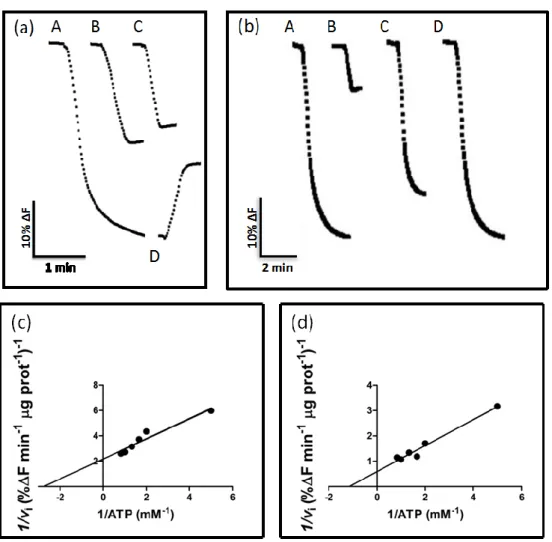
In order to assess the functionality of the vacuoles isolated, the V-H+-ATPase activity of this fraction was monitored. To that purpose, vacuoles were incubated with the pH-sensitive probe ACMA and fluorescence quenching was monitored by spectrofluorimetry. There was a fluorescence decrease in energized vacuolar suspensions indicative of V-H+-ATPase activity. Addition of CaCl2 led to dissipation of
the proton gradient and confirmed that the intra-vacuolar acidification was a consequence of the proton-pumping activity of V-H+-ATPase (Fig. 1a). This shows that the isolation protocol used in this study resulted in the purification of functional vacuoles. When the V-H+-ATPase inhibitor concanamycin A was added, only the unspecific effect of ATP addition on fluorescence quenching was detected, indicating that the V-H+-ATPase was the main proton pump responsible for the formation of the proton gradient (Fig. 1a). A similar result was obtained when H+ pumping activity was measured in vacuolar vesicles obtained from intact vacuoles (Fig. 1b). Moreover, in this fraction the mitochondrial F-ATPase inhibitor azide (NaN3; 100 M) decreased the activity only approximately 29%, and the plasma membrane P-ATPase inhibitor vanadate (Na3VO4; 100 M) did not affect proton pumping, suggesting the sample is not substantially contaminated with vesicles from the plasma membrane or from the internal mitochondrial membrane (Fig. 1b). This indicates that our samples have a purity degree suitable for single particle fluorescence analysis by flow cytometry.
The initial velocities of proton-pumping by V-H+-ATPase measured in intact vacuoles followed a Lineweaver-Burk kinetics, with an apparent Km of 0.6588 mM ATP
and a Vmax of 0.5908 ΔF min-1 µg-1 protein (Fig. 1c). A similar Km value (Km, 0.6448
mM ATP; Vmax, 1.457 ΔF min-1 µg-1 protein) was estimated when the V- H+-ATPase
36 Figure 1
Figure 1 –V-H+-ATPase activity in intact vacuoles (a, c) and vacuolar membrane vesicles (b, d) purified from yeast. (a) Typical fluorescence signal of the initial velocities of proton pumping by V-H+-ATPase in vacuolar suspension after adding 1 mM of ATP (A), and consequence of adding 1mM ATP to the fluorescence intensity of a sample without vacuoles (B), inhibition of proton pump activity by concanamycin A in intact vacuoles (C), dissipation of the proton gradient by 1 mM CaCl2 (D). (b) Fluorescence signal of the initial velocities of proton pumping by V-H+ -ATPase in vacuolar membrane vesicles after addition of 0.5 mM ATP (A) and, 0.05 M cancanamycin A (B), 100
M azide (C) or 100 M vanadate (D). Corresponding Lineweaver-Burk plots with the initial velocities of proton pumping by V-H+-ATPase in vacuolar suspensions (c) and vacuolar membrane vesicles (d).
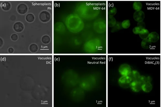
37 Characterization of yeast vacuoles by fluorescence microscopy
Isolated vacuole suspensions were also characterized by phase contrast and fluorescence microscopy, before and after staining with specific fluorescent probes. In spheroplasts, large intracellular vacuoles were observed by phase contrast microscopy (Fig. 2a), and the vacuolar membrane and intra-vacuolar membrane structures could be detected by staining with 10 µM of the structural marker MDY-64 (Fig. 2b). Though this structural marker has been used to visualize the vacuolar membrane in whole cells, it also stained isolated vacuoles (Fig. 2c). Both vacuoles and intra-vacuolar membrane structures could also be visualized by differential interference contrast (Fig. 2d). These intra-vacuolar structures were not present in all vacuoles, indicating they can present different degrees of complexity.
To ascertain whether the isolated vacuoles maintained their acidic pH and electrochemical potential, neutral red and DiBAC4(3) were used, respectively. Neutral
red accumulates in acidic compartments and can be visualized by bright field and fluorescent microscopy. DiBAC4(3) is a membrane potential indicator and accumulates
on positively charged membranes. Purified vacuoles exhibited intense fluorescence with both probes, indicating that their membrane integrity and function was preserved (Fig. 2e, f).
38 Figure 2
Figure 2 - Microscopy analysis of spheroplasts and vacuoles isolated from yeast. Spheroplasts were observed by phase contrast microscopy (a) and fluorescence microscopy after staining with the structural dye MDY-64 (b). Fluorescence microscopy analysis of vacuoles stained with MDY-64 (c), Neutral Red (e) and DibaC4(3) (f). Isolated vacuoles were also observed by differential interferential contrast (d).
Structural and functional analysis of intact vacuoles and vacuolar membrane vesicles by flow cytometry
Since isolated vacuoles exhibited positive staining with both structural and functional probes, flow cytometry was explored to further characterize and quantitatively analyze populations of isolated yeast vacuoles and vacuolar membrane vesicles. Biparametric histograms (FS log vs SS log) of mixtures of samples clearly revealed differences of complexity and size between cells, vacuoles, and vacuolar membrane vesicles (Fig. 3a). Cells appeared more complex and larger than vacuoles and vacuolar membrane vesicles. The latter appeared to be similar in size to the smallest vacuoles but with lower complexity. Vacuoles were the more heterogeneous population, but purification by the Silicone Oil centrifugation technique resulted in a more homogeneous population (Fig. 3b).
39 The positive staining of vacuoles with the structural vacuolar membrane probe MDY-64 observed by fluorescence microscopy was quantified by flow cytometry. The stained vacuolar population exhibited a green positive staining completely discriminated from the autofluorescence (Fig. 3c). Identical staining was obtained with the non-specific membrane fluorescent dye FM1-43 (Fig. 3d). These results indicate that flow cytometry allows for structural characterization of vacuolar suspensions stained with these two probes. However, this staining does not allow assessing whether the isolation procedure used compromises vacuoles integrity. Therefore, vacuoles from a strain expressing Pep4p-EGFP were isolated and analyzed by flow cytometry. This vacuolar suspension exhibited higher level of green fluorescence than vacuoles isolated from the wild type strain (Fig. 3e), confirming they retained Pep4p-EGFP and thus that vacuolar structure integrity was preserved during the purification procedure.
Flow cytometry also revealed useful to monitor the contamination of the vacuole suspension with other sub-cellular fractions. As shown in (Fig. 3f), the vacuole sample was not substantially contaminated with intact mitochondria or mitochondrial vesicles, since the fluorescence intensity after staining with NAO, which stains the mitochondrial membrane-specific lipid cardiolipin, was approximately ten times lower than that of isolated mitochondrial suspensions. These results are in accordance with those reported above showing only residual activity of the F-ATPase in the vacuole samples.
40 Figure 3
Figure 3 - Flow cytometry analysis of mixed samples of intact cells, intact vacuoles and vacuolar membranes vesicles. Scattergrams with distinguishable populations of cells, vacuoles, and vesicles (a) and of purified vacuoles obtained by the Silicon Oil method (b). Overlay of green fluorescence histograms of vacuole autofluorescence and vacuoles stained with the structural dyes MDY-64 (c) and FM1-43 (d). Overlay of green fluorescence histograms of vacuoles isolated from a control yeast strain and a strain expressing Pep4p-EGFP (e). Overlay of red fluorescence histograms of vacuolar membrane vesicles (A), vacuolar suspensions (B) and yeast mitochondria (C) stained with NAO (f).
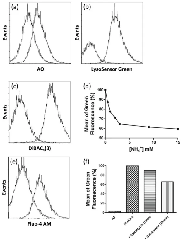
Incubation of vacuole samples from wild-type cells with several functional dyes resulted in a clear positive staining (Fig. 4). AO is a weak base that accumulates in a pH-dependent manner in acidic cellular compartments by an ion-trap mechanism (Cools and Janssen, 1986) and has been used to monitor vacuolar pH (Cohen et al., 1999). LysoSensor Green is another probe that becomes more fluorescent in acidic environments. Vacuoles were highly stained with AO and LysoSensor Green, confirming the acidic nature of the vacuole lumen (Fig. 4a, b). The same results were obtained by fluorescence microscopy using neutral red (Fig. 2e).
Flow cytometric analysis of DiBAC4(3)-stained vacuoles confirmed the presence
of an electric potential across the vacuole membrane (Fig. 4c), also detected by fluorescence microscopy (Fig. 2f). Addition of NH4Cl, which dissipates the electrical
41 potential, led to a concentration-dependent decrease in fluorescence intensity of DiBAC4(3) stained vacuoles, as assessed by flow cytometry (Fig. 4d). This suggests that
changes in fluorescence intensity reflect changes in vacuolar membrane potential, and validates DiBAC4(3) staining to monitor electrical potential of the isolated organelles.
Vacuoles are important calcium reservoirs in the cell. The calcium-sensitive probe Fluo-4 AM was therefore used to monitor vacuolar calcium content in isolated vacuoles. Results showed that isolated vacuoles were highly stained with Fluo-4 AM, indicating that they store high amounts of calcium (Fig. 4e). In agreement, incubation with the calcium ionophore calcymycin caused a 30% reduction in fluorescence intensity 25 min after treatment (Fig. 4f), and the addition of calcymycin 30 min prior to Fluo-4 AM prevented positive staining (not shown).
42 Figure 4
Figure 4 - Histograms of a vacuole-gated population stained with functional dyes. Staining with AO (a). Staining with LysoSensor Green (b). Staining with DiBAC4(3) (c). Mean green fluorescence intensity measured by flow cytometry of non-treated vacuoles, or treated with increasing amounts of NH4Cl (d). Staining with Fluo4-AM, (e). Mean green fluorescence intensity measured by flow cytometry of vacuole population non-stained and stained with Fluo4-AM non-treated or treated with 0.3 mM calcymicin (f).
43
DISCUSSION
In order to understand the function and structure of yeast vacuoles, several studies have been performed over the last decades using intact cells, purified vacuoles or vacuolar membrane vesicles (Reviewed by Li and Kane, 2009). In particular, purification of yeast vacuoles has been a valuable tool to characterize the V-H+-ATPase (Arata et al., 2002), determine the vacuole proteome (Wiederhold et al., 2009), study the vacuolar fusion and fission mechanisms, and characterize its involvement in different cell processes (Wickner, 2002). Like lysosomes, yeast vacuoles have recently been implicated in programmed cell death (Sousa et al., 2011), and thus the use of isolated vacuoles may also contribute to unveil their role in this process. Until now, the study of these organelles relied on biochemical, spectrofluorimetric, spectrophotometric, light and fluorescence microscopy techniques associated with the use of several fluorescent probes. However, these techniques only allow determining mean values and disregard the possible heterogeneity of the samples. Results from standard biochemical techniques used to functionally characterize these fractions are also often unreliable, since the contribution of soluble contaminants, particularly enzymes, is impossible to eliminate.
In the present study, we took advantage of the wide variety of fluorescent probes now available to monitor distinct structural and functional vacuolar features to perform a quantitative and statistically robust analysis of the structure and function of isolated yeast vacuoles under different experimental conditions by flow cytometry. The main advantage of this technique is the possibility of performing single particle fluorescence analysis to assay functional features of intact vacuolar membrane vesicles and vacuolar suspensions without interference from soluble contaminants.
The protocol used in this study for the isolation of yeast vacuoles resulted in highly purified vacuolar samples, in line with previous studies based on a similar procedure (Wiederhold et al., 2009). Inhibition studies with concanamycin A, NaN3 and Na3VO4 suggested that the vacuole membrane V-H+-ATPase is the main proton
pump operating in the sample. This is in accordance with flow cytometry and fluorescence microscopy studies showing that a purified fraction of intact vacuoles was obtained. In particular, the introduction of an additional centrifugation step with silicon