DOI: 10.5935/2359-4802.20170063
Mailing Address: Adson Renato Leite
Avenida Nossa Senhora de Copacabana, 1150, Apto 703. Postal Code 22070-012, Copacabana, Rio de Janeiro, RJ – Brazil. E-mail: [email protected]
Risk Correlation between Obstructive Sleep Apnea and Heart Failure in Primary Care
Adson Renato Leite, Erica de Abreu Macedo, Antonio José Lagoeiro Jorge, Maria Luiza Garcia Rosa
Universidade Federal Fluminense, Niterói, Rio de Janeiro, RJ – Brazil
Manuscript received August 25, 2016; revised February 12, 2017; accepted March 08, 2017.
S l e e p A p n e a , O b s t r u c t i v e ; H e a r t F a i l u r e ; polysomnography; Echocardiography.
Keywords
Introduction
Obstructive sleep apnea syndrome (OSAS), a chronic, progressive disease, with high mortality and morbidity, which has been associated with cardiovascular diseases, including heart failure (HF).1
The pathophysiological changes related to OSAS and its contribution to cardiovascular risk are consequences of increased sympathetic activity, increased oxidative stress, pro-inflammatory changes and endothelial dysfunction.2
In addition to the polysomnography, considered the gold standard for OSAS diagnosis, there are scales that do not diagnose the disorder but indicate individuals at risk, among which is the Berlin Questionnaire (BQ). Individuals classified as high risk for the syndrome showed a rate five times higher than the others in the Respiratory Disturbance Index (RDI). BQ showed a sensitivity of 86% and specificity of 77%, compared to that found by polissonografia.3
Doppler echocardiography is a method that can early identify cardiac structural and functional abnormalities in patients at risk for developing HF.4
Because diastolic dysfunction is commonly found in patients with OSAS, the routine assessment in these patients is required. This change is presented as an independent risk predictor, even in the absence of respiratory severity variables.5
Investigating the association of the risk for OSAS, with identification of cardiac abnormalities on echocardiography in patients without heart failure symptoms (stage A and B) can help in understanding the relationship between both syndromes.
Methodology
Observational, cross-sectional, and part of DIGITALIS study,6 involving 633 randomly selected, 45-99 aged
individuals, registered in the Niterói’s Programa Médico de Família program, with data obtained from August 2011 to December 2012. The Berlin Questionnaire was used to classify the high risk for OSAS and cardiac structural and functional abnormalities, using transthoracic echocardiography.
The units and individuals at each unit was selected through computer-generated random sequence, where the weight of each unit was proportional to the number of assisted individuals.
All individuals selected in the study underwent an evaluation in a single day, which consisted of the following: blood and urine collection, consultation and clinical examination, filling out of a questionnaire that included the Berlin Questionnaire, nutritional assessment, 12-lead electrocardiogram (ECG) and tissue Doppler echocardiogram (TDE).
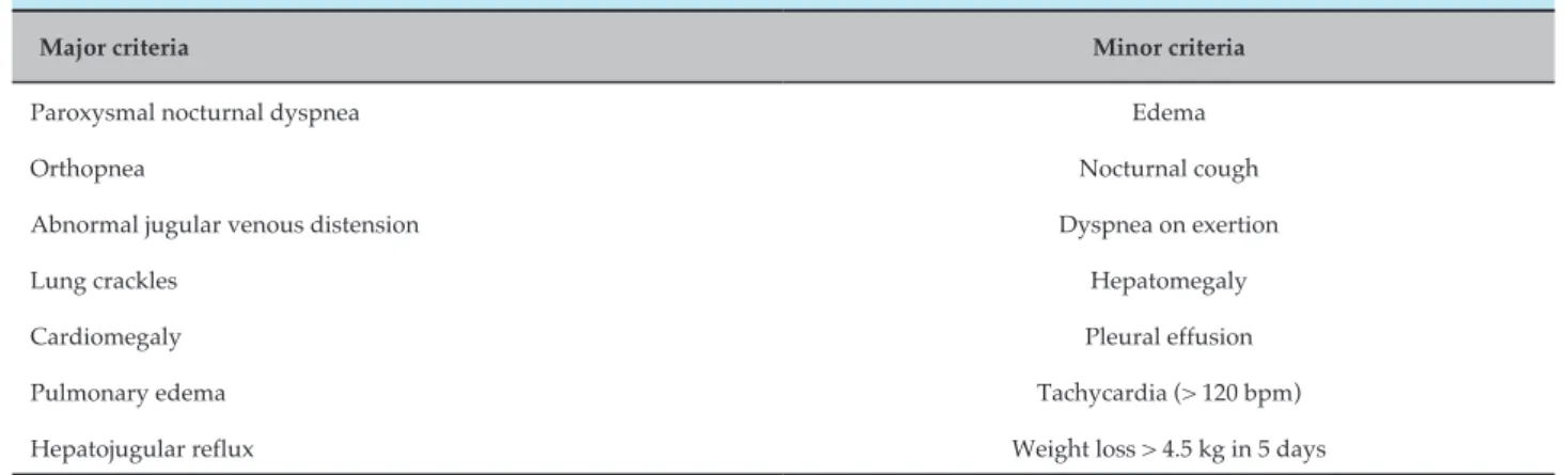
Inclusion criteria were: age from 45 to 99 years and signed Informed Consent Form. Individuals with clinical heart failure (using the major and minor clinical criteria – Table 1) and those impeded to carry out the necessary procedures for the evaluation were excluded.
Two devices were used to perform the echocardiogram and tissue Doppler imaging: Cypress 20 Acuson (Siemens, Mountain View, EUA) and AU-3 Partner (Esaote, Florence, Italy). The examinations were performed by two experienced echocardiographers without prior knowledge of the results of other clinical and laboratory tests. Three repeated measurements were obtained for each parameter and the resulting mean was used in the study. The tests were performed according to the recommendations for the quantification of chambers of the American Society of Echocardiography (ASE) and
Systolic function was assessed by measurement of left ventricular ejection fraction (LVEF) using Simpson's method and the longitudinal axis stretching (S’). Diastolic function parameters were obtained using tissue Doppler (TDE).
Diastolic diameter in women of 3.9-5.3 cm, and man, 4.2-5.9 cm, indexed to 2.4-3.2 cm/cm2 and 2.2-3.1 cm/cm2,
and systolic diameter of 2.1-4.0 cm, indexed to 1.4-2.1 cm/2.8
The left atrial volume (LAV) was obtained using the biplane area-length method, wherein the areas are obtained in the apical positions, excluding the left atrial appendage and the confluence of the pulmonary veins; the perpendicular length is measured between the MV annulus plane and the upper portion of the LA. LAV reference value for both sexes is 28 mL/m2.9 In the study, increase in LAV-I
from 29 mL/m2 was considered. LAV-I increase was not
used to characterize patients with stage B HF.
All valves and their flux patterns were assessed for valvular diseases.
Systolic dysfunction was defined by measuring LVEF using the Simpson's method, and resulting values smaller than 50% were considered abnormal.
Diastolic dysfunction has been defined as the presence of ventricular relaxation abnormalities, assessed by the measurement of the septal E’ wave less than 8 cm/s and/ or the presence of increased filling pressures of the left ventricle, using E/E’ greater than or equal to 15 and the increase in the indexed left atrial volume greater than or equal to 34 mL/m2.10
The TDE measurements for the mitral annulus velocity in early diastole in the septal wall (E’ wave)
reflects left ventricular relaxation and, together with the measurements for transmitral flux in early diastole (E wave), E/E’ ratio, it can be used to predict LV filling pressures. Using the measurements obtained for the septal wall, an E/E’ ratio <8 is usually associated with normal ventricular filling pressures, while an E/E’ > 15 ratio is associated with high filling pressures.10
DD grades were established according to the following criteria:
Grade I DD (mild) - presence of E’ < 8 and/or LAV-I ≥ 34 ml/m2 with E/E’ ratio < 8.
Grade II DD (moderate) - presence of E’ < 8 and/or LAV-I ≥ 34 ml/m2 with E/E’ ratio ≥ 8 and < 15.
Grade III DD (severe) - presence of E/E’ ratio ≥15. Cardiac structural abnormalities used in this study were:
Increasing mass and LV dilation (LVM-i).
The presence of left ventricular hypertrophy was defined on echocardiography by the presence of indexed LV mass > 95 g/m2 in women, and > 115 g/m2 in men.
Left ventricular dilation was defined by the presence of indexed EDV ≥ 97 mL/m2.
Individuals with moderate or severe lesions in mitral, aortic, pulmonary or tricuspid cardiac valves were deemed to have valve disease
Data for LV wall abnormalities were achieved through wall motion-related abnormalities (hypokinesia, dyskinesia or akinesia).
Interest exposure definition: high risk for OSAS. Berlin Questionnaire (BQ) and its scale score.
Table 1 – Clinical criteria for the diagnosis of heart failure - modified Framingham
Major criteria Minor criteria
Paroxysmal nocturnal dyspnea Edema
Orthopnea Nocturnal cough
Abnormal jugular venous distension Dyspnea on exertion
Lung crackles Hepatomegaly
Cardiomegaly Pleural effusion
Pulmonary edema Tachycardia (> 120 bpm)
Hepatojugular reflux Weight loss > 4.5 kg in 5 days
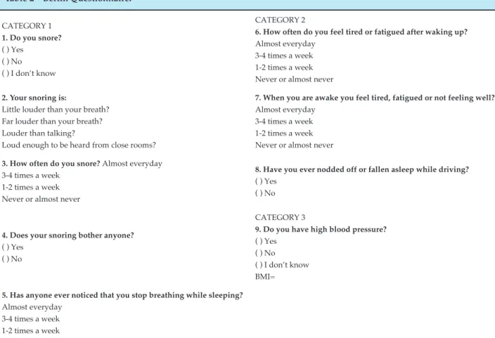
Table 2 – Berlin Questionnaire.12
CATEGORY 1
1. Do you snore?
( ) Yes ( ) No ( ) I don’t know
CATEGORY 2
6. How often do you feel tired or fatigued after waking up?
Almost everyday 3-4 times a week 1-2 times a week Never or almost never
2. Your snoring is:
Little louder than your breath? Far louder than your breath? Louder than talking?
Loud enough to be heard from close rooms?
7. When you are awake you feel tired, fatigued or not feeling well?
Almost everyday 3-4 times a week 1-2 times a week Never or almost never
3. How often do you snore? Almost everyday 3-4 times a week
1-2 times a week Never or almost never
8. Have you ever nodded off or fallen asleep while driving?
( ) Yes ( ) No
4. Does your snoring bother anyone?
( ) Yes ( ) No
CATEGORY 3
9. Do you have high blood pressure?
( ) Yes ( ) No ( ) I don’t know BMI=
5. Has anyone ever noticed that you stop breathing while sleeping?
Almost everyday 3-4 times a week 1-2 times a week Never or almost never
Question scoring: any ticked response is considered positive. Category scoring: Category 1 is positive with 2 or more positive responses to questions 1-5; Category 2 is positive with 2 or more positive responses to questions 6-8; Category 3 is positive if the response to question 9 is positive or BMI > 30. Results: 2 or more positive categories indicate high risk for OSAS.
The BQ includes 10 items organized into 3 categories related to snoring and witnessed apnea (5 items), daytime sleepiness (4 items) with a sub-question of drowsiness at driving (episodical naps while driving a motor vehicle) and hypertension (SAH)/obesity (1 item). Information on genre, age, height, weight, neck circumference and ethnicity is also required (Table 2). The determination of high or low risk for OSAS is based on responses to each category of items.
In category 1, high risk was defined as persistent symptoms (> 3-4 times/week) in two or more questions about snoring.
In category 2, high risk was defined as persistent symptoms (> 3-4 times/week), when with excessive daytime sleepiness, drowsiness while driving a motor vehicle or both.
In category 3, the high risk for sleep apnea was defined as the presence of high blood pressure history or body mass index over 30 kg/m2.
For a patient to be considered as high risk for sleep apnea, she had to score for at least two classes of symptoms. Those who have denied persistent symptoms or who scored for only one category were placed in the low risk group for sleep apnea.
Item 9 (Do you have high blood pressure?) is analyzed separately, as the response already predicts whether there is or not a risk.3,11
Statistical analysis
1. Gottlieb DJ, Yenokyan G, Newman AB. Prospective study of obstructive sleep apnea and incident coronary heart disease and heart failure: the Sleep Heart Health Study. Circulation. 2010;122(4):352-60.
2. Lévy P, Ryan S, Oldenburg O, Parati G. Sleep apnoea and the heart. Eur Respir Rev. 2013;22(129):333-52.
3. Netzer NC, Stoohs RA, Netzer CM, Clark K, Strohl KP. Using the Berlin Questionnaire to identify patients at risk for the sleep apnea syndrome. Ann Intern Med. 1999;131(7):485-91.
4. Carerj S, Carrubba S, Antonini-Canterin F, Di Salvo G, Frlicher A, Linguori E, et al.The incremental prognostic value of echocardiography in asymptomatic stage A heart failure. J Am Soc Echocardiogr. 2010;23(10):1025-34.
5. Bodez D, Lang S, Meuleman , A, Boyer-Chatenet L, Nguyen XL, Soulat-Dufour L, et al. Left ventricular diastolic dysfunction in obstructive sleep apnoea syndrome by an echocardiographic standardized approach: An observational study. Arch Cardiovasc Dis. 2015;108(10):480-90.
6. Jorge AJ, Rosa ML, Fernandes LC, Freire MD, Rodrigues RC, Correia DM, et al. Estudo da prevalência de insuficiência cardíaca em indivíduos
cadastrados no Programa Médico de Família - Niterói. Estudo DIgItalIs: desenho e método. Rev Bras Cardiol. 2011;24(5):320-5.
7. Lang MR, Bierig M, Devereux RB, Flachskampf FA,Guillebert TC, Marino PN, et al. Recommendations for chamber quantification. Eur J Echocardiogr; 2006;7:79-108.
8. Otto CM. Fundamentos de ecocardiografia clínica. 4ªed. Tradutor Laianza AC. Rio de Janeiro (RJ): Elsevier; 2010.p.61-62.
9. Sousa AC. Volume atrial esquerdo como indice de função diastólica. . Arq Bras Cardiol. 2006;87(3):e27-33.
10. Nagueh SF, Appleton CP, Gillebert TC, Marino PN, Oh JK, Smiseth AO, et al.Recommendations for the evaluation of left ventricular diastolic function by echocardiography. J Am Soc Echocardiogr. 2009;22(2):107-33.
11. Netzer NC, Hoegel JJ, Loube D, Netzer CM, Hay B, Alvarez-Sala R, et al.Prevalence of symptoms and risk of sleep apnea in primary care. Sleep in Primary Care International Study Group. Chest. 2003;124(4):1406-14.
12. Vaz AP, Drummond M, Mota PC, Severo M, Almeida J, Winck JC. Translation of Berlin Questionnaire to Portuguese language and its application in OSA identification in a sleep disordered breathing clinic. Rev Port Pneumol. 2011;7(2):59-65.
References
dysfunction, diastolic dysfunction, mass increase and LV wall thickness. Groups will be compared using the Pearson chi-square test for categorical variables and the t-Student test for continuous variables with normal distribution and the Mann Whitney test for the others. When comparing continuous variables for more than two groups, ANOVA and Kruskal-Wallis test will be used. Only variables showing, on univariate analysis, statistical significance up to 0.05 will remain in the multiple model, estimated by logistic regression. A p-value ≤ 0.05 was considered a statistical significance indicator.
Conclusion
The study of the association of obstructive sleep apnea syndrome (OSAS) and the emergence of cardiac structural and functional abnormalities may contribute to the discussion about the adoption of one more criteria for the selection of individuals at risk of developing HF in primary care.
Potential conflicts of interest
No relevant conflicts of interest.
Sources of funding
The study was funded by the researchers, the Research Development Program (FOPESq/UFF) and research fellowships granted by CNPq.
Laboratory tests were donated by Sergio Franco Laboratories and Abbott Laboratory (BNP).
Echocardiography and electrocardiography tests were conducted by researchers of the DIGITALIS Study.
Academic association
This manuscript is part of the Master’s dissertation of Adson Renato Leite from Universidade Federal Fluminense.
Ethical considerations
This study will be conducted in accordance with the principles set out in the Declaration of Helsinki and revised in 2000 (Scotland 2000).