Fátima Alexandra Meira Machado
November 2013
Development of a Liposomal Formulation
for Peptide Delivery to Serve as Vaccine
against Chronic Myeloid Leukemia
UMinho|20 13 Fátima Ale xandr a Meir a Mac hado De velopment of a Liposomal F ormulation for P ep tide Deliver y to Ser ve as V accine agains t Chronic My eloid Leuk
Escola de Ciências
Supervised by:
Prof. Doutora Maria Elisabete C. D. Real Oliveira
Prof. Doutora Andreia C. Gomes
Prof. Doutor Steffen Peterson
Fátima Alexandra Meira Machado
Mss Thesis
Master in Biophysics of Bionanosystems
Development of a Liposomal Formulation
for Peptide Delivery to Serve as Vaccine
against Chronic Myeloid Leukemia
Nome: Fátima Alexandra Meira Machado
Endereço electrónico: [email protected]
Número do Bilhete de Identidade: 13441552
Título dissertação:
- Development of a liposome formulation for peptide delivery to serve as vaccine against chronic myeloid leukemia
Orientador(es): Professora Doutora Elisabete Oliveira, Professora Doutora Andreia Gomes
Ano de conclusão: 2013
Designação do Mestrado: Mestrado em Biofísica e Bionanossistemas
É AUTORIZADA A REPRODUÇÃO INTEGRAL DESTA DISSERTAÇÃO APENAS PARA EFEITOS DE INVESTIGAÇÃO, MEDIANTE DECLARAÇÃO ESCRITA DO INTERESSADO, QUE A TAL SE COMPROMETE;
Universidade do Minho, ___/___/______
ACKNOWLEDGEMENTS
Agradeço a todos que de uma forma ou de outra colaboraram para a realização deste trabalho, em especial:
À Professora Doutora Maria Elisabete Real Oliveira e à Professora Doutora Andreia Gomes, pela orientação ao longo deste trabalho. Agradeço a constante disponibilidade, o incomensurável apoio e dedicação, a partilha de conhecimento e o estímulo transmitido durante a elaboração desta dissertação. Estou muito grata por me proporcionarem esta valiosa oportunidade de aprendizagem.
To Doctor Steffen Petersen and Doctor Teresa Petersen for sharing their knowledge. This was crucial for the elaboration of my thesis. Thank you for your interest and willingness to guide this work.
Ao João Neves, Artur Ribeiro e Raul Machado pela importante colaboração, pelas valiosas sugestões e muitos conhecimentos transmitidos durante a realização deste trabalho que permitiram ultrapassar muitos obstáculos. O seu apoio e boa vontade foram admiráveis. Um obrigado especial ao João Neves pelas imagens fornecidas para esta tese.
Aos meus colegas de laboratório pelo apoio constante, pelo seu espírito hospitaleiro, pela força e pela disponibilidade e grande paciência em ajudar no laboratório.
Aos colegas do Laboratório de Biologia Molecular, em especial ao André Costa, pela paciência e longos dias de apoio no laboratório, muito além do que o dever imporia.
Aos meus amigos e colegas que conheci neste longo percurso académico, Cátia Costa, Tiago Barbosa, Hugo Gonçalves e Juan Arroyo pelo enorme apoio, pelos momentos de descontracção e entreajuda e por tornarem este percurso menos custoso.
Ao Rui Pinto, pela motivação, otimismo, elogios, ajuda e alegrias que me deu ao longo destes anos, e acima de tudo pela paciência nos momentos difíceis. Um grande obrigado por tudo. Por último, mas não menos importante, à minha Mãe, ao meu Pai e à minha querida irmã por sempre terem acreditado que era possível chegar até aqui, e por estarem sempre ao meu lado. Por todo o amor, paciência e estrutura que sempre me proporcionaram não só durante este percurso, mas durante toda a minha vida. Não há palavras que possam expressar a minha gratidão.
ABSTRACT
The main goal of this work was to characterize and explore the potential of Dioctadecyldimethylammonium Chloride (DODAC) / Monoolein (MO) liposomes in a 1:2 proportion and identify the formulations that could be used in the development of an immunoprotective protocol for Chronic Myeloid Leukemia (CML).
CML has long been recognized as one of the most responsive leukemic disorder to immunotherapy. CML is potent model for immune therapy in humans because there is a specific gene rearrangement, BCR/ABL, which product, P210bcr/abl, can be the target antigen.
The loading of drugs into particles at the nanometer size range is a recognized technique for the optimization of controlled drug delivery. In its use in vaccines, liposomes have the advantage of being able to maintain antigens present in the organism for long enough to obtain an immune response.
Different methods of preparation and distinct peptide/lipid molar ratios were used to prepare P210bcr/abl / DODAC:MO (1:2) nanoparticles. This thesis describes results for biophysical characterization of the peptide/lipid system, encapsulation efficiency and exposure of THP-1 cells to the nanoparticles.
The lipid content was essential to achieve the desired nanoparticles. The highest lipid concentration showed higher encapsulation, however, a lower lipid content induced a more efficient cell response. The peptide/lipid system was capable of inducing a stronger cell response than the peptide by itself, emphasizing the potential of this system in vaccine development for the treatment of CML.
RESUMO
O objetivo principal deste trabalho foi caracterizar e explorar o potencial dos lipossomas de cloreto de Dioctadecildimetilamónio (DODAC) / Monooleina (MO) numa proporção de 1:2 e identificar as formulações que poderão ser usadas no desenvolvimento de um tratamento imunoprotetor para a Leucemia Mieloide Crónica (LMC).
A LMC é desde há muito tempo conhecida como uma das desordens imunológicas mais responsivas à imunoterapia. A LMC é um poderoso modelo para imunoterapia em humanos devido à existência de um gene específico BCR/ABL, cujo produto, P210bcr/abl, pode ser usado como antigene-alvo.
A incorporação de fármacos em partículas a uma escala nanométrica é uma técnica reconhecida para a optimização da entrega controlada de fármacos. No seu uso em vacinas, os lipossomas possuem a vantagem de ser capazes de manter os antigenes presentes no organismo o tempo suficiente para se obter uma resposta imune.
Diferentes métodos de preparação e várias razões molares de péptido/lipido foram usadas para preparar nanoparticulas de P210bcr/abl / DODAC:MO(1:2). Esta tese descreve os resultados obtidos da caracterização biofísica do sistema péptido/lípido, eficiência de encapsulação e exposição das células THP-1 às nanopartículas.
O conteúdo lipídico foi essencial para obter nanopartículas desejáveis. A concentração mais alta de lípido demonstrou maior eficiência de encapsulação, no entanto, uma concentração de lipído mais baixa mostrou-se mais eficiente em induzir uma resposta por parte das células. O sistema péptido/lipido foi capaz de induzir uma resposta mais forte do que o pétido por si só, enfatizando o seu potencial no desenvolvimento de uma vacina para o tratamento da LMC.
CONTENTS
LIST OF FIGURES ... xi
LIST OF TABLES ... xvii
SYMBOLS AND ABREVIATIONS ... xix
1. CHAPTER 1 BACKGROUND ... 1
1.1. Goals ... 1
1.2. Research motivation and contribution ... 1
1.3. Dissertation organization ... 2
2. CHAPTER 2 STATE OF THE ART ... 5
2.1. Liposomes ... 5
2.1.1. Particle Size ... 6
2.1.2. Liposome preparation ... 7
2.1.3. Liposome-based delivery systems ... 11
2.1.4. DODAC:MO-based liposomes ... 16
2.2. Chronic Myeloid Leukemia ... 19
2.2.1. Vaccination with BCR-ABL ... 20
2.2.2. The b2a2 breakpoint derived peptide ... 21
2.3. Liposomal vaccines ... 26
2.3.1. Role of adjuvants in vaccine development ... 30
3. CHAPTER 3 METHODOLOGIES ... 35
3.1. Materials ... 35
3.2. BCR-ABL peptide analyses ... 35
3.3. Nanoparticles preparation ... 36
3.3.1. Preparation of liposomes for hydration and injection method ... 37
3.3.2. Preparation of nanoparticles by post-insertion and direct-insertion ... 38
3.4. Biophysical Characterization ... 40
3.4.1. Dynamic Light Scattering assays ... 40
3.4.2. Zeta (ζ) Potential assays ... 42
3.4.3. Electrophoresis and Electrophoretic Mobility... 44
3.4.4. Mean diameter and zeta potential measurements ... 45
3.5. Encapsulation Efficiency ... 46
3.6. Delivery of antigenic BCR-ABL junctional peptide ... 47
3.6.2. LPS Extraction assay ... 48
3.6.3. LPS Activation assay ... 48
3.6.4. Peptide and Peptide/DODAC:MO(1:2) Activation assay ... 49
3.6.5. ELISA assay procedure ... 49
3.6.5.1. Experimental work ... 50
4. CHAPTER 4 RESULTS AND DISCUSSION ... 54
4.1. Effect of pH, peptide concentration and sonication ... 55
4.2. Incorporation of BCR_ABL peptide into DODAC:MO(1:2) liposomes ... 64
4.2.1. Citrate-phosphate versus HEPES ... 64
4.2.2. Incubation time ... 66
4.2.3. Effect of the preparation methods on nanoparticles behavior in solution ... 68
4.2.4. Effect of MLV liposomes and LUV liposomes in the final nanoparticle – Method A versus method E. ... 83
4.3. Encapsulation efficiency ... 90
4.4. Delivery of antigenic BCR-ABL junctional peptide ... 98
4.4.1. Optimization of the control LPS Activation ... 98
4.4.2. Peptide Activation assay ... 99
4.4.3. Peptide/DODAC:MO(1:2) Activation assay ... 101
5. CHAPTER 5 CONCLUSIONS AND FUTURE DIRECTION ... 108
5.1. Conclusions ... 108 5.2. Future work ... 110 REFERENCES ... 113 APPENDIX I ... 123 APPENDIX II ... 127 APPENDIX III ... 133
LIST OF FIGURES
Figure 2.1 – Representation of the steric organization of a liposome (up) and lipid bilayer
(bottom) (adapted from (Bitounis, Fanciullino, Iliadis, & Ciccolini, 2012)). ... 5
Figure 2.2 – Liposomes classification based on size and lamellarity (Laouini et al., 2012). ... 7
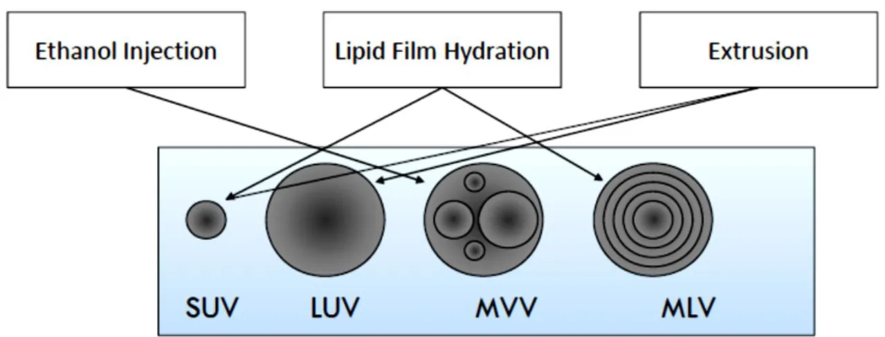
Figure 2.3 – Different types of liposomes produced by ethanol injection, lipid film hydration and extrusion: Small Uni-lamellar Vesicles (SUV); Large Uni-lamellar Vesicles (LUV); Multi-vesicular Vesicles (MVV); Multi-lamellar Vesicles (MLV). ... 8
Figure 2.4 – Representation of MLV preparation by lipid hydration method (adapted from Lopes et al., 2013). ... 9
Figure 2.5 – Representation of the extrusion process (Zhua, Xueb, Guob, & Marchant, 2007). ... 11
Figure 2.6 – Multi-functions of liposomes as nanocarriers. ... 13
Figure 2.7 – Liposome-cell interaction processes. ... 14
Figure 2.8 – Membrane destabilization of liposomes and endosomes. ... 15
Figure 2.9 – Chemical structure of DODAC (Thiele, Merkle, & Walter, 2003). ... 17
Figure 2.10 – Chemical structure of monoolein (Kulkarni, Wachter, Iglesias-Salto, Engelskirchen, & Ahualli, 2011) ... 18
Figure 2.11 – Representation of CMLb2a2-25mer peptide: A – Chemical bonds are denoted for Carbon (grey), Nitrogen (blue), Oxygen (red) and Hydrogen (white); B – molecule’s solvation area. The molecular properties of this peptide were attained by ChemBioOffice 13 program, developed by Cambridge Software. ... 22
Figure 2.12 – Amino acid structure (adapted from Hambly, 2013). ... 22
Figure 2.13 – Representation of the molecular mechanism of peptide-conjugated liposomes on cancer therapy (Wu & Chang, 2010). ... 29
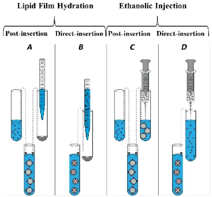
Figure 3.1 – Representation of the different methodologies applied in this work to attach peptide molecules to lipid vesicles: A - Preparation of liposomes by lipid film hydration before adding peptide solution; B - Preparation of liposomes by lipid film hydration with peptide solution being added before lipid vesicles are formed; C - Preparation of liposomes by ethanolic injection before adding peptide solution; D - Preparation of liposomes by ethanolic injection hydration with peptide solution being added before lipid vesicles are formed. ... 38 Figure 3.2 – Schematic representation of methods: A (left), preparation of liposomes by lipid
film hydration (1st) before adding peptide solution (2nd) followed by extrusion (3rd); and
method E (right), preparation of liposomes by lipid film hydration (1st) followed by extrusion
(2nd) before adding peptide solution (3rd). ... 39
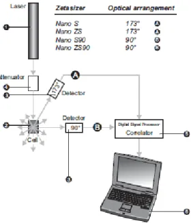
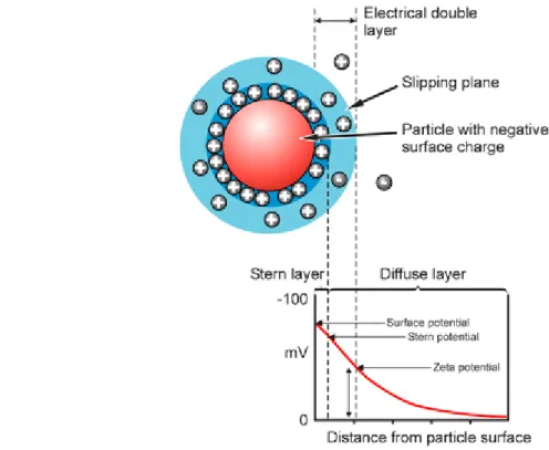
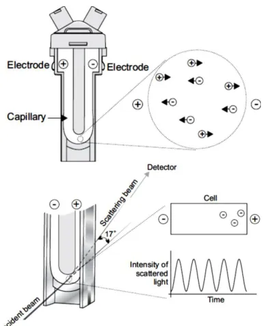
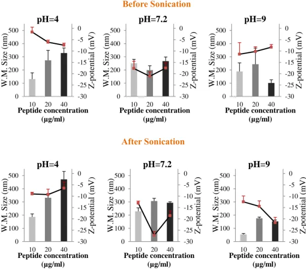
Figure 3.3 – Optical configurations of the Zetasizer Nano series for dynamic light scattering measurements (Malvern, 2005). ... 42 Figure 3.4 – Schematic representation of the double layer surrounding a particle in suspension (Malvern, 2005). ... 43 Figure 3.5 – The technique used to measure this velocity in Malvern’s Zetasizer Nano series(Malvern, 2005). ... 45 Figure 3.6 – The ELISA technique illustrated (“Mabtech,” 2013). ... 50 Figure 4.1 – Results of weighted mean size (nm) (bars - left axis) and z-potential (mV)
(▬■▬ - right axis) for preparations of 10 µg/mL, 20 µg/mL and 40 µg/mL of BCR-ABL
peptide, in citrate-phosphate buffer at pH=4, 7.2 and 9, before and after sonication. ... 56 Figure 4.2 – Results of weighted mean size (nm) (bars - left axis) and z-potential (mV)
(▬■▬ - right axis) for preparations of 10 µg/mL, 20 µg/mL and 40 µg/mL of BCR-ABL
peptide, in HEPES buffer and citrate-phosphate buffer at pH=7.2, before and after sonication. ... 62 Figure 4.3 – Photography of DODAC:MO (1:2) liposomes prepared by ethanolic injection in citrate-phosphate buffer at pH=9. ... 64
Figure 4.4 – At the left side are presented results of weighted mean size (nm) (bars - left axis)
and z-potential (mV) (▬■▬ - right axis) for lipid vesicles (control) and peptide/DODAC:MO
nanoparticles prepared by method A at 1/500 molar ratio. At the right side are presented the distribution of intensity profiles of the respective mean size and z-potential. ... 66 Figure 4.5 – At the left side are presented results of weighted mean size (nm) (bars - left axis)
and z-potential (mV) (▬■▬ - right axis) for lipid vesicles (control) and peptide/DODAC:MO
nanoparticles prepared by method C at 1/500 molar ratio. At the right side are presented the distribution of intensity profiles of the respective mean size and z-potential. ... 67 Figure 4.6 – At the left side are presented results of weighted mean size (nm) (bars - left axis)
and z-potential (mV) (▬■▬ - right axis) for peptide/DODAC:MO nanoparticles prepared by
method A and B at 1/100 molar ratio, after extrusion. At the right side are presented the distribution of intensity profiles of the respective mean size and z-potential. ... 69 Figure 4.7 – At the left side are presented results of weighted mean size (nm) (bars -left axis)
and z-potential (mV) (▬■▬ - right axis) for peptide/DODAC:MO nanoparticles prepared by
method A and B at 1/200 molar ratio, after extrusion. At the right side are presented the distribution of intensity profiles of the respective mean size and z-potential. ... 70 Figure 4.8 – At the left side are presented results of weighted mean size (nm) (bars - left axis)
and z-potential (mV) (▬■▬ - right axis) for peptide/DODAC:MO nanoparticles prepared by
method A and B at 1/300 molar ratio, after extrusion. At the right side are presented the distribution of intensity profiles of the respective mean size and z-potential. ... 71 Figure 4.9 – At the left side are presented results of weighted mean size (nm) (bars - left axis)
and z-potential (mV) (▬■▬ - right axis) for peptide/DODAC:MO nanoparticles prepared by
method A and B at 1/500 molar ratio, after extrusion. At the right side are presented the distribution of intensity profiles of the respective mean size and z-potential. ... 72 Figure 4.10 – At the left side are presented results of weighted mean size (nm) (bars - left
axis) and z-potential (mV) (▬■▬ - right axis) for peptide/DODAC:MO nanoparticles
prepared by method C and D at 1/100 molar ratio, after extrusion. At the right side are presented the distribution of intensity profiles of the respective mean size and z-potential. ... 75
Figure 4.11 – At the left side are presented results of weighted mean size (nm) (bars - left
axis) and z-potential (mV) (▬■▬ - right axis) for peptide/DODAC:MO nanoparticles
prepared by method C and D at 1/200 molar ratio, after extrusion. At the right side are presented the distribution of intensity profiles of the respective mean size and z-potential. .. 76 Figure 4.12 – At the left side are presented results of weighted mean size (nm) (bars - left
axis) and z-potential (mV) (▬■▬ - right axis) for peptide/DODAC:MO nanoparticles
prepared by method C and D at 1/300 molar ratio, after extrusion. At the right side are presented the distribution of intensity profiles of the respective mean size and z-potential. .. 77 Figure 4.13 – At the left side are presented results of weighted mean size (nm) (bars - left
axis) and z-potential (mV) (▬■▬ - right axis) for peptide/DODAC:MO nanoparticles
prepared by method C and D at 1/500 molar ratio, after extrusion. At the right side are presented the distribution of intensity profiles of the respective mean size and z-potential. .. 78 Figure 4.14 – At the left side are presented results of weighted mean size (nm) (bars - left
axis) and z-potential (mV) (▬■▬ - right axis) for nanoparticles prepared by method A and E
at 1/100 (up) and 1/200 (bottom) molar ratio. At the right side are presented the distribution of intensity profiles of the respective mean size and z-potential. ... 84 Figure 4.15 – At the left side are presented results of weighted mean size (nm) (bars - left
axis) and z-potential (mV) (▬■▬ - right axis) for nanoparticles prepared by method A and E
at 1/300 (up) and 1/500 (bottom) molar ratio. At the right side are presented the distribution of intensity profiles of the respective mean size and z-potential. ... 86 Figure 4.16 – Distribution of intensity profiles of the respective mean size (nm) and z-potential (mV) for peptide/DODAC:MO nanoparticles prepared by method E at 1/500 (left) and 1/250 (right) molar ratio. ... 89 Figure 4.17 – Photography of sample tubes after lyophilization, containing: free peptide fraction (1) and peptide encapsulated fraction in which it lipid was not separated from peptide (2) – first experiment; free peptide fraction (3) and peptide encapsulated fraction after separation from lipid (4) – second experiment. ... 92 Figure 4.18 – Gel stained with silver. Samples showed are relative to method E and method C and to peptide/lipid molar ratios of 1/300 and 1/500. ... 93
Figure 4.19 – Gels stained with silver. Samples showed are relative to method E and method C and to peptide/lipid molar ratios of 1/300 and 1/500. ... 94 Figure 4.20 – Gels with comassie blue staining. Samples showed are relative to method E and method C and to peptide/lipid molar ratios of 1/300 and 1/500. ... 96 Figure 4.21 – Quantitative results for TNF-α (pg/mL) produced in response to 4h, 12h and 24h of incubation with four LPS conditions: 0%, 25%, 50% and 100%. ... 98 Figure 4.22 – Quantitative results for TNF-α (pg/mL) produced by THP-1 cells in response to 4h of incubation with increasing peptide concentrations: 0 (cells control), 0 (HEPES control), 5, 10, 20, 50, 80 and 100 µg/mL. ... 100 Figure 4.23 – Quantitative results for TNF-α (pg/mL) produced by THP-1 cells in response to 4h of incubation with DODAC:MO (1:2) vesicles and peptide/DODAC:MO(1:2) nanoparticles. Cells without any treatment, HEPES at 10 mM, LPS at 100% and peptide at 23.8 µg/mL were used as controls. ... 102 Figure 4.24 – THP-1 cells after 4h, without any treatment (40x magnification). ... 103 Figure 4.25 – THP-1 cells after 4h of incubation with a peptide concentration of 100 µg/mL (40x magnification). ... 104
LIST OF TABLES
Table 2.1 – Amino acids properties. ... 23
Table 2.2 – Buried and surface amino acids, and the respective pka and pI, included in the
peptide CMLb2a2 peptide. ... 24 Table 2.3 – Positively and negatively charged amino acids for CMLb2a2 peptide at acidic
(pH=4), neutral (pH=7) and alkaline (pH=9) pH conditions, according to pKa. Amino and
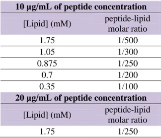
carboxyl groups placed at the two extremes of the peptide are also considered. ... 26 Table 3.1 – Total lipid concentration (mM) peptide/lipid molar ratios, for 10 µg/mL and 20µg/mL of peptide concentration. ... 40
SYMBOLS AND ABREVIATIONS
AP – Accelerated phase
APC – Antigen Presenting Cell BP – Blast phase
CML – Chronic myeloid leukemia CP – Chronic phase
CTL – Cytotoxic T-lymphocyte DC – Dendritic Cell
DLS – Dynamic Light Scattering
DODAC – Dioctadecyldimethylammonium Chloride EPR – Enhanced Permeability and Retention
EPR – Enhanced Permeability and Retention FBS – Fetal Bovine Serum
HEPES – 4-(2-hydroxyethyl)-1-piperazineethanesulfonic acid
HLA – Human Leukocyte Antigen
LAA – Leukemia-associated Antigens LMC – Leucemia Mielóide Crónica LPS – Lipopolysaccharide
LUV – Large Uni-lamellar Vesicles MHC – Major histocompatibility Complex MLV – Multi-lamellar Vesicles
MO – 1-monooleoyl-rac-glycerol (Monoolein) MVV – Multi-vesicular Vesicles
OLV – Oligo-lamellar Vesicles PDI – Polydispersity index Ph – Philadelphia chromosome pI – Isoelectric point
RES – Reticulum-endothelial System SUV – Small Uni-lamellar Vesicles.
Tm – Transition temperature
TNF-α – Tumor Necrosis Factor-alpha
Chapter 1
1. CHAPTER 1
BACKGROUND
Nowadays, knowledge about the properties of nanoparticles and how to handle them is still limited, opening a wide field of work and scientific research in nanotechnology to be explored. New technology related to the treatment of cancer has become a major focus in the world and new systems that could not have been developed before are now being accomplished. Nanobiotechnology permits bringing together, targeting, therapeutic and imaging compounds condensed in single liposome-based delivery systems. Liposomes provide several benefits, and therefore, are ideal candidates for controlled drug release in the affected region.
1.1. Goals
The use of liposomes as carriers of biomolecules has been widely reported in the international literature as an important step in the production of vaccines or drug-delivery systems. The main goal of this work is to characterize and explore the potential of the system DODAC/MO (1:2) for peptide delivery, enhancing the imunopotentiating action of a CML specific peptide. This increased antigen immunization will activate tumor-specific T cells and consequently increase the therapeutic action of the molecule.
For this purpose distinct approaches have been used throughout this work in order to fulfill three main stages: (i) inclusion of junctional peptide p210 from CML-specific oncoprotein BCR-ABL into liposomes (ii) encapsulation efficiency (iii) delivery of antigenic BCR-ABL junctional peptide to cells in vitro.
1.2. Research motivation and contribution
With the new nanotechnology instruments developed in the latest years and understanding the pathology involved, it is absolutely possible to develop an innovative strategy that could be extrapolated to other treatments. Indeed, considering the possibility of building combined systems with nano-sized particles and bioactive molecules, conditions are gathered to develop new vaccines with important therapeutic action. Furthermore, the interaction between
DODAC and MO is still poorly investigated, in spite of the potential application of such DODAC/MO mixture, which has also motivated the present investigation.
Moreover, the possibility of benefiting from the interaction between the Centers of Physics and Environmental Biology from University of Minho, as well as with the BioPhotonics Group from the International Iberian Nanotechnology Laboratory (INL), makes this project a gratifying multidisciplinary experience.
1.3. Dissertation organization
This dissertation encompasses all the stages of the experimental work, beginning with the finding of the most suitable formulation of lipid/peptide in order to test its capability on triggering immunological responses. The work has been organized into five main chapters. Chapter 1 describes the main goals of this work. It also explains the research motivation and contribution as well as the dissertation organization.
Chapter 2 provides a general overview on key factors related to physicochemical and biological parameters of liposome-based delivery systems for drug delivery and vaccine development. Detailed information about liposomes preparation is also presented. Moreover, principles and current status concerning treatment options for CML are discussed.
Chapter 3 presents a detailed description of the methodology used to create an innovative system to treat CML patients.
Chapter 4 presents data analysis that characterizes the peptide incorporation into the nano-delivery system. Furthermore, data analysis regarding the system’s ability to induce an immune response is also discussed.
In Chapter 5 conclusions and future directions are presented. Bibliography referenced in the text is listed at the end of the work.
Chapter 2
State of the art
"There are known knowns. There are things we know we know. We also know there are known unknowns. That is to say we know there are some things we do not know. There are also unknown unknowns, the ones we don't know we don't know." Donald Rumsfeld (2002)
2. CHAPTER 2
STATE OF THE ART
2.1. Liposomes
Liposomes are nano-sized artificial vesicles of spherical shape in which an aqueous volume is entirely enclosed by a membrane composed of lipid molecules, usually phospholipids (Vemuri & Rhodes, 1995). These structural units are amphipathic molecules that have a polar or hydrophilic head group (has affinity for water molecules) and a nonpolar hydrophobic tail consisting of fatty acid chains (that repeals water molecules), as shown in Figure 2.1. Phospholipids are characterized by its solubility in organic solvents and low solubility in water.
Figure 2.1 – Representation of the steric organization of a liposome (up) and lipid bilayer (bottom) (adapted from (Bitounis, Fanciullino, Iliadis, & Ciccolini, 2012)).
Liposomes were first introduced in the 60’s when Alec Bangham observed that phospholipids in aqueous solutions could form closed bilayer structures (Bangham, Standish, & Watkins, 1965). In fact, when phospholipids are combined with water they immediately form a bi-layered sphere, a process commonly referred as “self-assembly” by which a disordered system forms an organized structure due to the local interactions between the system units (monomers). Thus, they can be prepared so that they entrap materials both within their aqueous compartment and/or within the membrane.
A model referred to as “fluidic mosaic” introduced in 1972 by Singer and Nicholson, proposes that biological membranes are composed of lipids, proteins and carbohydrates (Singer & Nicolson, 1972). Their biological structure is very similar to that of normal human cellular membranes. Therefore, liposomes are good study models for biological membranes. The properties and structure of the lipid bilayer can be affected by the gel or fluid state (Ryhänen, 2006). With increasing temperature, lipid vesicles constituted by one type of phospholipid goes through a transition from a gel state into a fluid “liquid crystalline” state.
Each lipid has its own transition temperature (Tm) point, above which, in liquid crystalline
state, lipid bilayer becomes more fluid and elastic with increasing diffusion of the individual lipid molecules (Ryhänen, 2006). The fluid state of lipids facilitates liposome production and manipulation.
2.1.1. Particle Size
Particle size affects drug release. Smaller particles have larger surface area, therefore, most of the drug associated would be at or near the particle surface, leading to fast drug release. However, smaller particles also have higher risk of aggregation. On the other hand, larger particles have larger cores which allow higher quantity of drug to be encapsulated and slowly diffuse out. The ability to produce nanoparticles of desired size with great precision (narrow size distribution and small variation) is the key factor of producing the nano-suspensions (Silva, Little, Ferreira, & Cavaco-Paulo, 2008).
Liposomes are classified on the basis of different structural parameters and they are produced according to the purpose for which they are more suitable. Figure 2.2 presents different types of liposomes according to size and lamellarity. Multi-lamellar vesicles (MLV) are particles
that are usually up to 0,5 µm. The liposomes containing encapsulated vesicles are called multi-vesicular vesicles (MVV) and their size is up to 1 µm. Uni-lamellar vesicles ranging from 20-100 nm are referred to as SUV, whereas LUV are uni-lamellar vesicles bigger than 100 nm. There are also liposomes of very large size that are called giant liposomes (>1µm) which can be either uni-lamellar or multi-lamellar. OLV are oligo-lamellar vesicles ranging from 100-500 nm.
Figure 2.2 – Liposomes classification based on size and lamellarity (Laouini et al., 2012).
2.1.2. Liposome preparation
Different methods of liposome preparation allow the production of lipid vesicles with distinct structural parameters (Dua et al., 2012), as Figure 2.3 illustrates.
Figure 2.3 – Different types of liposomes produced by ethanol injection, lipid film hydration and extrusion: Small Uni-lamellar Vesicles (SUV); Large Uni-lamellar Vesicles (LUV); Multi-vesicular Vesicles (MVV); Multi-lamellar Vesicles (MLV) (courtesy from João Neves).
Liposomes self-close to form large, multi-lamellar vesicles (MLV) which prevents interaction of water with the hydrocarbon core of the bilayer. Once these particles have formed, reducing the size of the particle requires the use of mechanical treatments such as extrusion through polycarbonate membranes that can transform the MLV suspension into LUV or SUV. Sonication is another alternative to reduce liposome size and produces SUV, and it can be applied to the other methods of preparation.
Methods to prepare liposomes used in this work will be described in the following paragraphs. For further information, there are several reviewed methods (Dua et al., 2012; Laouini et al., 2012; Ulrich, 2002).
A) Multi-lamellar Liposomes (MLV) Lipid hydration method
First the lipids are thoroughly mixed in the organic solvent. After drying the lipid a thin film is formed at the bottom of round bottom flask. A suspension of MLV is readily obtained by hydrating the thin film with aqueous buffer dispersion. For larger volumes, the organic solvent should be removed by rotary evaporation. After adding aqueous buffer, the solution
lipid or above the Tm of the highest melting component in the lipid mixture. The compounds
to be encapsulated are added either to aqueous buffer or to organic solvent containing lipids depending upon their solubility. Spinning the round bottom flask in the warm water bath
maintained at a temperature above the Tm of the lipid suspension allows the lipid to hydrate in
its fluid phase.
This is the most widely used method for the preparation of MLV (Figure 2.4), making it simple to prepare and a variety of substances can be encapsulated in these liposomes. As for a disadvantage of this method, the resulting size distribution and lamellarity of the MLV is very heterogeneous. Still, sophisticated procedures have been developed to produce uniformly sized liposomes (Frézard, 1999; Lasic, 1997; New, 1994).
Alternatively, for small volumes of organic solvent (<1mL), the solvent may be evaporated using a nitrogen gas or argon stream over the mixture in a fume hood. After the removal of organic solvent an aqueous solution is added to hydrate and MLV are formed immediately in this aqueous phase. The content is then emulsified by vigorous vortexing and/or sonication.
Figure 2.4 – Representation of MLV preparation by lipid hydration method (adapted from Lopes et al., 2013).
B) Multi-vesicular Liposomes (MVV) Ethanol injection method
Another approach relies on injecting, drop by drop, the lipid dissolved in the organic solvent
to a vast excess of buffer pre-heated to the Tm of the lipids, under vigorous vortexing. A
heterogeneous mixture of SUV, LUV or MLV is immediately formed. Therefore, one of the drawbacks of the method is that the population is heterogeneous. Furthermore, liposomes are very diluted, it is difficult to remove all ethanol because it forms an azeotrop with water, and various biologically active macromolecules may be inactivated in the presence of even low amounts of ethanol (Batzri & Korn, 1973).
C) Large Uni-lamellar Liposomes (LUV) and Small Uni-lamellar Liposomes (SUV) (i) Sonication method
Disruption of MLV suspensions (produced by lipid film hydration method) using ultra-sonic energy (sonication) typically produces SUV. The most widely used instrumentation for preparation of SUV is a bath sonicator. Sonication of MLV dispersion is accomplished by placing a test tube containing the suspension in a bath sonicator for a certain amount of time. Sonication can be applied to the other methods to increase efficiency in the formation of hydrated lipid vesicles of the smallest size.
Mean size and its distribution are influenced by temperature, sonication time and power, volume, composition and concentration, and sonicator tuning. Thus, it is understandable that it is nearly impossible to reproduce the conditions of sonication, meaning that size variation between samples produced at different times is common. Moreover, due to the high degree of curvature of these membranes, SUV are unstable and have a tendency to undergo aggregation and fusion, forming larger vesicles when stored below their phase transition temperature.
(ii) Extrusion method
This process, showed in Figure 2.5 consists in submitting a suspension of liposomes through a small orifice, repeatedly and sequentially, through polycarbonate membranes filter of well-defined pore-size, under conditions of elevated pressure and temperature above the transition temperature of the lipid. LUV with a diameter near the pore size of the filter used, are
produced. Attempts to extrude below the Tm will be unsuccessful as the rigid membranes
cannot pass through the pores. Interestingly, extruded vesicles have been reported to retain significantly elongated elliptical shapes, which have to be taken in to account when evaluating their size and entrapped volume (Jin, Huster, Gawrisch, & Nossal, 1999). The method has some advantages over sonication method, being simple and rapid, reproducible and involving gentle handling of unstable materials. The resulting vesicles are somewhat larger than sonicated SUV.
Figure 2.5 – Representation of the extrusion process (Zhua, Xueb, Guob, & Marchant, 2007).
2.1.3. Liposome-based delivery systems
Soon after Alec Bangham observed that phospholipids in aqueous solutions could form closed bilayer structures (Bangham, Standish, & Watkins, 1965), the capture of liposomes by macrophages was recognized as the main mechanism by which liposomes potentiate immune responses to entrapped antigens, which was followed by many immunization studies (Alving, 1991; Gregory Gregoriadis, 1990; Kersten & Crommelin, 1995; V. Torchilin, 2003). Liposomes were first proposed as drug delivery system more than 30 years ago (Gregoriadis, 1973).
Since then, they have been extensively investigated for their potential as drug carriers. Advances in our understanding of the behavior of liposomes at the cellular and subcellular level have allowed the construction of bionanodevices for use in the treatment and prevention of a number of diseases.
Allison and Gregoriadis (Allison & Gregoriadis, 1974), conducted a pioneer work that demonstrated the immunoadjuvant properties of liposomes. In the early 80s, the involvement and investment of several companies, parallel to great achievements in liposome technology, led to the design and licensing of liposome formulations for the treatment of certain cancers, and the first liposome-based vaccine for use in humans. Finally, liposomes as adjuvants became an important attraction with the first liposome-based vaccine (against hepatitis A) having been licensed for use in humans (Gregoriadis, 1995).
However, the first results demonstrated that liposomes were physico-chemically and biologically unstable and drug encapsulation was not efficient. Fortunately, great advances in liposome technology allowed researchers to make significant improvements in its stability, as well as in the understanding of its characteristics and how they interact with biological fluids. Several factors have shown direct influence on the behavior of liposomes in a biological environment, such as preparation, vesicle size, composition, rate stability and drug encapsulation (Lasic, 1998). Thus, methods of characterization and controlling these factors became of extreme importance to produce these nanocarriers for drug delivery purposes. Similarities between the lipid bilayer structure and the cell membrane, make liposomes capable of interacting with the cells, allowing the targeting of the drug to reach the specific site, and therefore, with less toxicity than free drugs (McPhail, Tetley, Dufes, & Uchegbu, 2000).
Various peptides are used as highly specific and effective therapeutic agents. However, their use is complicated by their instability and side effects. Several peptide drugs have their therapeutic targets inside cells. Thus, it is important to bring these drugs into target cells without subjecting them to lysosomal degradation.
The use of liposomes as carriers of biomolecules is presented as an important step in the production of vaccines or drug-delivery systems, used for controlled delivery of drugs, markers for diagnosis, among other applications. In its use in vaccines, liposomes have the advantage of being able to maintain antigens (e.g. nucleic material, or small peptides) present
in the organism for long enough to obtain an immune response. Otherwise, these biomolecules easily degrade, or are rapidly removed from the circulatory system through the phagocytic cells of the reticulum-endothelial system (RES) (Takeuchi, Kojima, Yamamoto, & Kawashima, 2000), and there is not enough time to obtain an immune response. The antigenic materials can be retained on the surface of liposomes, or else could be encapsulated or embedded within the membrane.
Technology related to controlled release of drugs represents one of the frontiers of science. Nanocarriers are multifunctional and can contribute significantly to the improvement of human health. Drug delivery systems offer a number of advantages when compared to other conventional dosage forms. Various applications of nanocarriers have shown positive results. Figure 2.6 highlights the major functions of nanocarriers in general and, in particular, advantages of liposome-based delivery systems are summarized, to understand why they are candidates to this study (Mohanraj & Chen, 2006; Rawat, Singh, Saraf, & Saraf, 2006; Solaro, Chiellini, & Battisti, 2010).
Figure 2.6 – Multi-functions of liposomes as nanocarriers (adapted from Mohanraj & Chen, 2006; Rawat, Singh, Saraf, & Saraf, 2006; Solaro, Chiellini, & Battisti, 2010).
Understanding liposome-cell interaction processes may facilitate potentiating the desired effect of a drug. Many liposomes are made of certain components (e.g. pH-sensitive components) so that drug release can occur only in the target site. Figure 2.7 highlights some liposome-cell interaction processes already known (Torchilin, 2005).
Figure 2.7 – Liposome-cell interaction processes (Torchilin, 2005).
Liposomes can be specifically (a) or nonspecifically adsorbed onto the cell surface. Alternatively, they also fuse with the cell membrane (c), and release their content into the cell cytoplasm, or can be destabilized by certain cell membrane components when adsorbed on the surface (d) so that the released drug can enter cell via micropinocytosis. Direct or transfer-protein-mediated exchange of lipid components with the cell membrane is another process that liposomes can undergo (e) or, instead, be subjected to a specific or nonspecific endocytosis (f). In the case of endocytosis, a liposome can be delivered by the endosome into the lysosome (g) or, en route to the lysosome, the liposome can provoke endosome destabilization (h), which results in drug liberation into the cell cytoplasm (Figure 2.7).
Figure 2.8 highlights membrane destabilization of liposomes and endosomes (Torchilin, 2005).
Figure 2.8 – Membrane destabilization of liposomes and endosomes (Torchilin, 2005).
After accumulation in required sites in the body, liposomes containing stimuli sensitive components, such as lipids (a) in the membrane and drug (b) inside, after being subjected to a certain stimulus (such as pH or temperature), liposomes undergo local membrane destabilization, consequently drug efflux occurs from the liposome into surroundings (A). Destabilization of endosomal membrane (B) occurs after being endocytosed by the cell and taken inside the endosome, the liposome containing stimuli (pH)-sensitive components, such as lipids (a) in the membrane and drug (b) inside, can undergo pH-dependent membrane
destabilization and initiate the destabilization (Figure 2.8) of the lysosomal membrane, and
consequently drug efflux occurs into the cell cytoplasm.
Besides being prepared entirely synthetically, liposomes have also the benefit of being biodegradable, nontoxic and can be administrated in several forms. When immunostimulants are incorporated within these tiny particles, the effect of the resulting system will not only be an increase of their immunological action, but also a reduction of their toxic side effects. Liposomes effect is not only to improve drug action. In fact, many drugs and current classes
of therapeutics cannot even cross cell membranes to gain access to their intracellular site of action. Liposomes can easily overcome this obstacle.
Liposomes are versatile structures, in which many characteristics can be manipulated with high level of accuracy. Consequently, their immunoadjuvant properties can be handled as well. To name some structural characteristics, vesicle size, surface charge, the lipid to antigen mass ratio, bilayer fluidity, and the mode of antigen association with lipid vesicles, are all factors that can significantly influence adjuvanticity. Marketed liposomal and lipid-based products, plus a selection of products in clinical development have been recently reviewed (Allen & Cullis, 2013).
2.1.4. DODAC:MO-based liposomes
There is a class of surfactants suitable to form vesicles in aqueous solutions, and DODAC (Figure 2.9) is one example. Dioctadecyldimethylammonium chloride is a synthetic cationic surfactant suitable to form vesicles in aqueous solutions. Surfactant concentration, vesicle preparation method and solvent condition are aspects to take into account. Optically clear dispersions of dioctadecyldimethylammonium chloride are capable of forming LUV by simply warming the aqueous surfactant solution (typically 1mM) to 50 °C, above the gel to
liquid crystalline phase transition temperature, Tm = 48.9 °C (Feitosa, Barreleirob, &
Olofsson, 2000), and gently shaking it. Cryo-transmission electron microscopy micrographs show that DODAX vesicles are unilamellar and polydisperse (Feitosa, Karlsson, & Edwards, 2006). Feitosa et al, also demonstrated that these vesicles are stable for at least 1 month according to the ageing time-dependence of the turbidity and molar absorption coefficient. Figure 2.9 shows the structure of DODAC (Eloi Feitosa & Alves, 2008).
The cationic nature of certain liposomes is an attractive characteristic for drug-delivery and gene delivery (Zuhorn, Engberts, & Hoekstra, 2007). Cationic liposomes remain for a longer time in circulatory system than negative and neutral liposomes, because cationic formulations are able to escape phagocytosis. This ability of cationic liposomes is related to their interaction with blood cells (Aoki, Tottori, Sakurai, Fujib, & Miyajima, 1997). The positive charge of cationic liposomes exhibit high affinity for the negative charge of cell membrane, which facilitates cell uptake (Wiethoff, Smith, Koe, & Middaugh, 2001), and may be used for the release of exogenous genetic material intracellularly (Sharma & Sharma, 1997).
Apparently, cationic microparticles are optimal for uptake into macrophages and dendritic cells (DC) (Thiele, Merkle, & Walter, 2003).
Figure 2.9 – Chemical structure of DODAC (Thiele, Merkle, & Walter, 2003).
Despite promising candidates to effectively enhance immune responses (Christensen et al., 2007; Nakanishi et al., 1999), cationic liposomes may have an immunotoxic effect (Kedmi, Ben-Arie, & Peer, 2010; Lv, Zhang, Wang, Cui, & Yan, 2006), limiting their safety for clinical use. This brings to mind the importance of an ideal liposome/antigen formulation, with characteristics that have to be tuned to reach the most effective and harmless formulation as possible.
Monoolein, 1-monooleoil-rac-glycerol (MO), is a natural amphiphilic neutral single tail unsaturated lipid that assembles in water, as it has the particularity to form two non-lamellar
inverted bicontinuous cubic phases (QIIG and QIID ) even in excess H2O (Ericsson, Larsson, &
Fontell, 1983; Geil et al., 2000). Since the 1960s there has been a steady increase in publications, industrial applications and related patents (Kulkarni, Wachter, Iglesias-Salto, Engelskirchen, & Ahualli, 2011).
From the molecular point of view, despite being a simple molecule, it shows amphiphilic properties as it contains a polar head group and a nonpolar hydrocarbon chain. It is composed of a hydrocarbon chain, which is attached to a glycerol backbone by an ester bond. The remaining two hydroxyl groups of the glycerol moiety confer polar characteristics to this part of the molecule commonly referred as the head group. Thus, they may form hydrogen bonds
with water in aqueous solutions. In contrast, the C18 hydrocarbon chain (usually referred as
the ‘tail’), featuring a cis double bond at the 9, 10 position, is strongly hydrophobic (Kulkarni et al., 2011). Consequently, this allows monoolein molecules to self-assemble into different liquid crystalline structures under varying conditions of temperature and solvent composition (Ganem-Quintanar, Quintanar-Guerrero, & Buri, 2000).
Figure 2.10 shows the chemical structure of monoolein.
Figure 2.10 – Chemical structure of monoolein (Kulkarni, Wachter, Iglesias-Salto, Engelskirchen, & Ahualli, 2011)
Cubic phases are known to play an important role in many cell processes, such as membrane fusion (Luzzati, 1997), protein function (Epand, 1998) and ultra-structural organization (Lipowsky & Sackmann, 2004), and DNA condensation in lipoplexes (Silva, Coutinho, & Oliveira, 2008, 2011).
Therefore, MO has become a preferential model for the study of a broad range of applications. MO is a non-bilayer-forming surfactant that favors vesicle formation. New reported results on this subject indicate that both temperature and MO tendency to form non-bilayer structures largely influence the self-assembly process, affecting the structure of the final aggregates (Oliveira et al., 2012).
Overall, this study may provide further insight on the relationship between delivery efficiency and structural organization of peptide/DODAC:MO complexes. Different formulations of peptide/DODAC:MO will certainly affect the structural organization of the final particle. This will all be extensively studied. The potential of the system DODAC/MO has motivated this investigation. Monoolein is a nontoxic, biodegradable, and biocompatible material classified as GRAS (generally recognized as safe). Its biodegradability is due to the fact that monoolein is subject to lipolysis because of different kinds of esterase activity in different tissues. This remarkable molecule is particularly interesting due to its nature and physicochemical behavior, which makes it an attractive alternative in relation to other conventionally used materials. The most significant advantages of monoolein are probably its solubilizing capability, rheological behavior, and low toxicity. Furthermore, the versatility of monoolein makes possible to include it in very different systems and cubic phase reveals great flexibility since drugs of very different polarity and size may be accommodated within it.
2.2. Chronic Myeloid Leukemia
Chronic myeloid leukemia (CML) is a malignant disorder that originates in a single abnormal hematopoietic stem cell. The anomalous clone originated from this cell expands and infiltrates the medullar parenchyma, slowly but progressively, over the proliferation of normal cells. The disease is associated with a specific cytogenetic abnormality, the Philadelphia chromosome (Ph), resulting from a reciprocal translocation between the long arms of chromosomes 9 and 22 t(9;22)(q34;q11) that leads to the formation of a new leukemia-specific gene, BCR-ABL, generated by the fusion of the c-abl oncogene 1 (ABL1, from chromosome 9) with the breakpoint cluster region gene (BCR, from chromosome 22). In CML, the second or third exon of the BCR gene is usually spliced into the second exon of the ABL gene, creating B2A2 or B3A2 transcripts. Once translated, each B2A2 or B3A2 mRNA generate a 210-kDa BCR-ABL protein. The BCR-ABL fusion protein shows tyrosine kinase activity and is essential and sufficient for leukemia transformation and progression. In fact, the junctional sequences of BCR-ABL are only expressed in leukemia cells (Pinilla-Ibarz et al., 2000). This constitutively up regulated tyrosine kinase activity of the chimeric BCR-ABL1 protein affects several intracellular signaling pathways that promote proliferation and survival of cells and thus contribute to their malignant transformation (Guilhot et al., 2008; Quintás-Cardama & Cortes, 2009; Smahel, 2011). Under normal conditions, the BCR gene expressed on chromosome 22 encodes a protein whose function is related to cell cycle regulation, whereas gene ABL expressed on chromosome 9 encodes a protein tyrosine quinase (Druker et al., 2001). The reciprocal gene resulting from translocation ABL-BCR on chromosome 9q+, though active, plays no role in any kind of disease. The hybrid gene BCR-ABL produces a chimeric protein with elevated tyrosine kinase activity. The disease begins with a chronic phase (CP) that can last for 3 to 5 years, and if untreated, it progresses into accelerated phase (AP) and within a year, blast phase (BP). Survival at this point is less than 1 year.
CML patients were once regarded as incurable, but more recent understanding of the molecular anatomy and pathophysiology of the disease provides important insights into the targeting of treatment to a specific molecular abnormality. CML has been recognized as a potent model for immune therapy in humans because there is a specific gene rearrangement, BCR/ABL, which product, P210bcr/abl can be the target antigen for immune therapy. Peptides spanning the junction between BCR and ABL in P210bcr/abl are specific to CML
cells; they are not present in other normal cells neither in CML patients nor in cells in normal individuals without CML (Guilhot et al., 2008).
Despite the therapeutic advances that made possible the significant increase in the perspective of life in patients with CML, several biological mechanisms that favor the selection of malignant cells over normal cells have been responsible for treatment failure in many cases (Bergantini, Castro, Souza, & Fett-conte, 2005).
Because the preeminent mutation driving CML is BCR-ABL, therapies targeting this gene are the logical choice for disease-specific therapy directed at the BCR-ABL tyrosine kinase. However, even in the best responders residual leukemic cells may persist. Since these therapies fail to eradicate the CML stem cells, much work still has to be done and improvement or development of new and more effective strategies would be useful.
2.2.1. Vaccination with BCR-ABL
The breakpoint in the bcr gene occurs either between bcr exon 2 (b2) and 3 (b3) or between bcr exon 3 (b3) and 4 (b4). Hence, 2 alternative chimeric p210 bcr-abl proteins, comprising either a b3a2 or a b2a2 junction, can result from this fusion gene (Shtivelman, Lifshitz, Gale, Roe, & Canaani, 1986). The cellular processing of the products of these two fusion proteins can originate peptides capable of being presented in the cell surface, and can be recognized by cytotoxic T-lymphocytes (CTL) in the context of human leukocyte antigen (HLA) class I molecules (Falkenburg, Smit, & Willemze, 1997; Melief & Kast, 1995). Thus, despite the intracellular location of p210, those peptides can be recognized by T cells within the cleft of HLA. For this reason, and considering that the junction between the fused BCR and ABL genes produces a novel peptide sequence that is unique to leukemic cells, it is a reasonable target for leukemia specific immunotherapy. A list of BCR-ABL peptides used in vaccine trials in patients with chronic myeloid leukemia has been reviewed (Dao & Scheinberg, 2008). Furthermore, there are three different forms of the Bcr-Abl oncogene p185, p210, and p230 (Melo, 1996), which may represent alternative potential targets for immunotherapy approaches (Volpe et al., 2007).
In Bocchia et al. (2010) documented clinical trial, one patient was treated with a target immune approach receiving a therapeutic vaccine. This vaccine consisted of a 25-mer b2a2
breakpoint derived peptide (CMLb2a2-25) with binding properties for several HLA-DR
molecules and was able to elicit a consistent peptide-specific CD4+ T-cell response. This
study shows, for the first time, that these peptide vaccinations were able to reduce and even eradicate minimal residual disease in a patient with CML.
Hereupon, the present work emerges in an attempt to find an alternative target approach that can be added to CML currently used therapy to eradicate minimal residual disease through immunotherapy. The approach described in this thesis consists of a liposomal peptide vaccine. Because several peptides can be encapsulated in lipid vesicles, one day this strategy may be used as a multitherapeutic therapy. This concept provides a new paradigm for the treatment of CML, and CMLb2a2-25 peptide used in Bocchia et al. (2010) trial will be studied in the present work.
2.2.2. The b2a2 breakpoint derived peptide
A therapeutic vaccine consisted of a 25-mer b2a2 breakpoint derived peptide (CMLb2a2-25) tested in a 63-year old woman with chronic myeloid leukemia was able to elicit a consistent
peptide-specific CD4+ T-cell response (Bocchia, Defina, & Aprile, 2010). This resulted in a
reduction and even eradication of minimal residual disease.
CMLb2a2 peptide has sequence of 25 amino acids (3-letter code),
Thr-Val-His-Ser-Ile-Pro-Leu-Thr-Ile-Asn-Lys-Glu-Glu-Ala-Leu-Gln-Arg-Pro-Val-Ala-Ser-Asp-Phe-Glu-Pro-NH2,
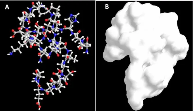
whose properties can be seen in Table 2.1 and Table 2.2. Figure 2.11 shows a representation of the CMLb2a2-25-25mer peptide that is going to be used in the present study.
Figure 2.11 – Representation of CMLb2a2-25mer peptide: A – Chemical bonds are denoted for Carbon (grey), Nitrogen (blue), Oxygen (red) and Hydrogen (white); B – molecule’s solvation area. The molecular properties of this peptide were attained by ChemBioOffice 13 program, developed by Cambridge Software.
The present thesis comprises a study of this peptide in different pH conditions. In order to
support and interpret results, few properties as charge, hydrophobicity, pKa and isoelectric
point (pI) should be elucidated (HubPages, 2013; Publishing, 2013). First we must know the amino acid structure. Figure 2.12 shows a representation of an amino acid.
Figure 2.12 – Amino acid structure (adapted from Hambly, 2013).
The alpha-carbon (center) in an amino acid:
- Is bonded to an amino group
- Is bonded to a carboxyl group
- Is bonded to a hydrogen
Peptide bonds allow amino acids to be linked, consequently forming peptides. This peptide bond consists of a carbonyl group's carbon atom directly bound to the nitrogen atom of a secondary amine. After the peptide formation, the peptide chain will have an unbound amino group free at one end, called the N-terminus, and a single free carboxylate group at the other end, named the C-terminus.
Amino acids can be classified, among other aspects, according to the side chain group (R).
Since amino acids, peptides, and proteins have different pKa values, there is the possibility
that they can have different charges at a given pH.
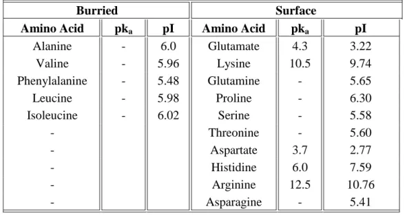
Table 2.1 shows the properties of the amino acids included in CMLb2a2 peptide, and the
respective pka and pI are shown in Table 2.2.
Table 2.1 – Amino acids properties.
Amino Acid 3-letter code Properties Amino Acid 3-letter code Properties
Alanine Ala aliphatic
hydrophobic neutral
Leucine Leu aliphatic
hydrophobic neutral
Arginine Arg polar
hydrophilic charged (+)
Lysine Lys polar
hydrophilic charged (+)
Asparagine Asn polar
hydrophilic neutral Phenylala-nine Phe aromatic hydrophobic neutral
Aspartate Asp polar
hydrophilic charged (-)
Proline Pro hydrophobic
neutral
Glutamine Gln polar
hydrophilic neutral
Serine Ser polar
hydrophilic neutral
Glutamate Glu polar
hydrophilic charged (-)
Threonine Thr polar
hydrophilic neutral
Histidine His aromatic
polar hydrophilic charged (+)
Valine Val aliphatic
hydrophobic neutral
Isoleucine Ile aliphatic
hydrophobic neutral
Table 2.2 – Buried and surface amino acids, and the respective pka and pI, included in the
peptide CMLb2a2 peptide.
Burried Surface
Amino Acid pka pI Amino Acid pka pI
Alanine - 6.0 Glutamate 4.3 3.22 Valine - 5.96 Lysine 10.5 9.74 Phenylalanine - 5.48 Glutamine - 5.65 Leucine - 5.98 Proline - 6.30 Isoleucine - 6.02 Serine - 5.58 - Threonine - 5.60 - Aspartate 3.7 2.77 - Histidine 6.0 7.59 - Arginine 12.5 10.76 - Asparagine - 5.41
An atom group in a molecule may lose or gain a proton when the molecule is placed in an aqueous solution. The exact probability that a molecule will be protonated or deprotonated
depends on the pKa of the molecule and the pH of the solution. Half of molecules will lose
protons if they are in a solution with pH = pKa. The higher the pH value, the more likely a
molecule will lose a proton (Publishing, 2013). Furthermore, from the definition of pH, a high
concentration of HO- ions is present at higher pH, thereby being capable of accepting more
protons which results in the neutralization of positive charges.
As for the isoelectric point, when the pH is lowered far below the pI, the protein will lose its negative charge and will contain only positive charges, but if the pH is adjusted to the isoelectric point of the protein, its net charge will be zero. Most proteins at physiological pH are above their pI, thereby having an overall negative charge. This information, along with table 2, suggests that at neutral pH, BCR-ABL peptide has a net negative charge.
By observing table 2 it can be noted that pKa is not shown for certain amino acids. The pKa
essentially refers to the tendency for H+ to dissociate from a molecule, and thus is a measure
of the extent of H+ dissociation under equilibrium conditions. So, if a molecule's hydrogen has
no tendency to dissociate, then it will not have a pKa. The R groups of such amino acids have
no dissociable hydrogens - these groups are neither acids nor bases. It should be noted that we
the backbone of amino and carboxyl groups. The carboxyl group has a pKa ranging from 1.8
to 2.4 and is most likely negatively charged at neutral pH, whereas the amino group has a pKa
ranging from 8 to 11 and is most likely positively charged.
From Table 2.2 we can also infer that the peptide charged amino acids are characterized as being surface amino acids. Therefore, all these charges will be considered when predicting and analyzing the surface charge of the BCR-ABL peptide at different pH conditions.
From Table 2.1 and Table 2.2 we can observe that four negatively charged (Glu (3x) and Asp) and three positively charged (Lys, Arg and His) amino acids are included in this peptide. Thus, it is expected that this peptide will exhibit a negative overall charge which will be attracted for the positively charged membranes of liposomes (Friede, Vanregenmortel, & Schuber, 1993; Gregory Gregoriadis, 2007a). Additionally, the amphipathic character of this peptide may increase its affinity for biological membranes (Bessalle et al., 1993; Wimmer et al., 2006).
CMLb2a2 peptide contains two amino acids with beta strands, Val and Ile, and two amino acids with alpha helix, Ala and Leu. The special conformation of these amino acids may play an important role in the interaction with lipid membranes and affect peptide incorporation into liposomes. However, the interaction with liposomes can stabilize the alpha helix structure (Bessalle et al., 1993; Wimmer et al., 2006).
This peptide has a preference for secondary structures and contains 48% of hydrophobic amino acids, 16%, 12% and 24% of acidic, basic and neutral amino acids, respectively.
Peptide molecular formula is C124H200N34O39, has a molecular weight of 2790,47 g/mol and a
molecular volume of 3377,307 Å3.
From the information above we can predict the amino acids charge and, consequently, the overall surface charge of BCR-ABL peptide at acidic, neutral and alkaline pH (Table 2.3). It should be noted that histidine (His) is very sensitive to pH change in the physiological range.
The R group of histidine (pKa = 6.0) has only 10% probability to become positively charged
at pH = 7, but the probability increases to 50% at pH = 6. Therefore, histidine will not be considered as positively charged at neutral and alkaline pH.
Table 2.3 – Positively and negatively charged amino acids for CMLb2a2 peptide at acidic
(pH=4), neutral (pH=7) and alkaline (pH=9) pH conditions, according to pKa. Amino and
carboxyl groups placed at the two extremes of the peptide are also considered.
pH condition 4 7 9
Charge Negative Positive Negative Positive Negative Positive
Amino Acids
- Lys Glu Lys Glu Lys
- Arg Glu Arg Glu Arg
- His Glu - Glu -
Asp - Asp - Asp -
Carboxyl/Amino
groups carboxyl - carboxyl Amino carboxyl -
Total 2 3 5 3 5 2
Considering the information from table 3, and considering that at acid pH the excess of H+
ions is capable of neutralizing peptide negative charges and that at alkaline pH the excess of
HO- is capable of neutralizing peptide positive charges, we can expect that the peptide
negative surface charge will be stronger at pH 7 and 9, than at pH 4.
Amino acids properties described above are of major importance since factors affecting peptide–liposome membrane interactions include effects of peptide length, charge, hydrophobicity, secondary structure, and topology (Strömstedt, Ringstad, Schmidtchen, & Malmsten, 2010).
CMLb2a2 peptide incorporation into liposomes will be analyzed using specific techniques described in chapter 3.
2.3. Liposomal vaccines
Extensive information about the use of liposomes as immunological adjuvants for protein and peptide antigens is available since the early 90s (G. Gregoriadis, Florence, & Patel, 1993; Philippot & Schuber, 1995). When peptide antigens are encapsulated in lipid vesicles, the liposome-peptide complexes formed are phagocytosed by macrophages. Eventually, liposome-peptide systems accumulate in lysosomes, where the encapsulated peptides are presented to the MHC class II complex. At this stage, the system is capable of stimulating