Síndrome antifosfolipídica
Antiphospholipid syndrome
Ricard Cervera, Gerard Espinosa
Resumo
A síndrome antifosfolipídica (SAF) carateriza-se pelo desen-volvimento de tromboses venosas e/ou arteriais, muitas vezes múltiplas, e pela morbidade gestacional (por perdas fetais re-correntes), na presença de anticorpos antifosfolipídicos. As estimativas indicam que a incidência da SAF é de cerca de 5 novos casos por 100 000 pessoas por ano e a prevalência é de cerca de 40-50 casos por 100 000 pessoas. Os aPL são positivos em aproximadamente 13% dos pacientes com acidente vascular cerebral, 11% com enfarte do miocárdio, 9,5% dos pacientes com trombose venosa profunda e 6% dos pacientes com morbidade gestacional.
Atualmente, há consenso no tratamento de pacientes com SAF com trombose com anticoagulação oral de longa du-ração e para evitar manifestações obstétricas com o uso de aspirina e heparina. Esta revisão resume os principais conhecimentos sobre os aspectos clínicos e terapêuticos desta síndrome.
Palavras-chave: Acidente Vascular Cerebral; Anticorpos Anti-cardiolipina; Anticorpos Antifosfolipídeos; Síndrome Antifosfo-lipídica
Department of Autoimmune Diseases, Hospital Clínic, Barcelona, Catalonia, Spain
http://revista.spmi.pt - DOI: 10.24950/rspmi/revisao/4/2018;
Abstract
The antiphospholipid syndrome (APS) is defined by the de-velopment of venous and/or arterial thromboses, often mul-tiple, and pregnancy morbidity (mainly, recurrent fetal loss-es), in the presence of antiphospholipid antibodies (aPL). Some estimates indicate that the incidence of the APS is around 5 new cases per 100 000 persons per year and the prevalence around 40-50 cases per 100 000 persons. The aPL are positive in approximately 13% of patients with stroke, 11% with myocardial infarction, 9.5% of patients with deep vein thrombosis and 6% of patients with pregnancy morbidity.
Currently, there is consensus in treating APS patients with thrombosis with long-term oral anticoagulation and to pre-vent obstetric manifestations with the use of aspirin and heparin. This review summarizes the main knowledge on the clinical and therapeutic aspects of this syndrome. Keywords: Antibodies, Anticardiolipin; Antibodies, Antiphos-pholipid; Antiphospholipid Syndrome; Stroke
Introduction
The antiphospholipid syndrome (APS) is characterized by the development of venous and/or arterial thromboses, of-ten multiple, and pregnancy morbidity (mainly, recurrent fe-tal losses), in the presence of antiphospholipid antibodies (aPL), namely lupus anticoagulant (LA), anticardiolipin
anti-bodies (aCL), or anti-β2glycoprotein-I (β2GPI) antibodies.1-4
The APS can be found in patients having neither clini-cal nor laboratory evidence of another definable condition (primary APS) or it may be associated with other diseases, mainly systemic lupus erythematosus (SLE), but
occasion-ally with other autoimmune conditions,1 infections,2 drugs,1
and malignancies.3 A minority of APS patients can develop
a devastating variant termed catastrophic APS.5
Epidemiology
The aPL can appear in different scenarios, such as asymp-tomatic aPL carrier patients, thrombotic APS with recurrent venous and/or arterial thrombosis, obstetric APS affecting otherwise healthy women with recurrent pregnancy loss, patients with non-APS classification criteria manifestations (i.e, thrombocytopenia, hemolytic anemia or livedo
reticula-ris) or with catastrophic APS.5
Prevalence of the aPL in the general population ranges be-tween 1% - 5%. However, only a minout together all the pub-lished case reports as well as the new diagnosed cases from all over the world, an international registry of patients with catastrophic APS ("CAPS Registry") was crerity of these individuals develop the APS. Some estimates indicate that the incidence of the APS is around 5 new cases per 100 000 persons per year and the prevalence around 40-50 cases
per 100 000 persons.6 Specifically, aPL are positive in
ap-proximately 13% of patients with stroke, 11% with myocar-dial infarction (MI), 9.5% of patients with deep vein
throm-bosis (DVT) and 6% of patients with pregnancy morbidity.7
The prevalence of the catastrophic APS is scarce (less
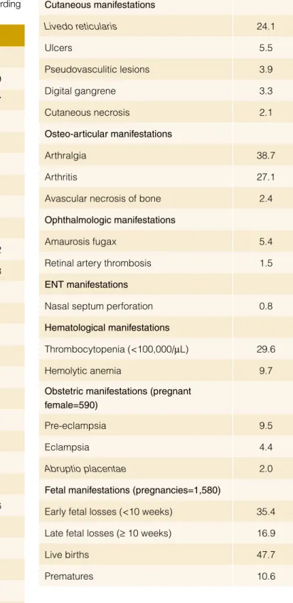
Table 1: Most common manifestations in the APS, according
to the “Euro-Phospholipid Project”.12
Manifestations %
Peripheral thrombosis
Deep vein thrombosis 38.9
Superficial thrombophlebitis in legs 11.7
Arterial thrombosis in legs 4.3
Venous thrombosis in arms 3.4
Arterial thrombosis in arms 2.7
Subclavian vein thrombosis 1.8
Jugular vein thrombosis 0.9
Neurologic manifestations
Migraine 20.2
Stroke 19.8
Transient ischemic attack 11.1
Epilepsy 7.0 Multiinfarct dementia 2.5 Chorea 1.3 Acute encephalopathy 1.1 Pulmonary manifestations Pulmonary embolism 14.1 Pulmonary hypertension 2.2 Pulmonary microthrombosis 1.5 Cardiac manifestations Valve thickening/dysfunction 11.6 Myocardial infarction 5.5 Angina 2.7 Myocardiopathy 2.9 Vegetations 2.7
Coronary by-pass rethrombosis 1.1
Intraabdominal manifestations
Renal manifestations (glomerular thrombosis, renal infarction, renal artery thrombosis, renal
vein thrombosis) 2.7
Gastrointestinal manifestations (esophageal
or mesenteric ischemia) 1.5
Splenic infaction 1.1
the European Forum on Antiphospholipid Antibodies. Currently, it documents the entire clinical, laboratory and therapeutic data of
more than 500 patients.5
Pathogenesis
Autoantibodies associated with APS are directed against a number of proteins of the plasma or expressed on, or bound to, the surface of vascular endothelial cells or platelets. The involvement of aPL in clinically important normal procoagulant
Cutaneous manifestations Livedo reticularis 24.1 Ulcers 5.5 Pseudovasculitic lesions 3.9 Digital gangrene 3.3 Cutaneous necrosis 2.1 Osteo-articular manifestations Arthralgia 38.7 Arthritis 27.1
Avascular necrosis of bone 2.4
Ophthalmologic manifestations
Amaurosis fugax 5.4
Retinal artery thrombosis 1.5
ENT manifestations
Nasal septum perforation 0.8
Hematological manifestations
Thrombocytopenia (<100,000/µL) 29.6
Hemolytic anemia 9.7
Obstetric manifestations (pregnant female=590)
Pre-eclampsia 9.5
Eclampsia 4.4
Abruptio placentae 2.0
Fetal manifestations (pregnancies=1,580)
Early fetal losses (<10 weeks) 35.4
Late fetal losses (≥ 10 weeks) 16.9
Live births 47.7
and anticoagulant reactions and on certain cells altering the expression and secretion of various molecules are the basis for possible mechanisms by which aPL may develop thrombotic events in patients with APS. In depth reviews of these
mecha-nisms can be found elsewhere.10,11
Clinical Manifestations
The clinical picture of the APS is characterized by venous and arterial thromboses, pregnancy morbidity (mainly, fetal losses) and moderate thrombocytopenia. Single vessel involvement or multiple vascular occlusions may give rise to a wide variety of presentations. Any combination of vascular occlusive events may occur in the same individual and the time interval between them also varies considerably from weeks to months or even
years.12-14 The prevalence of the main manifestations in a cohort
of 1000 patients with APS (“Euro-Phospholipid Project”) are
col-lected in Table 1.12
Several attempts have been made in order to identify the
in-dividual risk of thrombosis in aPL positive patients.15,16 A study
of pregnant women with APS reported that patients with triple
aPL positivity (ie, positivity for LA, aCL, and anti-β2GPI) and/or
previous thromboembolism had an increased likelihood of poor neonatal outcomes than patients with double or single aPL
pos-itivity and no thrombosis history.15 More recently, a global APS
score (GAPSS) was developed in a cohort of 211 SLE from a single centre after the combination of several independent risk factors for both thrombosis and pregnancy loss. The final score includes 6 factors with different weights: IgG/IgM aCL (5 points),
IgG/IgM anti-β2GPI antibodies (4 points), LA (4 points), IgG/
IgM anti-phosphatidylserine–prothrombin complex antibodies (3
Table 2: Revised classification criteria for the APS.18
Clinical criteria
1. Vascular thrombosis1
One or more clinical episodes of arterial, venous, or small vessel thrombosis, in any tissue or organ. Thrombosis must be confirmed by imaging or Doppler studies or histopathology, with the exception of superficial venous thrombosis. For histopathologic confirmation, thrombosis should be present without significant evidence of inflammation in the vessel wall
2. Pregnancy morbidity
(a) One or more unexplained deaths of a morphologically normal fetus at or beyond the 10th week of gestation, with normal fetal morphology documented by ultrasound or by direct examination of the fetus, or
(b) One or more premature births of a morphologically normal neonate before the 34th week of gestation because of: (a) eclampsia
or severe preeclampsia defined according to standard definitions, or (b) recognised features of placental insufficiency2, or
(c) Three or more unexplained consecutive spontaneous abortions before the 10th week of gestation, with maternal anatomic
or hormonal abnormalities and paternal and maternal chromosomal causes excluded.
In studies of populations of patients who have more than one type of pregnancy morbidity, investigators are strongly encouraged to stratify groups of subjects according to a, b, or c above.
Laboratory criteria3
1. Anticardiolipin antibody of IgG and/or IgMisotype in serum or plasma, present in medium or high titer (i.e. >40 GPL or MPL, or >the 99th percentile, or >mean + 3SD of 40 healthy controls), on 2 or more occasions, at least 12 weeks apart, measured by
a standardized enzyme-linked immunosorbent assay.
2. Lupus anticoagulant present in plasma, on 2 or more occasions at least 12 weeks apart, detected according to the guidelines of the International Society on Thrombosis and Hemostasis (Scientific Subcommittee on Lupus Anticoagulants/Phospholipid-Dependent Antibodies). 3. Anti-β2 glycoprotein-I antibody of IgG and/or IgMisotype in serum or plasma, present on 2 or more occasions, at least 12 weeks apart,
measured by a standardized enzyme-linked immunosorbent assay, according to recommended procedures.
o Definite APS is present if at least one of the clinical criteria and one3 of the laboratory criteria are met, with the first measurement
of the laboratory test performed at least 12 weeks from the clinical manifestation.4
1Coexisting inherited or acquired factors for thrombosis are not reason for excluding patients from APS trials. However, two subgroups of APS patients should be
recognized, according to: (a) the presence, and (b) the absence of additional risk factors for thrombosis. Indicative (but not exhaustive) such cases include: age (>55 in men, and >65 in women), and the presence of any of the established risk factors for cardiovascular disease (hypertension, diabetes mellitus, elevated LDL or low HDL cholesterol, cigarette smoking, family history of premature cardiovascular disease, body mass index ≥ 30 kg/m2, microalbuminuria,
estimated GFR <60 mL/min), inherited thrombophilias, oral contraceptives, nephrotic syndrome, malignancy, immobilization, surgery. Thus, patients who fulfill criteria should be stratified according to contributing causes of thrombosis.
2Generally accepted features of placental insufficiency include: (1) abnormal or non-reassuring fetal surveillance test(s), e.g., a non-reactive non-stress test,
suggestive of fetal hypoxemia, (2) abnormal Doppler flow velocimetry waveform analysis suggestive of fetal hypoxemia, e.g., absent end-diastolic flow in the umbilical artery, (3) oligohydramnios, e.g., an amniotic fluid index of 5 cm or less, or (4) a post natal birth weight less than the 10th percentile for the gestational
age.
3Investigators are strongly advised to classify APS patients in studies into one of the following categories:
I: More than one Laboratory criteria present (any combination) IIa: Anti-cardiolipin antibody present alone
IIb: Lupus anticoagulant present alone IIc: Anti-β2 glycoprotein-I antibody present alone
points), hyperlipidemia (3 points) and arterial hypertension (1 point). A GAPSS cut-off value higher than 10 points
ap-pears to have the best prognostic yield.16
Classification Criteria
A preliminary classification set of criteria was established after an
expert workshop held in Sapporo, Japan, in 1999.17 These
clas-sification criteria were updated in another workshop was held in Sydney, Australia, in 2006 in which the experts proposed the
in-clusion of anti-β2GPI antibodies. Although no new clinical criteria
were added, some particular features were remarked on, such as associated APS features, including cardiac valve involve-ment, livedo reticularis, thrombocytopenia, APS nephropathy, and non-thrombotic central nervous system manifestations (i.e.
cognitive dysfunction)18 (Table 2).
This revised APS classification criteria19 provide a more
uni-form basis for selecting patients for APS research by emphasis-ing risk stratification. They strongly recommend investigatemphasis-ing co-existing inherited and acquired thrombosis risk factors in patients
with APS, especially in those who are included in clinical trials. An assessment of these 2006 revised APS classification criteria has shown that only 59% of the patients meeting the 1999 APS
Sapporo classification criteria met the revised criteria.19
The preliminary classification criteria for catastrophic APS were formulated at a workshop in Taormina, Italy, in 2002, during the
10th International Congress on aPL (Table 3).20 A validation study
showed that they have a sensitivity of 90.3%, a specificity of 99.4%, a positive predictive value of 99.4% and a negative
pre-dictive value of 91.1%.21 A recent evidence-based set of
guide-lines for the diagnosis and management of catastrophic APS has formulated the recommendation that these classification criteria
can be also used as diagnostic criteria.22
Treatment
Treatment of thrombosis in APS patients is based on long-term oral anticoagulation and treatment of obstetric manifestations on
the use of aspirin and heparin.23 These recommendations are
based on randomized controlled trials and observational studies
Table 3: Preliminary criteria for the classification of catastrophic APS. Clinical criteria
1. Evidence of involvement of three or more organs, systems and/or tissues* 2. Development of manifestations simultaneously or in less than a week.
3. Confirmation by histopathology of small vessel occlusion in at least one organ or tissue**.
4. Laboratory confirmation of the presence of antiphospholipid antibodies (lupus anticoagulant and/or anticardiolipin antibodies)*** * Usually, clinical evidence of vessel occlusions, confirmed by imaging techniques when appropriate. Renal involvement is defined by a 50% rise in serum creatinine, severe systemic hypertension (>180/100 mmHg) and/or proteinuria (>500 mg/24 h).
** For histopathological confirmation, significant evidence of thrombosis must be present, although vasculitis may coexist occasionally.
*** If the patient had not been previously diagnosed as having an APS, the laboratory confirmation requires that presence of antiphospholipid antibodies must be detected on two or more occasions at least 6 weeks apart (not necessarily at the time of the event), according to the proposed preliminary criteria for the classification of definite APS.
Definite catastrophic APS: All 4 criteria Probable catastrophic APS:
- All 4 criteria, except for only two organs, systems and/or tissues involvement.
- All 4 criteria, except for the absence of laboratory confirmation at least 6 weeks apart due to the early death of a patient never tested for aPL before the catastrophic APS.
- 1, 2 and 4
- 1, 3 and 4 and the development of a third event in more than a week but less than a month, despite anticoagulation.
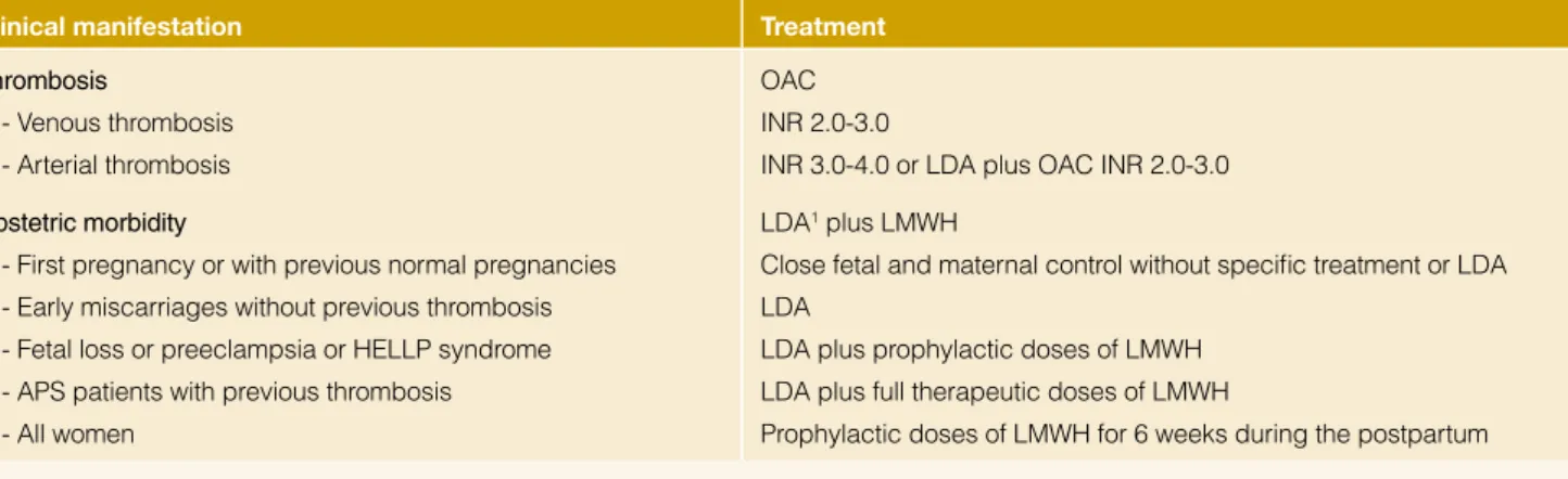
Table 4: Our treatment strategies of patients with definite antiphospholipid syndrome. Clinical manifestation Treatment
Thrombosis
- Venous thrombosis - Arterial thrombosis
OAC INR 2.0-3.0
INR 3.0-4.0 or LDA plus OAC INR 2.0-3.0 Obstetric morbidity
- First pregnancy or with previous normal pregnancies - Early miscarriages without previous thrombosis - Fetal loss or preeclampsia or HELLP syndrome - APS patients with previous thrombosis - All women
LDA1 plus LMWH
Close fetal and maternal control without specific treatment or LDA LDA
LDA plus prophylactic doses of LMWH LDA plus full therapeutic doses of LMWH
Prophylactic doses of LMWH for 6 weeks during the postpartum
1Start aspirin before conception in all cases
Abbreviations: APS: antiphospholipid syndrome; HELLP: hemolysis, elevated liver enzymes, low count platelets; INR: international randomized ratio; LDA: low dose aspirin; LMWH: low-molecular-weight hepari
(Table 4). In detail, patients with definite APS with first venous thrombosis have to be treated with prolonged oral anticoagu-lation at a target international normalized ratio (INR) of 2.0-3.0. Anticoagulation at INR of 3.0-4.0, isolated antiaggregation, anti-coagulation at INR 2.0-3.0 or antianti-coagulation at INR 2.0-3.0 plus antiaggregation have been proposed for definite APS patients with arterial thrombosis. Regarding obstetric APS, although com-bined therapy with low-dose aspirin and low-molecular-weight heparin is the mainstay of treatment in women with obstetric APS,
the strength of evidence of its efficacy is under discussion.24
Otherwise, in the field of APS there are grey areas where the evidence is scarce and where the management of cer-tain patients is difficult. Some examples are patients with
“seronegative” APS, those who do not display formal (clin-ical or laboratory) classification criteria for APS, those with refractory APS despite optimal treatment (recurrent thrombot-ic events despite optimal antthrombot-icoagulation or recurrent fetal losses despite the combination of aspirin and low molecular weight heparin), and the treatment of clinical manifestations not included in the classification criteria such as hematologic manifestations (thrombocytopenia and haemolytic anemia), neurologic manifestations (chorea, myelitis or multiple sclero-sis-like lesions), nephropathy and heart valve disease asso-ciated with antiphospholipid antibodies. In these cases, the recommendations are based on the common sense since the
published evidence is scarce or it does not exist.23
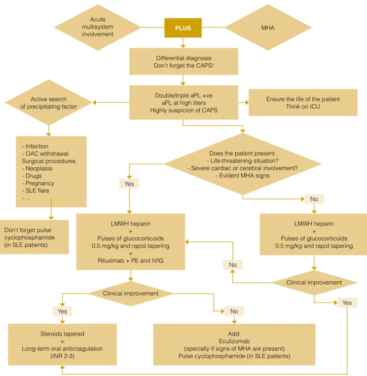
Differential diagnosis Don't forget the CAPS!
Ensure the life of the patient Think on ICU Double/triple aPL +ve
aPL at high titers Highly suspicion of CAPS
LMWH heparin +
Pulses of glucocorticoids 0.5 mg/kg and rapid tapering
+
Rituximab + PE and IVIG
LMWH heparin +
Pulses of glucocorticoids 0.5 mg/kg and rapid tapering
Steroids tapered +
Long-term oral anticoagulation (INR 2-3)
Add: Eculizumab
(specially if signs of MHA are present) Pulse cyclophosphamide (in SLE patients) - Infection - OAC withdrawal Surgical procedures - Neoplasia - Drugs - Pregnancy - SLE flare - ...
Don't forget pulse cyclophosphamide (in SLE patients)
PLUS Yes Yes No No No Yes
Figure 1: Algorithm for the management of catastrophic antiphospholipid syndrome.
Acute multisystem involvement Clinical improvement Clinical improvement Active search of precipitating factor MHA
Does the patient present: - Life-threatening situation? - Severe cardiac or cerebral involvement?
quired (Fig. 1). Therefore, early diagnosis is very important to start adequate therapy and decrease the high mortality rate of these patients. Once the diagnosis is made or suspected, searching and treating the precipitating factor, mainly infection, is the first step of treatment. The specific therapy of catastrophic APS is the combination of anticoagulation with heparin, and corticosteroids as first line of treatment. Additionally adding intravenous immu-noglobulins and/or plasma exchange have to be considered in life-threatening cases. In patients with associated SLE, intra-venous cyclophosphamide has demonstrated be beneficial. In
refractory cases, rituximab or eculizumab should be added.22,25
Outcome and Organ Damage
Given that APS affect predominantly young patients, asses-ment of organ damage is crucial but evidence in that field is limited. A retrospective analysis was published that focused in morbidity, mortality, and organ damage in 135 APS patients (89
primary APS and 46 with secondary APS).26 Patients were
clus-tered according to the initial event: arterial thrombosis, DVT or pregnancy morbidity. One-fourth of the patients progressed to organ damage in a mean time of 10 years from disease onset. The highest morbidity was attributed to neurologic damage, which was more common among patients with arterial throm-bosis as an initial manifestation.
During the follow-up study period of the “Euro-Phospholipid
Project”, a 10-year survival rate of 91% was reported.14 During
this follow-up period, 93 (9.3%) patients died. The main causes of death were thrombosis (36.5%) and infections (26.9%).
Patients with APS still develop significant morbidity and mor-tality despite current treatment (mainly oral anticoagulants and/ or antiaggregant agents); therefore, it is imperative to increase the effort in determining optimal prognostic markers and ther-apeutic measures to prevent these important complications of
the APS. ■
Conflitos de Interesse: Os autores declaram a inexistência de conflitos de interesse na realização do presente trabalho.
Conflicts of interest: The authors have no conflicts of interest to declare. Fontes de Financiamento: Não existiram fontes externas de financiamen-to para a realização deste artigo.
Financing Support: This work has not received any contribution, grant or scholarship.
Correspondence/Correspondência:
Ricard Cervera, MD, PhD, FRCP – [email protected] Hospital Clínic
Villarroel 170
08036-Barcelona, Catalonia, Spain. Received/Recebido: 09/09/2018 Accepted/Aceite: 30/09/2018 REFERENCES
1. Cervera R, Espinosa, Khamashta MA. Antiphospholipid syndrome in syste-mic autoimmune diseases. 2nd ed. Amsterdam: Elsevier; 2016.
2. Cervera R, Asherson RA, Acevedo ML, Gomez-Puerta JA, Espinosa G, De La Red G, et al. Antiphospholipid syndrome associated with infections: cli-nical and microbiological characteristics of 100 patients. Ann Rheum Dis. 2004;63:1312-7.
sals M, et al. Antiphospholipid antibodies associated with malignancies: cli-nical and pathological characteristics of 120 patients. Semin Arthritis Rheum. 2006;35:322-32.
4. Gómez-Puerta JA, Martin H, Amigo MC, Aguirre MA, Camps MT, Cuadrado MJ, et al. Long-term follow-up in 128 patients with primary antiphospholipid syndrome: do they develop lupus? Medicine. 2005;84:225-30.
5. Rodríguez-Pintó I, Moitinho M, Santacreu I, Shoenfeld Y, Erkan D, Espino-sa G, et al. Catastrophic antiphospholipid syndrome (CAPS): Descriptive analysis of 500 patients from the International CAPS Registry.Autoimmun Rev. 2016;15:1120-4. doi: 10.1016/j.autrev.2016.09.010.
6. Durcan L, Petri M. Epidemiology of the Antiphospholipid Syndrome. In: Cer-vera R, Espinosa, Khamashta MA, editors. Antiphospholipid syndrome in systemic autoimmune diseases. 2nd ed. Amsterdam: Elsevier; 2016.p. 17-30.
7. Andreoli L, Chighizola CB, Banzato A, Pons-Estel GJ, Ramire de Jesus G, Erkan D. Estimated frequency of antiphospholipid antibodies in patients with pregnancy morbidity, stroke, myocardial infarction, and deep vein thrombo-sis: a critical review of the literature.Arthritis Care Res. 2013; 65: 1869-73. doi: 10.1002/acr.22066.
8. Cervera R, Espinosa G, Bucciarelli S, Gomez-Puerta JA, Font J. Lessons from the catastrophic antiphospholipid syndrome (CAPS) registry. Autoimmun Rev. 2006;6:81-4.
9. Cervera R, Bucciarelli S, Plasin MA, Gomez-Puerta JA, Plaza J, Pons-Estel G, et al. Catastrophic antiphospholipid syndrome (CAPS): descriptive analysis of a series of 280 patients from the "CAPS Registry". J Autoimmun. 2009;32:240-5. doi: 10.1016/j.jaut.2009.02.008.
10. Espinosa G, Cervera R. Antiphospholipid syndrome: frequency, main cau-ses and risk factors of mortality. Nat Rev Rheumatol. 2010; 6: 296-300. doi: 10.1038/nrrheum.2010.47.
11. Giannakopoulos B, Krilis SA. The pathogenesis of the antiphospholipid syn-drome. N Engl J Med. 2013;368:1033-44. doi: 10.1056/NEJMra1112830. 12. Cervera R, Piette JC, Font J, Khamashta MA, Shoenfeld Y, Camps MT, et al.
Antiphospholipid syndrome: clinical and immunologic manifestations and pa-tterns of disease expression in a cohort of 1,000 patients. Arthritis Rheumatol. 2002;46:1019-27.
13. Cervera R, Khamashta MA, Shoenfeld Y, Camps MT, Jacobsen S, Kiss E, et al. Morbidity and mortality in the antiphospholipid syndrome during a 5-year period: a multicentre prospective study of 1000 patients. Ann Rheum Dis. 2009;68:1428-32. doi: 10.1136/ard.2008.093179.
14. Cervera R, Serrano R, Pons-Estel GJ, Ceberio-Hualde L, Shoenfeld Y, de Ramón E, et al. Morbidity and mortality in the antiphospholipid syndrome during a 10-year period: a multicentre prospective study of 1000 patients. Ann Rheum Dis. 2015; 74: 1011-8. doi: 10.1136/annrheumdis-2013-204838. 15. Ruffatti A, Calligaro A, Hoxha A, Trevisanuto D, Ruffatti AT, Gervasi MT, et al.
Laboratory and clinical features of pregnant women with antiphospholipid syndrome and neonatal outcome. Arthritis Care Res. 2010;62:302-7. doi: 10.1002/acr.20098.
16. Sciascia S, Sanna G, Murru V, Roccatello D, Khamashta MA, Bertolaccini ML. GAPSS: the Global Anti-Phospholipid Syndrome Score. Rheumatology. 2013;52:1397-403. doi: 10.1093/rheumatology/kes388.
17. Wilson WA, Gharavi AE, Koike T, Lockshin MD, Branch DW, Piette JC, et al. International consensus statement on preliminary classification criteria for de-finite antiphospholipid syndrome: report of an international workshop. Arthritis Rheumatol. 1999;42:1309-11.
18. Miyakis S, Lockshin MD, Atsumi T, Branch DW, Brey RL, Cervera R, et al. In-ternational consensus statement on an update of the classification criteria for definite antiphospholipid syndrome (APS). J Thromb Haemost. 2006;4:295-306.
19. Kaul M, Erkan D, Sammaritano L, Lockshin MD. Assessment of the 2006 revised antiphospholipid syndrome classification criteria. Ann Rheum Dis. 2007;66:927-30.
20. Asherson RA, Cervera R, de Groot PG, Erkan D, Boffa MC, Piette JC, et al. Catastrophic antiphospholipid syndrome: International consensus statement on classification criteria and treatment guidelines. Lupus. 2003;12:530-4. 21. Cervera R, Font J, Gomez-Puerta JA, Espinosa G, Cucho M, Bucciarelli S,
et al. Validation of the preliminary criteria for the classification of catastrophic antiphospholipid syndrome. Ann Rheum Dis. 2005;64:1205-9.
22. Legault K, Schunemann H, Hillis C, Yeung C, Akl EA, Carrier M, et al. McMas-ter RARE-Bestpractices clinical practice guideline on diagnosis and mana-gement of the catastrophic antiphospholipid syndrome. J Thromb Haemost. 2018 (in press). doi: 10.1111/jth.14192.
23. Espinosa G, Cervera R. Current treatment of antiphospholipid syndrome: lights and shadows. Nat Rev Rheumatol. 2015;11:586-96. doi: 10.1038/nr-rheum.2015.88.
24. Chighizola CB, Andreoli L, Gerosa M, Tincani A, Ruffatti A, Meroni PL. The treatment of anti-phospholipid syndrome: A comprehensive clinical approa-ch. J Autoimmun. 2018;90:1-27. doi: 10.1016/j.jaut.2018.02.003.
25. Cervera R, Rodríguez-Pintó I, Espinosa G. The diagnosis and clinical ma-nagement of the catastrophic antiphospholipid syndrome: A comprehensive review. J Autoimmun. 2018;92:1-11. doi: 10.1016/j.jaut.2018.05.007. 26. Grika EP, Ziakas PD, Zintzaras E, Moutsopoulos HM, Vlachoyiannopoulos PG.
Morbidity, mortality, and organ damage in patients with antiphospholipid syn-drome. J Rheumatol. 2012;39:516-23. doi: 10.3899/jrheum.110800.