Letters to the Editor
Radiol Bras. 2016 Mai/Jun;49(3):199–204
202
http://dx.doi.org/10.1590/0100-3984.2015.0082 extraplexus nerve transfer techniques, the areas of activation
mainly being located in the contralateral cortex(2–8).
This study has some limitations. We presented the patient data only in comparison with those of a single control participant, rather than with a group of control, and both data sets were ac-quired at only one time point. In addition, the patient did not un-dergo a pre-operative fMRI scan.
REFERENCES
1. Oberlin C, Béal D, Leechavengvongs S, et al. Nerve transfer to biceps muscle using a part of ulnar nerve for C5-C6 avulsion of the brachial plexus: anatomical study and report of four cases. J Hand Surg Am. 1994;19:232–7.
2. Malessy MJ, van der Kamp W, Thomeer RT, et al. Cortical excitability of the biceps muscle after intercostal-to-musculocutaneous nerve transfer. Neurosurgery. 1998;42:787–95.
3. Iwase Y, Mashiko T, Ochiai N, et al. Postoperative changes on functional mapping of the motor cortex in patients with brachial plexus injury: com-parative study of magnetoencephalography and functional magnetic reso-nance imaging. J Orthop Sci. 2001;6:397–402.
4. Malessy MJ, Bakker D, Dekker AJ, et al. Functional magnetic resonance
imaging and control over the biceps muscle after intercostal-musculocu-taneous nerve transfer. J Neurosurg. 2003;98:261–8.
5. Beaulieu JY, Blustajn J, Teboul F, et al. Cerebral plasticity in crossed C7 grafts of the brachial plexus: an fMRI study. Microsurgery. 2006; 26:303–10.
6. Sokki AM, Bhat DI, Devi BI. Cortical reorganization following neuro-tization: a diffusion tensor imaging and functional magnetic resonance imaging study. Neurosurgery. 2012;70:1305–11.
7. Hua XY, Liu B, Qiu YQ, et al. Long-term ongoing cortical remodeling after contralateral C-7 nerve transfer. J Neurosurg. 2013;118:725–9. 8. Dimou S, Biggs M, Tonkin M, et al. Motor cortex neuroplasticity
follow-ing brachial plexus transfer. Front Hum Neurosci. 2013;7:500.
Ana Caroline Siquara de Sousa1, José Fernando Guedes-Corrêa1
1. Hospital Universitário Gaffrée e Guinle – Universidade Federal do Estado do Rio de Janeiro (Unirio), Rio de Janeiro, RJ, Brazil. Mailing address: Dr. José Fernando Guedes-Corrêa. Hospital Universitário Gaffrée e Guinle – Departamento de Neurocirurgia. Rua Mariz e Barros, 775, Tijuca. Rio de Janeiro, RJ, Brazil, 20270-004. E-mail: neuroguedes@ yahoo.com.br.
Hughes-Stovin syndrome: an unusual cause of pulmonary artery aneurysms
Dear Editor,
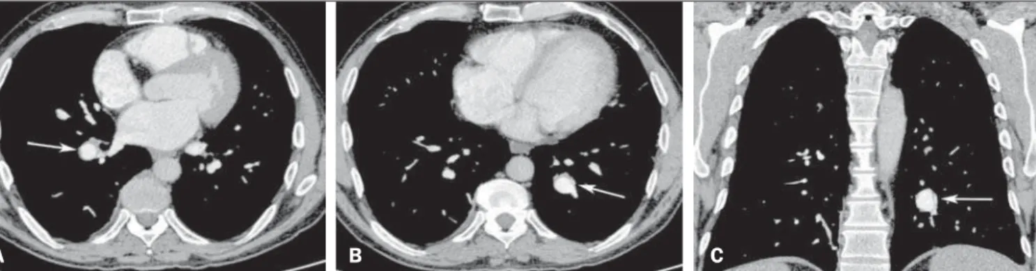
A 43-year-old male presented with a two-month history of persistent cough and fever, associated with recurrent episodes of superficial thrombophlebitis and venous thrombosis of the lower limbs. Physical examination revealed no evidence of oral or geni-tal ulcers. Ancillary tests showed negative blood culture; no thrombophilia or neoplasia; negative serology; mild normocytic, normochromic anemia; elevated C-reactive protein; and elevated erythrocyte sedimentation rate. Contrast-enhanced computed to-mography identified aneurysms in branches of the pulmonary arteries (Figure 1). The final diagnosis was Hughes-Stovin syn-drome.
Idiopathic and vascular diseases of the thorax have been the subject of recent publications in the radiology literature of Bra-zil(1–6). Hughes-Stovin syndrome is a rare condition, characterized by the combination of multiple pulmonary artery aneurysms and peripheral venous thrombosis, that mainly affects males (80–90% of cases) between the second and fourth decades of life(7–11). Al-though the lesions often affect arteries and veins simultaneously
(in 68% of cases), isolated arterial or venous impairments are re-ported at frequencies of 25% and 7%, respectively(9,11).
In its typical presentation, Hughes-Stovin syndrome occurs in three stages(7–9,11): in the first stage, there are signs and symp-toms of thrombophlebitis; the second stage includes the forma-tion and expansion of pulmonary artery aneurysms; and the third stage is characterized by aneurysmal rupture with massive hemop-tysis, progressing to death. The formation of pulmonary aneurysms has been attributed to the weakening of the vessel walls by an inflammatory process. Other hypotheses proposed to explain these changes include septic embolism and angiodysplasia of the bron-chial arteries(8,9,11). Aneurysms can be single or multiple, unilat-eral or bilatunilat-eral, and can even arise at other sites (in the iliac, femo-ral, popliteal, carotid, or hepatic arteries), although with a lower risk of rupture(9–11).
Some authors consider Hughes-Stovin syndrome an incom-plete form of Behcet’s disease, due to the similarity between the two in terms of the clinical, radiological, and pathological find-ings(7–11). Therefore, Behcet’s disease, which typically affects young males, is the main differential diagnosis(11). The major (and mandatory) criterion for a diagnosis of Behcet’s disease is oral ulcers that recur at least three times within 12 months, which should be Figure 1. Cortical activation during motor tasks in the patient. Only the cortex is represented, and the peak of activation in the coordinates were as follows: A: left hand, x = 38 mm, y = –22 mm, z = 65 mm; B: left elbow, x = 42 mm, y = –26 mm, z = 65 mm; C: right elbow, x = –33 mm, y = –18 mm, z = 70 mm. (R, right hemisphere; L, left hemisphere).
Letters to the Editor
Radiol Bras. 2016 Mai/Jun;49(3):199–204
203
accompanied by at least two of the minor criteria (not necessarily simultaneously), including recurrent genital ulcers, ocular lesions, skin lesions, and a positive pathergy test(12), none of which were observed in our patient. Other causes of pulmonary artery aneu-rysms are trauma, infection, pulmonary hypertension, and Marfan syndrome(8–11).
There is no standard treatment for Hughes-Stovin syndrome, the most widely used treatment option being immunosuppres-sion therapy involving a combination of glucocorticoids and cy-clophosphamide, which has the potential to stabilize aneurysms or even promote regression in some cases(11). The use of antico-agulants is controversial because of the risk of fatal hemoptysis, being allowed only in selected cases and provided jointly adminis-tered with immunosuppression therapy(7–11). Other possible treat-ments include surgical resection and arterial embolization, which are used in most cases in which there is massive hemoptysis(11).
REFERENCES
1. Batista MN, Barreto MM, Cavaguti RF, et al. Pulmonary artery sar-coma mimicking chronic pulmonary thromboembolism. Radiol Bras. 2015;48:333–4.
2. Yamanari MGI, Mansur MCD, Kay FU, et al. Bullet embolism of pul-monary artery: a case report. Radiol Bras. 2014;47:128–30. 3. Pessanha LB, Melo AMF, Braga FS, et al. Acute post-tonsillectomy
negative pressure pulmonary edema. Radiol Bras. 2015;48:197–8. 4. Francisco FAF, Rodrigues RS, Barreto MM, et al. Can chest
high-reso-Bruno Niemeyer de Freitas Ribeiro1, Renato Niemeyer Ribeiro1, Gláucia Zanetti2, Edson Marchiori2
1. Instituto Estadual do Cérebro Paulo Niemeyer, Rio de Janeiro, RJ, Brazil. 2. Universidade Federal do Rio de Janeiro (UFRJ), Rio de Janeiro, RJ, Brazil. Mailing address: Dr. Edson Marchiori. Rua Tho-maz Cameron, 438, Valparaíso. Petrópolis, RJ, Brazil, 25685-120. E-mail: [email protected].
lution computed tomography findings diagnose pulmonary alveolar microlithiasis? Radiol Bras. 2015;48:205–10.
5. Koenigkam Santos M. Diagnosis of pulmonary alveolar microlithiasis [Editorial]. Radiol Bras. 2015;48(5):ix–x.
6. Fernandes GL, Teixeira AA, Antón AGS, et al. Churg-Strauss syndrome: a case report. Radiol Bras. 2014;47:259–61.
7. Cruz VA, Muniz YA, Silva Torres PPT, et al. Síndrome de Hughes-Sto-vin. Rev Bras Reumatol. 2009;49:747–52.
8. Chung MP, Yi CA, Lee HY, et al. Imaging of pulmonary vasculitis. Radiology. 2010;255:322–41.
9. El Aoud S, Frikha F, Snoussi M, et al. Moderate hemoptysis caused by Hughes-Stovin syndrome. Clin Pract. 2014;4:647.
10. Silva R, Escobar A, Vega R, et. al. Síndrome Hughes-Stovin: caso clínico. Rev Med Chile. 2013;141:922–6.
11. Khalid U, Saleem T. Hughes-Stovin syndrome. Orphanet J Rare Dis. 2011;6:15.
12. Belczak SQ, Aun R, Valentim L, et. al. Tratamento endovascular de aneurismas da aorta em pacientes com doença de Behçet: relato de dois casos. J Vasc Bras. 2010;9:89–94.
Figure 1. Contrast-enhanced computed tomography of the chest, with axial slices (A,B) and coronal slices (C), showing aneurysms in branches of the pulmonary arteries (arrows).
A
B
C
http://dx.doi.org/10.1590/0100-3984.2015.0048
Differential diagnosis of anterior sacral meningocele during the evaluation of post-hysterectomy pelvic collections
Dear Editor,
Here, we present the case of a 34-year-old woman who suf-fered postoperative pain and fever after subtotal abdominal hys-terectomy. Conventional radiography of the pelvis showed unilat-eral sacral curvature (Figure 1A). Computerized tomography (CT) performed on the second postoperative day revealed a dense locu-lated collection, interspersed with small air bubbles, in the pelvic cavity and a cyst-like formation with hypodense liquid content in the presacral space, communicating with the spinal canal, dislo-cating the rectum to the right (Figure 1B). A diagnosis of anterior sacral meningocele (ASM) was made, and the surgical team was informed of its coexistence with the postoperative pelvic collections. A new procedure was carried out to drain the collections, care being taken to avoid the sacculation caused by the ASM, which was vis-ible and palpable. Magnetic resonance imaging (MRI) was car-ried out in order to monitor the postoperative drainage and to char-acterize the malformation in greater detail (Figure 1C).
Various conditions related to anomalies in central nervous system development have been reported in Brazil(1–3). ASM is a rare form of spinal dysraphism, in which the meningeal sac herni-ates into the presacral space(4,5). It accounts for approximately 5% of all retrorectal masses and is more prevalent in women(6).
ASM can occur in isolation or in association with other con-genital abnormalities, such as urocon-genital malformations, anorectal malformations, lipoma, teratoma, epidermoid tumor, and dermoid cyst(7,8). Due to its occult nature, it is generally diagnosed in the second or third decades of life. It can be asymptomatic or present with nonspecific symptoms, such as constipation, urological symp-toms, and, in rare cases, neurological symptoms(9). The diagnos-tic investigation can include conventional radiography, ultrasound, CT, and MRI.