Araujo IF et al. / Variations of the celiac trunk and hepatic artery
32 Radiol Bras. 2018 Jan/Fev;51(1):32–36
Original Article
Computed tomography angiography study of variations
of the celiac trunk and hepatic artery in 100 patients
Estudo angiotomográfico das variações do tronco celíaco e da artéria hepática em 100 pacientes
Ivelise Regina Canito Brasil1, Igor Farias de Araujo2, Adriana Augusta Lopes de Araujo Lima3, Ernesto Lima
Araujo Melo4, Ronaldo de Matos Esmeraldo5
Brasil IRC, Araujo IF, Lima AALA, Melo ELA, Esmeraldo RM. Computed tomography angiography study of variations of the celiac trunk and hepatic artery in 100 patients. Radiol Bras. 2018 Jan/Fev;51(1):32–36.
Abstract
Resumo
Objective: To describe the main anatomical variations of the celiac trunk and the hepatic artery at their origins.
Materials and Methods: This was a prospective analysis of 100 consecutive computed tomography angiography studies of the abdomen performed during a one-year period. The findings were stratified according to classification systems devised by Sureka et al. and Michels.
Results: The celiac trunk was “normal” (i.e., the hepatogastrosplenic trunk and superior mesenteric artery originating separately from the abdominal aorta) in 43 patients. In our sample, we identified four types of variations of the celiac trunk. Regarding the hepatic artery, a normal anatomical pattern (i.e., the proper hepatic artery being a continuation of the common hepatic artery and bifurcating into the right and left hepatic arteries) was seen in 82 patients. We observed six types of variations of the hepatic artery.
Conclusion: We found rates of variations of the hepatic artery that are different from those reported in the literature. Our findings
underscore the need for proper knowledge and awareness of these anatomical variations, which can facilitate their recognition and inform decisions regarding the planning of surgical procedures, in order to avoid iatrogenic intraoperative injuries, which could lead to complications.
Keywords: Anatomy; Computed tomography, Celiac artery; Hepatic artery; Liver transplantation.
Objetivo: Relatar as principais variações anatômicas do tronco celíaco e da artéria hepática em sua origem.
Materiais e Métodos: Foram analisadas, de forma prospectiva, 100 angiotomografias abdominais consecutivas realizadas em
serviço público no período de um ano. Os achados foram categorizados segundo a classificação de Sureka et al. e de Michels.
Resultados: De um total de 100 pacientes, 43 tiveram tronco celíaco normal, ou seja, tronco hepatogastroesplênico e artéria mesentérica superior originando-se separadamente da aorta abdominal. Quatro tipos de variação do tronco celíaco foram en -contrados em nosso trabalho. Oitenta e dois pacientes apresentaram o padrão de anatomia normal, ou seja, a artéria hepática originando-se da artéria hepática comum e bifurcando-se em artéria hepática direita e artéria hepática esquerda. Seis tipos de variação da artéria hepática foram encontrados em nosso estudo.
Conclusão: O nosso trabalho apresenta índices de variações que diferem dos artigos encontrados na literatura. Esses achados chamam a atenção para a necessidade do conhecimento das variações anatômicas no nosso meio, colaborando e facilitando o seu reconhecimento, sua utilização no planejamento técnico operatório e evitando lesões inadvertidas que poderiam comprometer o resultado dos procedimentos médicos, levando a complicações.
Unitermos: Anatomia; Tomografia computadorizada; Artéria celíaca; Artéria hepática; Transplante hepático.
Study conducted in the Department of Anatomy and Radiology, School of Medi -cine, Universidade Estadual do Ceará (UECE), and at the Hospital Geral de Fortaleza (HGF), Fortaleza, CE, Brazil.
1. PhD, Adjunct Professor of Clinical Surgery, School of Medicine, Universidade Estadual do Ceará (UECE), Head of the Liver Transplant Program at the Hospital Geral de Fortaleza (HGF), Fortaleza, CE, Brazil.
2. Medical Student at the Universidade Estadual do Ceará (UECE), Fortaleza, CE, Brazil.
3. PhD, Substitute Professor of Anatomy, School of Medicine, Universidade Esta -dual do Ceará (UECE), Fortaleza, CE, Brazil.
4. PhD, Adjunct Professor of Diagnostic Imaging, School of Medicine, Universidade Estadual do Ceará (UECE), Fortaleza, CE, Brazil.
5. MD, General Surgeon, Hospital Geral de Fortaleza (HGF), Fortaleza, CE, Brazil. Mailing address: Igor Farias Araujo. Rua Joaquim Lima, 1315, ap. 701, Papicu. Fortaleza, CE, Brazil, 60175-005. E-mail: [email protected].
Received October 3, 2016. Accepted after revision January 2, 2017.
INTRODUCTION
The trifurcation of the celiac trunk was first described by Haller in 1756. In 1955, Michels developed a system for classifying the anatomical pattern of the celiac trunk,
based on the dissection of 200 cadavers. In 1966, an in
-ternational classification system for anatomical variations
of the hepatic artery was proposed(1–5).
(OptiVantage; Mallinckrodt, Cincinnati, OH, USA), with an 18–20 gauge catheter for peripheral access in the arm, at a flow rate of 4 mL/s. None of the patients included in this study had a reaction to the use of contrast.
Radiological interpretation
The imaging data obtained were archived electronically and restored on a workstation (Advantage Workstation 4.4; General Electric Healthcare, Milwaukee, WI, USA),
after which they were reconstructed by multiplanar re
-construction, maximum intensity projection, and volume rendering. Those techniques were actively applied during the interpretation phase, the images being interpreted by a radiologist with 14 years of experience in abdominal and vascular imaging.
In this study, we analyzed the anatomy of the celiac trunk, as well as the origins of the common hepatic, splenic,
left gastric, and superior mesenteric arteries. We also ex
-amined the origins of the right hepatic artery, the left he
-patic artery, the gastroduodenal artery, and any accessory hepatic arteries.
RESULTS
Variations of the celiac trunk
The anatomy of the celiac trunk was categorized ac
-cording to the classification system devised by Sureka et al.(3), as detailed in Table 1. We identified the normal ana
-tomical pattern—the hepatogastrosplenic trunk and su
-perior mesenteric artery originating from the abdominal aorta—in 43% of the cases. We also identified five patterns of anatomical variations: a hepatosplenic trunk with the
left gastric artery emerging 0.4–2.5 cm above the bifurca
-tion of the celiac trunk and the superior mesenteric artery emerging from the abdominal aorta, in 47% of the cases; a gastrosplenic trunk with the common hepatic artery and
superior mesenteric artery originating from the abdomi
-nal aorta, in 2%; a gastrosplenic trunk with the hepatic artery emerging from the superior mesenteric artery, in 3%; a hepatosplenic mesenteric trunk with the left gastric
the celiac trunk trifurcates (into the splenic artery, com
-mon hepatic artery, and left gastric artery) slightly below
the point at which it emerges from the aorta(4,6–11).
It is now known that the abdominal vasculature has several common patterns of origin. Knowledge of the most
common anatomical variations is crucial in surgical plan
-ning and in interventional examinations(6,10,12–19).
With advances in the imaging techniques employed in angiography—computed tomography (CT) and magnetic
resonance—not only in data acquisition but also in im
-age post-processing on workstations, it is possible to ob
-tain information that facilitates the planning of a given surgical procedure, which can, ultimately, contribute to reducing the associated rates of morbidity and mortality.
For example, one important application that requires a de
-tailed knowledge of the vascular anatomy is the infusion of
chemotherapy via catheter for the treatment of unresect
-able malignant liver tumors. Laparoscopic surgery can be understood as a model of the importance of recognizing
vascular variations in order to avoid iatrogenic complica
-tions, given that the surgical field is limited(11,13,14,16,18–22). As it evolved, CT began to allow the acquisition of a
greater quantity of computerized images in a shorter pe
-riod of time, through the use of scanners with multiple rows of submillimeter detectors. Thus, CT facilitated the
acquisition of images of the standard abdominal vascula
-ture and its variations, as an aid in emergency situations, such as gastrointestinal bleeding(11,13,16,18,20,21,23).
The objective of the present study was to evaluate the patterns of anatomical variations of the celiac trunk and hepatic artery. To that end, we analyzed CT angiography examinations performed in multidetector scanners.
MATERIALS AND METHODS
The study was approved by the Research Ethics Com
-mittee of the General Hospital of Fortaleza, located in the city of Fortaleza, in the state of Ceará, Brazil. All of the patients included in the study had received a physician referral for the examination, due to causes unrelated to the research, and it was therefore unnecessary to obtain written informed consent. We prospectively analyzed 100 consecutive CT angiography examinations of the abdomen performed at the General Hospital of Fortaleza between June 2013 and June 2014. We excluded patients with a history of abdominal surgery.
CT examination
The examinations were performed on a 64-channel multislice CT scanner (Brilliance; Philips Healthcare, Eindhoven, The Netherlands). We acquired source images
with a thickness of 0.6 mm, reconstructed with a thick
-ness of 2 mm and an increment of 1 mm. The contrast medium used was iobitridol (300 mg/mL, Henetix 300; Guerbet Produtos Radiológicos, Rio de Janeiro, Brazil), which was administered intravenously by injection pump
Table 1—Anatomical variation of the celiac trunk in 100 patients, according to the classification system devised by Sureka et al.(3).
Anatomical pattern – celiac trunk
Normal anatomy (HGSpT + SMA) Anatomical variations
HSpT + LGA + SMA GSpT + CHA + SMA GSpT + HMT CMT
HMT + LGA + SpA HSpMT + LGA Ambiguous pattern
Number of patients
43 57 47 2 3 0 0 1 4
artery originating from the abdominal aorta, in 1%; and an ambiguous anatomical pattern, which did not meet the criteria for any of the other patterns, in 4%.
Variations of the hepatic artery
The anatomy of the hepatic artery was categorized ac
-cording to the classification system devised by Michels(5),
as detailed in Table 2. We identified the normal anatomi
-cal pattern—the hepatic artery emerging from the com
-mon hepatic artery and bifurcating into the right and left hepatic arteries, designated type I—in 82% of the cases. We also identified six patterns of anatomical variations: the left hepatic artery emerging from the left gastric artery (type II), as illustrated in Figure 1, in 1% of the cases;
the right hepatic artery emerging from the superior mes
-enteric artery (type III), in 10%; an accessory left hepatic artery emerging from the left gastric artery (type V), in 1%;
an accessory right hepatic artery emerging from the supe
-rior mesenteric artery (type VI), in 1%; a common hepatic artery emerging from the superior mesenteric artery (type IX), as illustrated in Figure 2, in 4%; and an unspecified anatomical pattern (type XI), in 1%. None of the patients in our study showed the Michels type IV, VII, VIII, or X anatomical pattern.
Figure 1. CT angiography reconstruction by maximum intensity projection showing the left hepatic artery (arrowhead) emerging from the left gastric ar-tery.
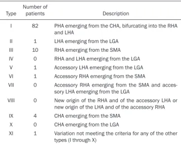
Table 2—Anatomical variation of the hepatic artery in 100 patients, according to the classification system devised by Michels(5).
Type
I
II III IV V VI VII
VIII
IX X XI
Number of patients
82
1 10
0 1 1 0
0
4 0 1
Description
PHA emerging from the CHA, bifurcating into the RHA and LHA
LHA emerging from the LGA RHA emerging from the SMA RHA and LHA emerging from the LGA Accessory LHA emerging from the LGA Accessory RHA emerging from the SMA
Accessory RHA emerging from the SMA and acces -sory LHA emerging from the LGA
New origin of the RHA and of the accessory LHA or new origin of the LHA and of the accessory RHA CHA emerging from the SMA
CHA emerging from the LGA
Variation not meeting the criteria for any of the other types (I through X)
PHA, proper hepatic artery; CHA, common hepatic artery; RHA, right hepatic artery; LHA, left hepatic artery; LGA, left gastric artery; SMA, superior mesen -teric artery.
Figure 2. CT angiography reconstruction by maximum intensity projection in the coronal plane, showing the common hepatic artery (arrowhead) emerging from the superior mesenteric artery (arrow).
DISCUSSION
We prospectively analyzed 100 consecutive CT angi
-ography examinations of the abdomen performed at the General Hospital of Fortaleza over a one-year period. The imaging evaluation of the rhino-orbital region has been
the object of a series of recent publications in the radiol
-ogy literature of Brazil(24–29).
In the standard definition of the visceral anatomy, the celiac trunk originates from the abdominal aorta and trifurcates into the left gastric artery, splenic artery, and
common hepatic artery(4,7,30). Although there are 15 pos
-sible patterns of anatomical variations of the celiac trunk, we detected, in our study, only six types, the same number
found in the study conducted by Sureka et al.(3). In our
study, the normal anatomic pattern of the celiac trunk was
found in 43% of the cases, compared with 89% in the dis
-section study conducted by Michels(5); 91% in the study
conducted by Sureka et al.(3); 86% in the study conducted
by Sankar et al.(8); 85.1%, 89.5%, and 95.4%, respectively,
in cadaver studies, imaging studies, and liver transplan -tation studies, as reported by Panagouli et al.(31); 89.1% in the study conducted by Song et al.(18); 89.8% in the
study conducted by Chen et al.(32), who analyzed a popu
reported by Sankar et al.(8), the 9.6% reported by Song et al.(18) , and the 10.2% reported by Chen et al.(32).
The most common anatomical pattern of variation
in our study—found in 47% of the cases—was a hepato
-splenic trunk with the left gastric artery emerging 0.4–2.5
cm above the bifurcation of the celiac trunk and the su
-perior mesenteric artery originating from the abdominal
aorta. That anatomical pattern was also observed by Su
-reka et al.(3), Michels(5), and Song et al.(18) , although in only 2.3%, 4%, and 4.42% of the cases, respectively. In the
study conducted by Araujo-Neto et al.(33), the most com
-mon anatomical pattern of variation—found in 8.3% of the cases—was a hepatosplenic trunk with the left gastric artery originating from the abdominal aorta.
Regarding the anatomy of the hepatic artery, the
most commonly seen pattern, according to the classifica
-tion system devised by Michels(5), is type I; that is, the hepatic artery emerging from the common hepatic artery
and bifurcating into the right and left hepatic arteries(5).
In our study, we found Michels type I anatomical pattern of the hepatic artery in 82% of the cases, whereas Gumus et al.(15), Sureka et al.(3), and Chen et al.(1) reported Mi -chels type I anatomical pattern in 66.8%, 55%, and 51%,
respectively. In a cadaver study conducted in Brazil, Seb
-ben et al.(2) identified Michels type I anatomical pattern in
73% of the cadavers dissected. In a liver transplantation study conducted in the Brazilian state of Paraná, Freitas
et al.(30)found the normal anatomical pattern in 76.82%
of the cases.
In our study, 18% of the patients had some varia -tion in the anatomy of the hepatic artery, compared with
33.2% reported by Gumus et al.(15), 45% reported by Su
-reka et al.(3), 49% reported by Chen et al.(1), 27% reported
by Sebben et al.(2), 23.18% reported by Freitas et al.(30),
10.2% reported by Chen et al.(32), and 21.7% reported
by Araujo-Neto et al.(33). The most common anatomical
pattern of variation in our study—found in 10% of the
cases—was the right hepatic artery emerging from the su
-perior mesenteric artery. In the studies analyzed, that was also the main pattern of anatomical variation, although in different proportions of the samples evaluated. Gumus et
al.(15), reported that same pattern in 10.1% of the patients
in their sample, comparable to the 11% reported by Su
-reka et al.(3), the 15% reported by Chen et al.(1), the 10%
reported by Sebben et al.(2), and the 11.38% reported by
Freitas et al.(30).
Our study provides an insight into the anatomical pat
-terns found in Brazil. In view of our findings, it is possible to conclude that, because of the extensive miscegenation
in Brazil, the indices of anatomical variation, despite shar
-ing points of similarity, also present patterns of variations
that differ considerably from those reported in the interna
-tional literature, especially when we compare the patterns of celiac trunk variation with those found in homogeneous populations(2,9,14,32).
It is noteworthy that some isolated studies conducted
in Brazil have analyzed the anatomical pattern of the he
-patic artery and its branches in cadavers and during liver
transplantations. However, studies dealing with the ana
-tomical variations of the celiac trunk are really quite rare in Brazil, this being the aspect of our study that distin -guishes it from others in the literature(2,9,14,22,30).
On the basis of our findings, we can state that studies like ours are scarce and should be encouraged, in order to improve understanding of the anatomical patterns in the population of Brazil. Further studies of this nature could lead to better technical planning of surgical procedures
and could avoid inadvertent injuries that might compro
-mise the results of medical procedures, leading to compli
-cations. Better knowledge of anatomical variations could ultimately contribute to reducing the rates of morbidity
and mortality in endovascular procedures, abdominal sur
-geries, and transplantations, especially those of the liver and pancreas(2,14,22,30).
REFERENCES
1. Chen H, Yano R, Emura S, et al. Anatomic variation of the celiac trunk with special reference to hepatic artery patterns. Ann Anat. 2009;191:399–407.
2. Sebben GA, Rocha SL, Sebben MA, et al. Variações da artéria hepática: estudo anatômico em cadáveres. Rev Col Bras Cir. 2012; 40:221–6.
3. Sureka B, Mittal MK, Mittal A, et al. Variations of celiac axis, com-mon hepatic artery and its branches in 600 patients. Indian J Radiol Imaging. 2013;23:223–33.
4. Venieratos D, Panagouli E, Lolis E, et al. A morphometric study of the celiac trunk and review of the literature. Clin Anat. 2013; 26:741–50.
5. Michels NA. Newer anatomy of the liver and its variant blood sup-ply and collateral circulation. Am J Surg. 1966;112:337–47. 6. Catalano OA, Singh AH, Uppot RN, et al. Vascular and biliary
variants in the liver: implications for liver surgery. Radiographics. 2008;28:359–78.
7. Nayak SR, Prabhu LV, Krishnamurthy A, et al. Additional branches of celiac trunk and its clinical significance. Rom J Morphol Em-bryol. 2008;49:247–9.
8. Sankar KD, Bhanu PS, Susan PJ. Variant anatomy of the celiac trunk and its branches. Int J Morphol. 2011;29:581–4.
9. Varma KS, Pamidi N, Vollala VR. Common celiacomesenteric trunk: a rare anatomic variation. J Vasc Bras. 2009;8:271–3.
10. Wang Y, Cheng C, Wang L, et al. Anatomical variations in the ori-gins of the celiac axis and the superior mesenteric artery: MDCT angiographic findings and their probable embryological mecha-nisms. Eur Radiol. 2014;24:1777–84.
11. Melo-Leite AF, Mota Jr A, Chagas-Neto FA, et al. Acquired portosys-temic collaterals: anatomy and imaging. Radiol Bras. 2016;49:251– 6.
12. Atasoy Ç, Ozyürek E. Prevalence and types of main and right portal vein branching variations on MDCT. AJR Am J Roentgenol. 2006; 187:676–81.
13. Winston CB, Lee NA, Jarnagin WR, et al. CT angiography for delin-eation of celiac and superior mesenteric artery variants in patients undergoing hepatobiliary and pancreatic surgery. AJR Am J Roent-genol. 2007;188:W13–9.
15. Gümüs H, Bükte Y, Özdemir E, et al. Variations of the celiac trunk and hepatic arteries: a study with 64-detector computed tomographic angiography. Eur Rev Med Pharmacol Sci. 2013;17:1636–41. 16. Iezzi R, Cotroneo AR, Giancristofaro D, et al. Multidetector-row
CT angiographic imaging of the celiac trunk: anatomy and normal variants. Surg Radiol Anat. 2008;30:303–10.
17. Saylisoy S, Atasoy Ç, Ersöz S, et al. Multislice CT angiography in the evaluation of hepatic vascular anatomy in potential right lobe donors. Diagn Interv Radiol. 2005;11:51–9.
18. Song SY, Chung JW, Yin YH, et al. Celiac axis and common hepatic artery variations in 5002 patients: systematic analysis with spiral CT and DSA. Radiology. 2010;255:278–88.
19. Reis FRS, Cardia PP, D’Ippolito G. Computed tomography angiog-raphy in patients with active gastrointestinal bleeding. Radiol Bras. 2015;48:381–90.
20. Erbay N, Raptopoulos V, Pomfret EA, et al. Living donor liver trans-plantation in adults: vascular variants important in surgical planning for donors and recipients. ARJ Am J Roentgenol. 2003;181:109–14. 21. Schmidt S, Demartines N, Soler L, et al. Portal vein normal anato-my and variants: implication for liver surgery and portal vein embo-lization. Semin Intervent Radiol. 2008;25:86–91.
22. Soares RV, Coelho JCU, Matias JEF, et al. Anatomia da artéria he-pática em doadores e receptores de transplante hepático intervivos. Rev Col Bras Cir. 2006;33:63–7.
23. Araujo-Neto SA, Mello-Júnior CF, Franca HA, et al. Multidetector computed tomography angiography of the celiac trunk and hepatic arterial system: normal anatomy and main variants. Radiol Bras. 2016;49:49–52.
24. Giardino A, Miller FH, Kalb B, et al. Hepatic epithelioid hemangio-endothelioma: a report from three university centers. Radiol Bras. 2016;49:288–94.
25. Cruz JF, Cruz MAF, Machado Neto J, et al. Prevalence and
sono-graphic changes compatible with fatty liver disease in patients re-ferred for abdominal ultrasound examination in Aracaju, SE. Radiol Bras. 2016;49:1–5.
26. Siqueira GRS, Guimarães MD, Franco LFS, et al. Exophytic hepa-tocellular carcinoma, simulating a mesenchymal tumor, in a non-cirrhotic liver. Radiol Bras. 2017;50:62–3.
27. Staziaki PV, Teixeira BC, Pedrazzani BM, et al. Hepatoblastoma with solid and multicystic aspect mimicking a mesenchymal hamartoma: imaging and anatomopathologic findings. Radiol Bras. 2017;50:68. 28. Ramalho M, Matos AP, AlObaidy M, et al. Magnetic resonance im-aging of the cirrhotic liver: diagnosis of hepatocellular carcinoma and evaluation of response to treatment – Part 1. Radiol Bras. 2017; 50:38–47.
29. Ramalho M, Matos AP, AlObaidy M, et al. Magnetic resonance im-aging of the cirrhotic liver: diagnosis of hepatocellular carcinoma and evaluation of response to treatment – Part 2. Radiol Bras. 2017; 50:115–25.
30. Freitas ACT, Coelho JCU, Matias JEF, et al. Anatomia arterial hep-ática: estudo em 150 transplantes hepáticos. Rev Col Bras Cir. 2000;28:13–6.
31. Panagouli E, Venieratos D, Lolis E, et al. Variations in the anatomy of the celiac trunk: a systematic review and clinical implications. Ann Anat. 2013;195:501–11.
32. Chen H, Yano R, Emura S, et al. Anatomic variation of the celiac trunk with special reference to hepatic artery patterns. Ann Anat. 2009;191:399–407.
33. Araujo-Neto SA, Franca HA, Mello-Júnior CF, et al. Anatomical variations of the celiac trunk and hepatic arterial system: an analy-sis using multidetector computed tomography angiography. Radiol Bras. 2015;48:358–62.
34. Özbülbül NI. CT angiography of the celiac trunk: anatomy, variants and pathologic findings. Diagn Interv Radiol. 2011;17:150–7.