A new technique, combined plication-incision (CPI), for
correction of penile curvature
_______________________________________________
Hamed Abdalla Hamed
1, Mohamed Roaiah
1, Ahmed M. Hassanin
1, Adham Ashraf Zaazaa
1, Mahmoud Fawzi
11 Department of Andrology, Faculty of Medicine, Cairo University, Cairo, Egypt
ABSTRACT
ARTICLE
INFO
______________________________________________________________ ______________________
Introduction: Penile curvature (PC) can be surgically corrected by either corporoplasty or plication techniques. These techniques can be complicated by post-operative: penile shortening, recurrent PC, painful/palpable suture knots and erectile dysfunction. Objective: To avoid the complications of corporoplasty and plication techniques using a new technique: combined plication-incision (CPI).
Materials and Methods: Two groups (1&2) were operated upon: group 1 using CPI and group 2 using the 16-dot technique. In CPI, dots were first marked as in 16 dot tech-nique. In each group of 4 dots the superficial layer of tunica albuginea was transverse-ly incised (3-6mm) at the first and last dots. Ethibond 2/0, passed through the interior edge of the first incision plicating the intermediate 2 dots and passed out of the interior edge of the last incision, was tightened and ligated. Vicryle 4/0, passed through the exterior edges of the incisions, was tightened and ligated to cover the ethibond knot. Results: Twelve (57.1 %) participants in group 2 complained of a bothering palpable knot compared to none in group 1 with statistically significant difference (P=0.005). Postoperative shortening (5mm) of erect penis, encountered in 9 participants, was dou-bled in group 2 but with insignificant difference (P>0.05). Post-operative recurrence of PC, was encountered in only 1 (4.8%) participant in group 2, compared to none in group 1, with insignificant difference (P>0.05). Post-operative erectile rigidity was normally maintained in all participants.
Conclusion: The new technique was superior to the 16-dot technique for correction of PC.
Keywords:
Penile Induration; Penis; Erectile Dysfunction
Int Braz J Urol. 2018; 44: 180-7
_____________________ Submitted for publication: October 21, 2016
_____________________ Accepted after revision: December 26, 2016 _____________________ Published as Ahead of Print: May 03, 2017
INTRODUCTION
Penile curvature (PC) is not an uncommon disorder in men. PC may be congenital or acqui-red, mostly caused by Peyronie’s disease (1). PC may cause difficulty and pain during intercourse, or even coital impairment, and severe psycholo-gical problems due to the cosmetic appearance of the affected person (2,3). Surgical correction of the penile curvature is required when coital func-tion is impaired (3).
Surgical correction of PC can be classi-fied into three main categories: tunical shortening procedures, tunical lengthening procedures, and penile prosthesis implantation (4). Tunical shor-tening is the commonly used surgical technique for correction of PC. Tunical shortening restricts the convex, longer, side of the penis to match the length of the oppositely shorter side (5).
corporoplasty, a tunical ellipse is excised or sin-gle (or multiple) longitudinal tunical incision(s) is (are) made and the remaining defect is closed horizontally (7-9). Plication of the tunica includes Essed & Schröder technique (10) and 16 dot tech-nique (11).
Tunical shortening complications include loss of length, recurrent or residual penile curva-ture, erectile dysfunction, change in penile sensa-tion, and painful or palpable suture knots. Many of these outcomes can be quite distressing to the patient (5).
In this study, we describe a new technique, where we combine the plication and the incisional techniques, without opening of the cavernous tis-sue, aiming to get the advantages and to avoid the disadvantages of either technique.
MATERIALS AND METHODS
This study was designed to compare a new technique: combined plication-incision (CPI), with the 16 dot technique for correction of penile cur-vature. The study was approved by the Andrology board and Ethics Committee, Faculty of Medicine, Cairo University.
In the period from Dec 2013 till Oct 2015 one hundred ninety men presented with a com-plaint of penile curvature to the Andrology ou-tpatient clinic at the University hospital. Seventy nine cases required surgery for correction of cur-vature and matched the inclusion and exclusion criteria. Thirty nine eligible participants agreed to participate in this research and to be available for follow-up. Informed consents were taken from participants in the study.
For potentially eligible participants of men presenting with a complaint of penile curvature, the following procedures were designed: history taking, physical examination, international index of erectile function questioning, intra-corporeal injection and penile duplex.
History taking included: age, history of general diseases (e.g. diabetes mellitus, hyper-tension), Dupuytren’s contracture, history of any accidents/genital trauma and medications as beta-blocker and Methotrexate. Any history of psychiatric disease and/or treatment was reported.
Sexual history included the presence of normal erectile rigidity, easy intromission, presen-ce/absence of painful erection, and/or painful in-tercourse to any partner. The patient was also asked about desire, orgasm and ejaculatory problems. Par-ticipants were asked to complete an abridged five--item version of the International Index of Erectile Function (IIEF) (12). During physical examination the penis was inspected for the size and the site of the urethral meatus and was palpated for tender-ness, chordae or plaques. An intracorporal injection of 20 micrograms PGE1 was given and the grade of erection was evaluated (13). During rigid erection (E4/E5) the penile length (Table-2) and the angle of penile deviation were measured, by the doctor, using metal seizer and protractor respectively.
Duplex evaluation: peak systolic velocity (PSV) >30cm/min., end diastolic velocity (EDV)<5 cm/min and arterial dilatation more than 70% were considered normal hemodynamics.
Inclusion criteria
Men aged >21 years presenting with con-genital or acquired penile curvature of >30º, who had normal erectile function, E4 or E5 response on intracorporal injection, and normal penile duplex parameters with stable course in case of Peyronie’s disease (more than one year).
Exclusion criteria
Men with hypospadias with chordee, epis-padias, poor response to PGE1 (response <E4) and/ or active phase of Peyronie’s disease (less than one year). Laboratory testing (blood glucose, lipid pro-file and routine preoperative labs) were done for included participants.
Selection of patients for either technique
CPI technique was used mainly for patients with congenital penile lateral or dorsal deviation.
In case with congenital ventral curvature: performing tunical incisions was avoided to pre-serve structures of neurovascular bundle. Howe-ver, in four patients only, removal of a segment of the deep dorsal vein allowed a space to perform adequate incisions to use the new technique. Re-maining patients with congenital ventral curvature together with acquired cases were operated upon using the 16 dot technique (11) and were named group 2. Participants operated upon by the CPI te-chnique were named group 1.
Surgical technique
All participants received spinal anaesthe-sia and lied in supine position. After scrubbing and draping, an artificial erection was induced by intra-corporeal injection of 20 micrograms of PGE1. Skin incision was made at the circumcision line. Blunt dissection was performed to enter Colle’s fascia. The entire penis was then degloved till the level of peni-le base. Buck’s fascia was dissected longitudinally from the coronal sulcus to the base of the penis un-til tunica albuginea and neurovascular bundle were visualized and identified. Strict care was employed to preserve all structures of neurovascular bundle dorsally and the corpus spongiosum/urethra
ven-trally. In all cases, the point of maximum curvature was identified during full rigid erection.
CPI technique
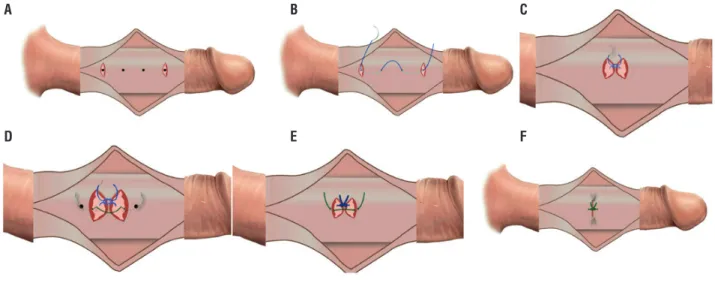
Sutures were applied on the convex side of the penis just proximal to the point of maxi-mal curvature passing through 8 dots. Those 8 dots were divided into two lines of 5mm spaced 4 dots, on either side of the middle line “in cases of ventral/dorsal deviation” and parallel to each other on right or left side in cases with lateral deviation. Transverse incisions 3-6mm were per-formed, with a fine scalpel, at the site of the first and last dots (of each group of 4 dots) through the superficial layer of tunica albuginea (Figures 1A and 2A), without cutting into the corpus ca-vernosum, to prepare a cavity for the knots. The 00 ethibond suture was passed through the inte-rior edge of the first incision, plicating the inter-mediate 2 dots to end out from the interior edge of the last incision (Figures 1B and 2B), leaving free exterior incisions edges (Figure-1C). Vicryl 4/0 suture was passed through the exterior edges of the incisions (Figures 1D and 2C). Tightening and ligation of the ethibond was first done (Fi-gure-1E) and the vicryl was then tightened and ligated over the ethibond suture knot to cover it (Figures 1F and 2D).
Figure 1 - Diagrammatic steps of CPI technique 1A) Two transverse incisions through outer layer of tunica albuginea at first and last dots. 1B and C) Ethibond suture passing through interior edges of the incisions and plicating the intermediate 2 dots. 1D) Vicryl suture passing through outer free edges of the incisions. 1E) Tightening and ligation of the Ethibond. 1F) Closure of outer edges over ethibond suture knot.
A
D
B
E
C
Sixteen dot technique was performed accor-ding to the description of Gholami and Lue (11).
In all cases, for proper correction during ti-ghtening of ethibond sutures, the penis should not be in full rigidity and the assistant should bend the dis-tal shaft in the direction of correction to allow pro-per tightening of the suture. Avoidance of crushing of the suture material with instruments during liga-tion is mandatory.
After ligation of the 1st set of sutures a full
rigid erection was induced to assess straightness of the penis. Usually there was a remaining deviation but with point of maximum deviation displaced dis-tally. The previous steps were repeated till reaching a complete straightness. Over-correction was avoided by measuring the degree of correction before liga-ting the sutures. This was done by traction of the two suture ends against each other simulating ligation.
By inspecting the degree of correction we decided the degree of tightening of the suture needed whe-ther completely tight or little looser. The second pla-ne of penile curvature was corrected using the same technique used for correction of the main plane.
Buck’s and superficial fascia were closed lon-gitudinally with continuous vicryl 3/0 or 4/0 sutures. Postoperative erected penile length was measured (Table-2) and rigidity was evaluated. Skin was closed with vicryl 5/0 in continuous manner but in three separate parts. Then, dressing was applied with mini-mal pressure on the penile shaft for bandage. Finally, 1cc of diluted phenylephrine was injected intracorpo-really if no spontaneous detumescence occurred.
Follow up
A weekly postoperative follow up was done to ensure complete wound healing and to
Figure 2 - Steps of CPI technique. 2A) Two transverse incisions through outer layer of tunica albuginea at first and last dots. 2B) Ethibond suture passing through interior edges of the incisions and plicating the intermediate 2 dots. 2C) Vicryl suture passing through outer free edges of the incisions. 2D) Tightening and ligation of the Ethibond sutures followed by closure of outer edges over ethibond suture knot.
A B
C
manage any post-operative complications if pre-sent. After 1 month, measurement of the postope-rative flaccid length was performed.
Examination for palpable suture knot along the shaft was performed by a doctor who was blind to the operative procedure (patients’ group). Furthermore, the patient was questioned about sensation of a knot and if it bothers him. However, we decided to rely on the patient words for assortment of cases positive for palpable sutu-re knot. After 3 months, after sutu-resuming sexual ac-tivity, participants were followed up by the IIEF-5. Furthermore, all participants were asked about any decrease in the erectile function noticed and about a recurrence of deviation. This was repeated every 3 months till the end of follow-up period that ranged from 12 to 31 months with mean a duration of 20.5±6.9 months.
Statistical analysis
Data were statistically described in ter-ms of range, frequencies, percentages, mean and standard deviation (SD). Comparison used Student t test, Mann Whitney U test, paired t test and Wil-coxon signed rank test. Correlation between va-rious variables was done using Spearman Rank correlation equation. For comparing categorical data, Chi square test was performed. Exact test was used instead when the expected frequency was less than 5. P values <0.05 were set as statis-tically significant. All statistical calculations were done using computer program SPSS (Statistical Package for the Social Science; SPSS Inc., Chi-cago, IL, USA) release 15 for Microsoft Windows (2006).
RESULTS
This study included 39 participants, 18 in group 1 and 21 in group 2. Mean age was 30.6±10.2, 28±7.3 and 32.8±11.8 for all parti-cipants, group 1 and group 2 respectively with insignificant difference (P=0.14). Sixteen (41%) participants were smokers, 2 (5.1%) were hyper-tensive and 1 (2.6%) was diabetic type 2 with in-significant difference between both groups regar-ding these data (P>0.05). Five (12.8%) participants
had acquired curvature for more than 1.5 year. All of them were assigned to group 2 to avoid exag-geration of the condition with incisions.
Thirty one (79.5%) participants had sin-gle plane deviation and eight (20.5%) had bidi-rectional deviation, 3 (16.7%) in group 1 and 5 (23.8%) in group 2. Table-1 presents the preo-perative direction of curvature. Deviation angle ranged 30-90 at the main plane and 30-35 at the second plane. The mean of the angles of the main plane was 43.3±18.1 and 53.1±18.1 for group 1 and 2 respectively with insignificant difference (P=0.1).
The number of dots marked during the surgeries ranged from 8-24 with a mean 15.28±5.79, 13.33±5.48 and 16.95±5.64 dots, for the total cases, group 1 and group 2 respecti-vely. Significant positive correlation was found between the number of dots and the degree of curvature (r=0.69 and P<0.001).
Post-operative follow up ranged from 12 to 31 months with a mean duration 20.5±6.9, 20.1±7.7 and 20.9±6.4 months for all cases, group 1 and group 2 respectively.
One participant in the group 1 develo-ped a hematoma. Post-operative pain was repor-ted by 3 cases in each group representing 14.3% and 16.7% of group 1 and 2 respectively with insignificant difference (P=0.53). Postoperati-ve shortening was encountered in nine (23.1%), three (16.7%) in group 1 and 6 (28.6%) in group 2 with insignificant difference (P=0.4). Shorte-ning was 0.5cm of the erected penile length in all cases. Twelve (57.1%) participants in group 2 complained of feeling a bothering palpable knot, compared with none in group 1, with statistically significant difference (P=0.005). By examination, palpable knot was found in 16 (76.2%) cases in group 2 and none in group 1. However, we con-sidered cases positive only when reported by the patients themselves after asking about it.
Residual curvature did not exceed 10 de-grees. This was encountered in 2 (11.1%) in group 1 and 1 (4.8%) in group 2. Post-operative recur-rence of curvature was recorded in only 1 (4.8%) participant in group 2 at the 18th month of his
Throughout the follow-up period, all parti-cipants retained a postoperative rigid erection and none of them reported a postoperative decrease of erectile rigidity. The pre and postoperative IIEF-5 score(s) were reported by 25 (64.1%) participants. The preoperative IIEF-5 score was 20.28±1.41, 20.55±1.38 and 20.04±1.43 for all participants, group 1 and group 2 respectively. The postope-rative IIEF-5 was 20.25±1.37, 20.5±1.29 and 20.04±1.43 for all participants, group 1 and group 2 respectively. Comparative studies of the pre and postoperative scores for each group were statisti-cally insignificant (P=0.579 and 1 for group 1 and 2 respectively).
DISCUSSION
Tunical corporoplasty and tunical pli-cation are two techniques used to correct PC. In tunical corporoplasty, permanent fusion of the tunical margins, by the healing process, adds to the strength of the sutures and allow for much
better results in terms of recurrence. However, the invasiveness of corporoplasty is great and the tourniquet, if used for long time, may be harmful to the sensory nerves and the erectile tissue (14). On the contrary, tunical plication procedures are less invasive (15-17) and separation of dorsal neu-ro–vascular bundles from the tunica albuginea is not done in 16 dot plication technique (11, 18). However, in plication, the strength depends only on the sutures and not on the healing process (11, 17, 19, 20). After tunical plication, the recurrences rates were high (10, 16, 19) and the presence of permanent palpable knots at the site of the tuni-cal sutures (15, 21) was noted causing discomfort or even pain (11, 22, 23). Furthermore, creation of a protruding bulk inside the cavernous cavity decreases its volume and compresses cavernosal tissue (23) and excessive folding may also lead to decreased distal rigidity (24).
For these reasons, we improved the tech-nique of penile straightening in order to avoid di-sadvantages and to preserve the most important
Table 1 - Direction of curvature preoperatively among both groups.
Main plane Second plane
Group 1 Group 2 Total Group 1 Group 2 Total
Ventral 4 (22.2%) 13 (61.9%) 17 (43.6%) 0 1 (4.8%) 1 (2.6%)
Dorsal 2 (11.1 %) 3 (14.3 %) 5 (12.8%) 0 1 (4.8%) 1 (2.6%)
Right 4 (22.2 %) 1 (4.8 %) 5 (12.8 %) 1 (5.6%) 1 (4.8%) 2 (5.1%)
Left 8 (44.4%) 4 (19.0%) 12 (30.8%) 2 (11.1%) 2 (9.5%) 4 (10.3%)
None 0 0 0 15 (83.3) 16 (76.2) 31 (79.5%)
Total 18 (100%) 21 (100%) 39 (100%) 18 (100%) 21(100%) 39 (100%)
Table 2 - Pre and postoperative flaccid and erected penile length.
Flaccid penile length (preoperative) Erected penile length (preoperative)
Erected penile length (postoperative)
Flaccid penile length (postoperative)
Range (cm) Mean±SD Range (cm) Mean±SD Range (cm) Mean±SD Range (cm) Mean±SD
Group 1 10–14 12±1.1 12–18 15.8±1.6 12–18 15.8±1.6 10–14 12±1.1
Group 2 10.5–17 12.4±1.7 13–20 14.9±1.7 12.5–20 14.7±1.7 10.5–17 12.4±1.7
advantages of corporoplasty and tunical plication. The new technique avoids complete incision of the tunica albuginea which may compromise the erec-tile rigidity postoperatively (25) and at the same time, the new technique, gets the advantage of the healing edges and avoid the mere dependence on the suture as in plication techniques. Furthermo-re, in this new technique the superficial layers of tunica albuginea was ligated by absorbable su-ture covering the ethibond susu-ture knots aiming to decrease the discomfort and/or pain caused the none-absorbable knots. However, in ventral curvature we preferred the 16 dot technique to avoid mobilization of neurovascular bundle and to maintain its integrity within the tunica albu-ginea. Also, we preferred the 16 dot technique for cases with Peyronie’s disease to avoid incision and healing in these cases with idiopathic abnormal healing.
Results of group 2 were comparable to other study that used the 16-dot technique (11). Furthermore, the correction of penile curvature, residual curvature and recurrences, using the new technique, was comparable to another study that used corporoplasty (19).
In the present study, the post-operative complications, penile shortening and recurrence of penile curvature, were higher, with the 16-dot technique compared to the new technique but the differences were statistically insignificant. Ho-wever, statistically significantly higher (P=0.005) post-operative complaint of feeling of a bothering palpable knot was reported by participants opera-ted upon by the 16-dot plication technique. Fur-thermore, the post-operative erectile function was normally maintained in all participants as indicated by having a postoperative E4/E5, a non-significant difference between the pre and postoperative IIEF-5 scores and all participants retained their rigid erec-tion during the whole period of follow-up. Those findings indicated the importance of using the new technique to correct the penile curvature.
CONCLUSIONS
The new technique is superior to the 16 dot plication technique regarding post-operative feeling of a bothering suture knot especially when
done for properly selected cases. It is better to be applied to cases with lateral or dorsal deviation of congenital penile curvature. Sixteen-dot tech-nique is better to be applied to congenital ventral curvature and for patients with Peyronie’s disease.
CONFLICT OF INTEREST
None declared.
REFERENCES
1. De la Peyronie F G. Sur Quelques Obstacles, qui S’Opposent à L’Ejaculation Naturelle de la Semence. Mem Acad R Chir. 1743; 1:425-34
2. Smith JF, Walsh TJ, Conti SL, Turek P, Lue T. Risk factors for emotional and relationship problems in Peyronie’s disease. J Sex Med. 2008;5:2179-84.
3. Tal R, Nabulsi O, Nelson CJ, Mulhall JP. The psychosocial impact of penile reconstructive surgery for congenital penile deviation. J Sex Med. 2010;7:121-8.
4. Tornehl CK, Carson CC. Surgical alternatives for treating Peyronie’s disease. BJU Int. 2004;94:774-83.
5. Segal RL, Burnett AL. Surgical Management for Peyronie’s Disease. World J Mens Health. 2013;31:1-11.
6. Mobley EM, Fuchs ME, Myers JB, Brant WO. Update on plication procedures for Peyronie’s disease and other penile deformities. Ther Adv Urol. 2012;4:335-46.
7. Nesbit RM. Congenital curvature of the phallus: report of three cases with description of corrective operation. J Urol. 1965;93:230-2.
8. Pryor JP, Fitzpatrick JM. A new approach to the correction of the penile deformity in Peyronie’s disease. J Urol. 1979;122:622-3.
9. Yachia D. Modified corporoplasty for the treatment of penile curvature. J Urol. 1990;143:80-2.
10. Essed E, Schroeder FH. New surgical treatment for Peyronie disease. Urology. 1985;25:582-7.
11. Gholami SS, Lue TF. Correction of penile curvature using the 16-dot plication technique: a review of 132 patients. J Urol. 2002;167:2066-9.
12. Rosen RC, Cappelleri JC, Smith MD, Lipsky J, Peña BM. Development and evaluation of an abridged, 5-item version of the International Index of Erectile Function (IIEF-5) as a diagnostic tool for erectile dysfunction. Int J Impot Res. 1999;11:319-26.
14. Perdzyński W, Adamek M. A new corporoplasty based on stratified structure of tunica albuginea for the treatment of congenital penile curvature - long-term results. Cent European J Urol. 2015;68:102-8.
15. Chen R, McCraw C, Lewis R. Plication procedures-excisional and incisional corporoplasty and imbrication for Peyronie’s disease. Transl Androl Urol. 2016;5:318-33.
16. Leonardo C, De Nunzio C, Michetti P, Tartaglia N, Tubaro A, De Dominicis C, et al. Plication corporoplasty versus Nesbit operation for the correction of congenital penile curvature. A long-term follow-up. Int Urol Nephrol. 2012;44:55-60. 17. Nyirády P, Kelemen Z, Bánfi G, Rusz A, Majoros A, Romics
I. Management of congenital penile curvature. J Urol. 2008;179:1495-8.
18. Ahmadnia H, Kamalati A, Younesi Rostami M, Imani MM, Asadpour AA, Hariri MK. The Therapeutic Effects of Intracavernosal Plaque Excision in Peyronie’s Disease: A None Grafting or Tunical Excising Procedure. World J Plast Surg. 2016;5:62-6.
19. Vicini P, Di Nicola S, Antonini G, De Berardinis E, Gentile V, De Marco F. Geometrical modified nesbit corporoplasty to correct different types of penile curvature: description of the surgical procedure based on geometrical principles and long-term results. Int J Impot Res. 2016;28:209-15.
20. Kuehhas FE, Egydio PH. Superficial tunica albuginea excision, using geometric principles, for the correction of congenital penile curvature. BJU Int. 2012;110:E949-53. 21. Baskin LS, Lue TF. The correction of congenital penile
curvature in young men. Br J Urol. 1998;81:895-9.
22. Schultheiss D, Meschi MR, Hagemann J, Truss MC, Stief CG, Jonas U. Congenital and acquired penile deviation treated with the essed plication method. Eur Urol. 2000;38:167-71. 23. Poulsen J, Kirkeby HJ. Treatment of penile curvature--a
retrospective study of 175 patients operated with plication of the tunica albuginea or with the Nesbit procedure. Br J Urol. 1995;75:370-4.
24. Perovic SV, Djordjevic ML, Djakovic NG. A new approach to the treatment of penile curvature. J Urol. 1998;160:1123-7. 25. Hatzichristodoulou G. Grafting techniques for Peyronie’s
disease. Transl Androl Urol. 2016;5:334-41.
_______________________ Correspondence address:
Ahmed M. Hassanin, MD, PhD Department of Andrology, Faculty of Medicine, Cairo University 1 Al-Saraya Street, Al-Manial, Cairo, 11559, Egypt