287
RADIOLOGY PAGE
A thirty-three-year-old male presented to an outside emergency department with scro-tal swelling and pain after intercourse. A scroscro-tal ultrasound revealed hematoma, with no other abnormalities and the patient was discharged. He then presented to our institution where ex-amination showed diffuse ecchymosis through the shaft of the penis, suprapubic region, and scrotum without a palpable cavernosal defect.
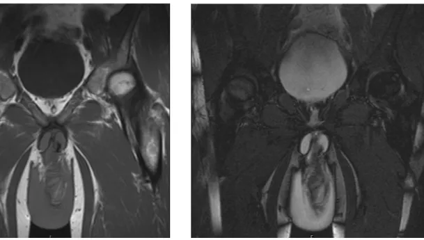
Magnetic resonance imaging (MRI) with-out contrast was obtained after the injection of 10 micrograms of intracavernosal alprostadil. The low signal tunica albuginea is easily
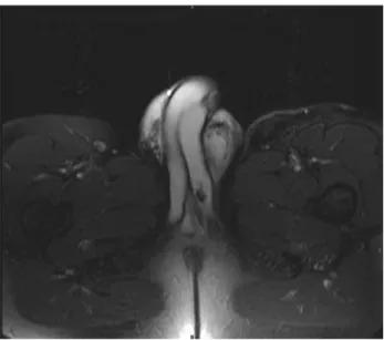
demar-cated compared to the high T2 and intermedi-ate T1 signal of the corpora cavernosum (Figures 1-3) (1,2). Hematoma shows heterogeneous in-termediate T1 and T2 signal (Figures 2 and 3) (1). Penile fracture is rupture of the corpus cavernosum from blunt trauma to the erect penis (3,4). Typical presentation is a pop during in-tercourse, immediate detumescence with edema, hematoma and penile deformity (3,4). In atypi-cal presentations, radiologiatypi-cal studies may be useful to determine the diagnosis. MRI provides the ability to identify disruption of the corpus cavernosum due to excellent tissue contrast and
Penile Fracture and Magnetic Resonance Imaging
_______________________________________________
Katie S. Murray, Michael Gilbert, Lawrence R. Ricci, Narendra Khare, Joshua Broghammer
University of Kansas Medical Center Department of Urology (KSM, JB), Kansas City, KS, University of Missouri-Kansas City Department of Urology (NK) and Department of Radiology (MG, LRR), Kansas City, MO, USA
_______________________________________________________________________________________ Vol. 38 (2): 287-288, March - April, 2012
Figure 1 - Coronal T1 image shows disruption of the low intensity inferior left tunica albuginea and associated low signal corpus cavernosal hematoma at the base of the penis.
288
IBJU|RADIOLOGY PAGEvisualization of soft tissue pathological process-es (5). MRI is an adjunctive tool in the evalua-tion of atypical presentaevalua-tions of suspected pe-nile fracture (3).
Figure 3 - Coronal T2 image shows disruption of the linear low signal left tunica albuginea at the base of the penis with associated heterogeneous hemorrhage medially.
REFERENCES
1. Fedel M, Venz S, Andreessen R, Sudhoff F, Loening SA: The value of magnetic resonance imaging in the diagnosis of suspected penile fracture with atypical clinical findings. J Urol. 1996; 155: 1924-7.
2. Choi MH, Kim B, Ryu JA, Lee SW, Lee KS: MR imaging of acute penile fracture. Radiographics. 2000; 20: 1397-405. Erratum in: Radiographics. 2000; 20: 1818.
3. Koifman L, Barros R, Júnior RA, Cavalcanti AG, Favorito LA: Penile fracture: diagnosis, treatment and outcomes of 150 patients. Urology. 2010; 76: 1488-92.
4. El-Assmy A, El-Tholoth HS, Abou-El-Ghar ME, Mohsen T, Ibrahiem el HI: False penile fracture: value of different diag-nostic approaches and long-term outcome of conservative and surgical management. Urology. 2010; 75: 1353-6. 5. Rahmouni A, Hoznek A, Duron A, Colombel M, Chopin DK,
Mathieu D, et al.: Magnetic resonance imaging of penile rupture: aid to diagnosis. J Urol. 1995; 153: 1927-8.
______________________ Correspondence address:
Dr. Katie Murray Department of Urology University of Kansas Medical Center