Cop
yright
© AE&M all rights r
eser
ved.
Association of serum thyrotropin
levels with coronary artery disease
documented by quantitative
coronary angiography:
a transversal study
Pedro D. Ortolani Jr.1, João H. Romaldini1, Ricardo A. Guerra1,
Evandro S. Portes1, George C. X. Meireles2,João Pimenta2
ABSTRACT
Objective: The association between coronary artery disease (CAD) and thyroid function remains controversial. We evaluated the thyroid function and graduated well-defined CAD as confirmed by quantitative coronary angiography (CA). Subjects and methods: We evaluated the serum TSH, free thyroxine, free triiodothyronine and thyroid antibody levels in 300 consecutive patients (age 61.6 ± 9.9 years and 54% were male) undergoing CAD diagnosis as confirmed by CA. Plaques with ≥ 50% stenosis being indicative of obstructive CAD, and patients were divided into groups according to main epicardial coronary arteries with plaques (0, 1, 2, 3). Lipid profiles and a homeostasis model assessment (HOMA-IR) were determined. Results: Serum median (25% and 75% percentile) TSH levels in patients with group 2 and 3 (2.25; 1.66-3.12 mU/L and 4.99; 4.38-23.60 mU/L, respectively) had significantly higher TSH concentrations (p < 0.0001) than the group 0 (1.82; 1.35-2.51 mU/L). Furthermore, patients of group 3 had higher TSH concentration (p < 0.0001) than those of group 1 (1.60; 0.89-2.68 mU/L). Group 3 were older (64 ± 8.5 vs. 59 ± 9.5, p = 0.001), had more patients with dyslipidemia (84% versus 58%, p < 0.001), male (54% versus 44%, p = 0.01), hypertension (100% versus 86%, p < 0.001), and smoking (61% versus 33%, p < 0.001) than group 0. Multivariate stepwise logistic analysis showed TSH, age, HbA1c, and HOMA-IR were the CAD associated variables. Conclusions: In this cohort, elevated TSH levels in the high normal range or above are associated with the presence and severity of CAD besides may represent a weak CAD risk factor. Arch Endocrinol Metab. 2018;62(4):410-5
Keywords
TSH; coronary artery disease; coronary angiography 1 Serviço de Endocrinologia,
Hospital do Servidor Público Estadual de São Paulo, Instituto de Assistência Médica ao Servidor Público Estadual (HSPE-IAMSPE), São Paulo, SP, Brasil 2 Serviço de Cardiologia, Hospital do Servidor Público Estadual de São Paulo, Instituto de Assistência Médica ao Servidor Público Estadual (HSPE-IAMSPE), São Paulo, SP, Brasil
Correspondence to:
Pedro Dirceu Ortolani Júnior Serviço de Endocrinologia, Hospital do Servidor Público Estadual Rua Pedro de Toledo, 1.800 04039-000 – São Paulo, SP, Brasil [email protected]
Received on Oct/3/2017 Accepted on Mar/10/2018
DOI: 10.20945/2359-3997000000054
INTRODUCTION
H
ypothyroidism is typically associated withelevated endothelial dysfunction and increased all-cause mortality in addition to cardiac death and/or hospitalization (1-3). Several studies have shown that coronary atherosclerosis in subjects with hypothyroidism is more frequent and severe than in normal subjects (4). In addition, there is some evidence that an elevated serum TSH in association with serum thyroid peroxidase antibody (TPOAb) may alter the traditional risk factors for coronary arterial disease (CAD), perhaps because of the accumulation of circulating levels of atherogenic total cholesterol (TC) and low-density lipoprotein (LDL) cholesterol particles (5-9). Consequently, the relation between thyroid dysfunction and CAD remains questionable (10), however, some authors have found a positive correlation between serum TSH levels and the
Cop
yright
© AE&M all rights r
eser
ved.
study was to yield greater insight into the assessment of thyroid function parameters and lipid concentrations in consecutive patients undergoing quantitative coronary angiography (CA).
SUBJECTS AND METHODS
Patients
Data were obtained from 343 consecutive patients in the Cardiology Outpatient Unit, Hospital do Servidor Público Estadual de São Paulo, Instituto de Assistência Médica ao Servidor Público Estadual (HSPE-IAMSPE) in Brazil that were referred for CA because of signs or symptoms relevant to CAD. All of the patients satisfied two inclusion criteria: 1) characteristic signs of myocardial ischemia on non-invasive tests and 2) clinical recommendation for further assessment. We used a questionnaire to assess classical risk factors for CAD such as diabetes, hypertension, dyslipidemia, smoking, and a family history of premature CAD (defined as ≤ 55 years old for males and ≤ 65 years old for females). Patients were excluded in cases with a history of thyroid diseases, documented CAD, acute CAD, or ischemic equivalent (such as dyspnea, sickness, confusion, stroke or pulmonary edema, compatible electrocardiographic signs, or increased creatine kinase-MB [CK-MB)] and/or troponin levels) (22). We excluded patients who were using amiodarone, levothyroxine, or antithyroid drugs (such as methimazole or propylthiouracil). Forty-three patients were excluded, and the remaining 300 patients were enrolled in the study. All of the patients included in the study received an explanation of the purpose of the study and signed a consent form. This work was submitted to the Research Ethics Committee of IAMSPE and approved without restrictions.
Assessment of CA
All of the patients were assessed via CA and further analyzed by an independent observer who was not involved in patients medical care. The quantification of the CA was performed manually. The CA was carried out using a femoral approach and a 6- or 7-F guiding catheter. Right and left CAs were performed in multiple projections using standard techniques and used to evaluate coronary artery stenosis (23). CAD was defined according to CA results, and significant stenosis was defined as a decrease in internal diameter of ≥ 50%, which could affect one or more of the main epicardial
coronary arteries (such as the right coronary, left anterior descending, or circumflex arteries) and classified as group 1, 2, or 3 according the number of vessels with significant stenosis (24). Patients were classified as group 0 if they had no CAD or when the main epicardial coronary arteries were < 50% of the internal diameter.
Biochemical analysis
Blood samples were drawn after overnight fasting prior to the CA. For the hormone assessment, patient samples were stored at -20°C until the time of measurement and analyzed in our laboratory. Blood glucose levels were estimated using the glucose oxidase method. Hemoglobin A1c (HbA1c), CK-MB, and cardiac troponin levels were determined by HPLC, electrochemiluminescent immunoassay, and two-site enzyme-linked immunosorbent assay methods, respectively. Serum TC, triglycerides (TG), and high-density lipoprotein (HDL) cholesterol levels were measured enzymatically. To determine LDL-cholesterol levels, the formula from Friedewald and cols. was used (25). Serum creatinine level was analyzed using a colorimetric method, and hepatic enzyme levels were determined enzymatically.
Hormonal analysis
Serum TSH, FT4, and FT3 levels were measured using direct chemiluminescent technology (Advia Centaur XP Immunoassay System; Siemens Healthcare Diagnostics, Deerfield, IL, USA) using reference values ranging from 0.3–4.1 mU/L for TSH, 0.8–2.0 ng/dL for FT4, and 1.6–4.1 pg/mL for FT3. The mean interassay CV was 3.1, 11.0, and 8.9 % for TSH, FT4, and FT3, respectively. TPOAb and TgAb serum concentrations were determined via a chemiluminescent immunometric assay (Diagnostic Products Co., Los Angeles, CA, USA), and values > 35 IU/L were considered to be positive. Serum insulin was analyzed using a chemiluminescent immunometric assay (Diagnostic Products Co.). The homeostasis model assessment of insulin resistance (HOMA-IR) index was determined using serum glucose and insulin values: fasting insulin (mU/mL)×fasting blood glucose (mg/dL)/405. Body mass index (BMI) was expressed as kg/m2.
Statistical analysis
Cop
yright
© AE&M all rights r
eser
ved.
comparison tests, and thereafter they were compared two-by-two using the Mann-Whitney rank test. Chi-square or Fischer’s exact tests were used to compare proportions Univariate and multivariate analyses with multinomial logistic regression analysis were used to evaluate the risks associated with CA results. For univariate analysis a less-restrictive alpha level with a 0.1 was used, and so a broad range of variables are considered for inclusion in the model. Thereafter, a backward stepwise multiple regression analysis was employed using CAD as dependent variable. A beta coefficient, standard error of beta coefficient (SE), and 95% confidence interval were obtained. SAS software version 9.4 (SAS Institute Inc., Cary, NC) was used for all analyses.
RESULTS
Characteristics of patients that were analyzed and patient demographics are reported in Table 1. All enrolled patients underwent all tests according to schedule. The mean age was of 61.6 ± 9.9, > 90% of patients had hypertension, 72% had dyslipidemia, and almost half of them were smokers. Mean serum TSH levels were 3.0 ± 2.7 (SD) mU/L, and 11% of patients had positive thyroid antibodies. Significant stenosis (≥ 50% in one or more main epicardial vessels)
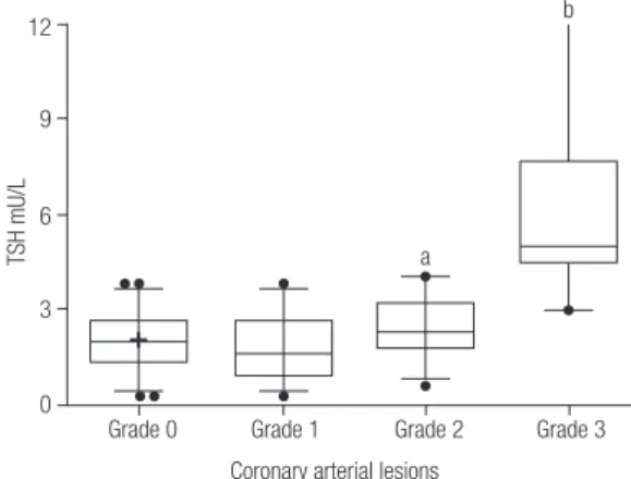
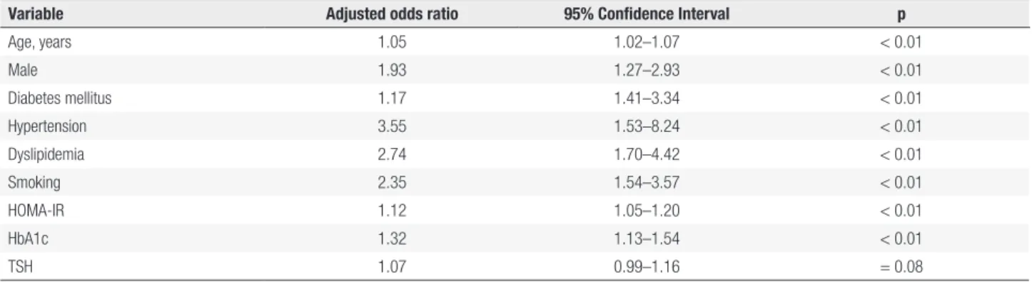
was observed in 62% (185) of patients. Arterial vessel stenosis was observed in 60 patients (group 1), 64 patients had two stenosed vessels (group 2), and 61 patients had three stenosed vessels (group 3). Figure 1 shows the results of serum TSH concentration in patients undergoing CA examination. Serum median and a 25% and 75% percentile of TSH levels in patients of group 2 (2.25; 1.66-3.12 mU/L) and group 3 (4.99; 4.38-23.60 mU/L) had significantly higher TSH concentrations (p < 0.0001) than the group 0 (1.82; 1.35-2.51 mU/L). Furthermore, patients of group 3 had higher TSH concentration (p < 0.0001) than those of group1 (1.60; 0.89-2.68 mU/L) as shown Figure 1. Moreover, group 3 had significant more patients with dyslipidemia, hypertension and smoking than group 0 patients. In group 3, the mean HbA1c levels were higher than other groups, and were older as shown Table 2. The group 2 had similar figure; had more male, smoking and diabetes patients as well as high HbA1c level, and older than group 0. Univariate logistical regression analysis revealed that male sex, diabetes mellitus, hypertension, dyslipidemia, smoking, older age and TSH were significant risk factors for CAD (Table 3). Thereafter, as shown in Table 4, the stepwise backward multiple regression model of CAD identified TSH, older age, HbA1c, and HOMA-IR as CAD risk factors (R2 = 0.377; Adjusted R2 = 0.369; SE = 0.922 F
= 42.526; p-value = 0.0001).
Table 1. Baseline characteristics of 300 patients participants in the study
Variables Number (%) or mean ± SD
Age, years 61.6 ± 9.9
Male 162 (54)
Diabetes mellitus 107 (36)
Hypertension 275 (92)
Smoking 147 (49)
Family history of CADa 70 (23)
Dyslipidemia 215 (72)
BMI, kg/m2 28.2 ± 4.5
Total cholesterolemia, mg/dL 178.7 ± 37.9
HDL-cholesterolemia, mg/dL 49.1 ± 14.1
Trygliceridemia, mg/dL 134 ± 73.1
HbA1c, % 6.3 ± 1.4
LDL-cholesterolemia, mg/dL 101 ± 31.9
Thyroid antibodiesb 34 (11)
TSH, mU/L 3.0 ± 2.7
FT4, ng/dL 1.2 ± 0.4
FT3, ng/dL 2.9 ± 0.6
SD: standard deviation.
a CAD coronary artery disease. b Presence of serum thyroglobulin antibody and thyroid
peroxidase antibody.
Figure 1. Box-and-whisker plot of the differences in the serum TSH
median observed between patients undergoing the coronary angiography. Group 0; absence of coronary artery disease (when the vessels were < 50% of the internal diameter). Group 1, 2, and 3; according to the number of vessels with significant stenosis. The median is the center line, the ends of the box represent the 25th and 75th percentiles, and the ends
of the lines extend to the 2.5th and 97.5th percentiles.
Differences between the groups were analyzed by the Kruskal-Wallis test. (H = 203.5; p < 0.0001).
a p < 0.02 vs. group 0; b p < 0.0001 vs. group 0, 1 and 2 (Verified by the
Mann-Whitney test was used). 12
9
TSH mU/L
6
a
b
3
0
Grade 0 Grade 1
Coronary arterial lesions
Cop
yright
© AE&M all rights r
eser
ved.
Table 2. Characteristics of patients according to the coronary artery angiography
Variables Group 0a Group 1 Group 2 pb Group 3 pc
number 115 60 64 61
Age, mean ± SD 59 ± 9.5 61.4 ± 10.1 63.8 ± 10.6 0.002 64.7 ± 8.5 0.001
Male, n (%) 51 (44) 31 (52) 36 (56) 0.015 33 (54) 0.01
Diabetes mellitus, n (%) 31 (27) 19 (32) 25 (39) 0.0001 33 (54) 0.107
Hypertension, n (%) 99 (86) 55 (92) 60 (94) 0.142 61 (100) 0.0001
Smoking, n (%) 38 (33) 32 (53) 39 (65) 0.001 37 (61) 0.0007
Familiar history of CAD 24 (21) 17 (28) 13 (20) 1.001 17 (28) 0.259
Dyslipidemia, n (%) 67 (58) 44 (73) 54 (84) 0.142 51 (84) 0.0001
BMI, Kg/m2, mean ± SD 29.0 ± 4.9 27.2 ± 4.0 27.5 ± 4.3 0.059 28.3 ± 3.9 0.097
Total cholesterolemia, mg/dL 185.2 ± 35.2 174.7 ± 39.1 174.4 ± 39.2 0.610 175.3 ± 38.9 0.088
HDL-cholesterolemia, mg/dL 50.3 ± 11.7 49.7 ± 12.4 48.0 ± 15.7 0.210 47.4 ± 12.7 0.130
Trygliceridemia, mg/dL 131 ± 70.6 136.3 ± 83.2 133.3 ± 65.4 0.450 137.6 ± 76.2 0.213
LDL-cholesterolemia, mg/dL 108 ± 32.5 97.7 ± 33.7 99.5 ± 29.8 0.750 100.5 ± 33.1 0.923
HbA1c, %, mean ± SD 6.0 ± 1.2 6.1 ± 1.3 6.5 ± 1.4 0.002 6.7 ± 1.8 0.018
Thyroid antibodiesd, n (%) 12 (10) 9 (15) 7 (10.9) 0.607 7 (11.4) 0.596
TSH, mU/L 2.7 ± 2.1 3.1 ± 2.6 2.9 ± 2.1 0.025 3.5 ± 3.8 0.0001
FT4, ng/dL 1.1 ± 0.2 1.1 ± 0.2 1.2 ± 0.2 0.139 1.1 ± 0.2 0.758
FT3, ng/dL 2.9 ± 0.5 2.9 ± 0.6 3.0 ± 0.5 0.254 2.9 ± 0.5 0.800
SD: standard deviation.
a Group was classified as the number of main epicardial vessels with stenosis: Group 0 when vessels without lesion or less than 50% of the internal diameter, Group 1 when one vessel with lesion
equal or more than 50% of the internal diameter, Group 2 when two vessels with lesion equal or more than 50% of the internal diameter, Group 3 when three vessels with lesion equal or more than 50% of the internal diameter.
b p-values significant between patients with Group 0 and Group-2, and no significant with Group 1.
c p-values significant between patients with Group 0 and Group 3, and no significant with Group 1 and Group 2.
d Presence of serum thyroglobulin antibody and thyroid peroxidase antibody Kruskal-Wallis was used (H = 7357.8; p < 0.00010. Mann-Whitney test was used for continuous variables between the
groups, and the Chi-Square or Fischer`s exact test was used for categorical variables.
Table 3. Risk factors associated with coronary arterial disease in patients undergoing coronary angiography by univariate logistic regression analysis
Variable Adjusted odds ratio 95% Confidence Interval p
Age, years 1.05 1.02–1.07 < 0.01
Male 1.93 1.27–2.93 < 0.01
Diabetes mellitus 1.17 1.41–3.34 < 0.01
Hypertension 3.55 1.53–8.24 < 0.01
Dyslipidemia 2.74 1.70–4.42 < 0.01
Smoking 2.35 1.54–3.57 < 0.01
HOMA-IR 1.12 1.05–1.20 < 0.01
HbA1c 1.32 1.13–1.54 < 0.01
TSH 1.07 0.99–1.16 = 0.08
Variables such as familiar history of CAD (p = 0.45), BMI (p = 0.21), thyroid antibodies (p = 0.78), FT4 (p = 0.58) and FT3 were not independent risk associated with CAD.
Table 4. Result from stepwise backward regression analysis for presence of coronary arterial disease in patients undergoing coronary angiography
Variables Coefficients SEa Beta t p
TSH 0.238544459 0.021348719 0.52981 11.173 0.0008
Age 0.018485266 0.005520596 0.159255363 3.348 0.0009
HbA1cb 0.454384746 0.121113561 0.17814673 3.752 0.0002
HOMA-IRc 0,026698419 0.11200489 0.112782451 2.38 0.017
a SE, the estimated standard deviation of the error in the model.
b Hemoglobin A1c.
c The homeostasis model assessment of insulin resistance.
Cop
yright
© AE&M all rights r
eser
ved.
DISCUSSION
In this study, we have demonstrated that within our study population, patients with two or three stenosed arterial vessels at the CA assessment had higher serum TSH levels than patients with no stenosis. This study shown that serum TSH level was associated with risk of stenosis, detected during the CA, especially for patients with older age, higher levels of HbA1c and presence of insulin resistance. Serum FT4 or FT3 levels had no effect on the presence of stenosis. Recent meta-analysis provided some important information about the role of thyroid autoimmunity as a possible risk factor for CAD and suggested that biomarkers of thyroid autoimmunity do not add independent prognostic information for CAD outcomes (26). In the present study population, there was no association between significant stenosis seen on the CA and the presence of circulating TPOAb or TgAb. Although our findings using stepwise backward multiple regression model of CAD identified TSH, older age, HbA1c, and HOMA-IR as CAD predictors besides TSH were a weak independent risk factor for CAD. These data are consistent with the hypothesis that serum TSH values, even within in the upper normal reference range, are associated with the presence of stenosis (10). Endothelial dysfunction has been detected in hypothyroid patients, and it has been shown to be associated with low-intensity chronic inflammation, arterial vasodilatation reduction, and the presence of circulating TPOAb or TgAb (27,28). A limitation of our study was the small number of enrolled patients, which reduced the strength of the reported associations. Furthermore, in an attempt to decrease the influence of acute coronary syndrome, we included only select patients without clinical symptoms of cardiac disease such as chest pain or patients with manifestations of ischemic heart disease. On the other hand, it seems that there was no clear agreement on the association of serum TSH, FT4, and FT3 values with CAD. A multicenter prospective study, including 55,000 patients, revealed that elevated serum TSH levels (particularly levels > 10 mU/L) were associated with an increased risk of cardiac events and mortality due to CAD, and Chaker and cols. found that high levels of TSH might increase the risk of stroke in patients < 65 years old (29,30). Auer and cols. identified a positive correlation between serum TSH levels and the presence and severity of CAD without excluding patients with previous thyroid disease (31). Miura and cols. studied angina patients found in CAD patients lower FT4 and
FT3 levels as compared to patients without coronary stenosis (18). On the contrary, Coceani and cols. assessed approximately 1,000 patients (excluding individuals with acute myocardial infarction) and found no correlation between serum TSH or FT4 levels and CAD and observed a negative correlation only with serum FT3 levels (17). We did not find any association between serum FT4 and/or FT3 levels and CAD. A direct correlation between TC and LDL cholesterol is well-known, even when serum TSH values are within the reference range, and is usually associated with insulin resistance and in smokers (8,32). Among the many factors associated with CAD, Tatar and cols. reported a direct effect of serum TSH in the upper normal reference range on an increase in arterial stiffness in dialysis patients with euthyroidism (33). On the other hand, serum TSH also increased inflammatory molecules and inhibited nitric oxide production due to oxidative stress as demonstrated by Desideri and cols. and Dardano and cols. (34,35). We were unable to find any evidence for an association between lipid profiles and serum TSH levels, most likely due to the fact that the majority of patients were taking dyslipidemia drugs. Despite the elevated frequency of hypertensive patients in our study population, no association with serum TSH values was noted.
Some paper found results similar to ours study: Yun and cols. used a serum TSH cutoff point of 2.1 mU/L; observed a significant correlation between serum TSH levels and coronary artery lesions in patients with unstable angina (36). Furthermore, using the median serum TSH value (1.73 mU/L) as a cutoff value, Yang and cols. studied the hospital-based diagnoses of myocardial infarction in 318 patients and found an association between elevated serum TSH values (still within the reference range) and CAD (37). Daswani and cols. found lower FT3 levels in patients with three stenosed vessels were more affected in CAD than in normal patients (38). Finally, the possibility that serum TSH levels is a cardiovascular risk factor has been a subject of debate, and a broad studies are needed for answer this point. In summary, our study suggests that in a population with multiple risk factors for CAD development, there is an association with high serum TSH, even in the upper normal range, with presence and severity of CAD as detected with CA. Furthermore, elevated serum TSH may represent a weak CAD risk factor.
Cop
yright
© AE&M all rights r
eser
ved.
REFERENCES
1. Sara JD, Zhang M, Gharib H, Lerman LO, Lerman A. Hypothy-roidism is associated with coronary endothelial dysfunction in women. J Am Heart Assoc. 2015;4(8):e002225.
2. Ning N, Gao D, Triggiani V, Iacoviello M, Mitchell JE, Ma R, et al. Prognostic role of hypothyroidism in heart failure: A Meta-Analy-sis. Medicine (Baltimore). 2015;94(30):e1159.
3. Cappola AR, Ladenson PW. Hypothyroidism and atherosclerosis. J Clin Endocrinol Metab. 2003;88(6):2438-44.
4. Cappola AR, Fried LP, Arnold AM, Danese MD, Kuller LH, Burke GL, et al. Thyroid status, cardiovascular risk, and mortality in old-er adults. JAMA. 2006;295(9):1033-41.
5. Hollowell JG, Staehling NW, Flanders WD, Hannon WH, Gunter EW, Spencer CA, et al. Serum TSH, T(4), and thyroid antibodies in the United States population (1988 to 1994): National Health and Nutrition Examination Survey (NHANES III). J Clin Endocrinol Metab. 2002;87(2):489-99.
6. Walsh JP, Bremner AP, Bulsara MK, O’Leary P, Leedman PJ, Fed-dema P, et al. Subclinical thyroid dysfunction as a risk factor for cardiovascular disease. Arch Intern Med. 2005;165(21):2467-72. 7. Biondi B. Heart failure and thyroid dysfunction. Eur J Endocrinol.
2012;167(5):609-18.
8. Canaris GJ, Manowitz NR, Mayor G, Ridgway EC. The Colorado thyroid prevalence study. Arch Intern Med. 2000;160(4):526-34. 9. Ochs N, Auer R, Bauer DC, Nanchen D, Gussekloo J, Cornuz J,
et al. Meta-analysis: subclinical thyroid dysfunction and the risk for coronary heart disease and mortality. Ann Intern Med. 2008;148(11):832-45.
10. Rodondi N, Bauer DC. Subclinical hypothyroidism and cardiovas-cular risk: how to end the controversy. J Clin Endocrinol Metab. 2013;98(6):2267-9.
11. Iervasi G, Molinaro S, Landi P, Taddei MC, Galli E, Mariani F, et al. Association between increased mortality and mild thyroid dysfunc-tion in cardiac patients. Arch Intern Med. 2007;167(14):1526-32. 12. Tseng FY, Lin WY, Lin CC, Lee LT, Li TC, Sung PK, et al.
Subclini-cal hypothyroidism is associated with increased risk for all-cause and cardiovascular mortality in adults. J Am Coll Cardiol. 2012;60(8):730-7.
13. Hak AE, Pols HA, Visser TJ, Drexhage HA, Hofman A, Witteman JC. Subclinical hypothyroidism is an independent risk factor for atherosclerosis and myocardial infarction in elderly women: the Rotterdam Study. Ann Intern Med. 2000;132(4):270-8.
14. Imaizumi M, Akahoshi M, Ichimaru S, Nakashima E, Hida A, Soda M, et al. Risk of ischemic heart disease and all-cause mortality in sub-clinical hypothyroidism. J Clin Endocrinol Metab. 2004;89(7):3365-70. 15. Rodondi N, Newman AB, Vittinghoff E, de Rekeneire N,
Satter-field S, Harris TB, et al. Subclinical hypothyroidism and the risk of heart failure, other cardiovascular events and death. Arch Intern Med. 2005;165(21):2460-6.
16. Asvold BO, Bjøro T, Platou C, Vatten LJ. Thyroid function and the risk of coronary heart disease: 12-year follow-up of the HUNT Study in Norway. Clin Endocrinol (Oxf). 2012;77(6):911-7. 17. Coceani M, Iervasi G, Pingitore A, Carpeggiani C, L’Abbate A.
Thy-roid hormone and coronary artery disease: from clinical correla-tions to prognostic implicacorrela-tions. Clin Cardiol. 2009;32(7):380-5. 18. Miura S, Iitaka M, Suzuki S, Fukasawa N, Kitahama S, Kawakami
Y, et al. Decrease in serum levels of thyroid hormone in patients with coronary heart disease. Endocr J. 1996;43(6):657-63. 19. Kim ES, Shin JA, Shin JY, Lim DJ, Moon SD, Son HY, et al.
As-sociation between low serum free thyroxine concentrations and coronary artery calcification in healthy euthyroid subjects. Thy-roid. 2012;22(9):870-6.
20. Razvi S, Shakoor A, Vanderpump M, Weaver JU, Pearce SH. The influence of age on the relationship between subclinical
hypothy-roidism in ischemic heart disease: a metaanalysis. J Clin Endocri-nol Metab. 2008;93(8):2998-3007.
21. Gussekloo J, van Exel E, de Craen AJ, Meinders AE, Frölich M, Westendorp RG. Thyroid status, disability and cognitive function, and survival in old age. JAMA. 2004;292(21):2591-9.
22. Cohn PF, Fox KM, Daly C. Silent ischemia. Circulation. 2003;108(10):1263-77.
23. Williams DO, Gruntzig A, Kent KM et al. Guidelines for the per-formance of percutaneous transluminal coronary angioplasty. Circulation. 1982;66(4):693-4.
24. Brown BG, Bolson E, Frimer M, Dodge HT. Quantitative coronary arteriography: estimation of dimensions, hemodynamic resis-tance, and atheroma mass of coronary artery lesions using the ar-teriogram and digital computation. Circulation. 1977;55(2):329-37. 25. Friedewald WT, Levy RI, Fredrickson DS. Estimation of the concen-tration of low-density lipoprotein cholesterol in plasma, without the use of preparative centrifuge. Clin Chem. 1972;18(6):499-502. 26. Collet TH, Bauer DC, Cappola AR, Asvold BO, Weiler S, Vittinghoff
E, et al. Thyroid antibody status, subclinical hypothyroidism, and the risk of coronary heart disease: an individual participant data analysis. J Clin Endocrinol Metab. 2014;99(9):3356-62.
27. Taddei S, Caraccio N, Virdis A, Dardano A, Versari D, Ghiadoni L, et al. Low-grade systemic inflammation causes endothelial dys-function in patients with Hashimoto’s thyroiditis. J Clin Endocri-nol Metab. 2006;91(12):5076-82.
28. Xiang GD, He YS, Zhao LS, Hou J, Yue L, Xiang HJ. Impairment of en-dothelium-dependent arterial dilation in Hashimoto’s thyroiditis pa-tients with euthyroidism. Clin Endocrinol (Oxf). 2006;64(6):698-702. 29. Rodondi N, den Elzen WP, Bauer DC, Cappola AR, Razvi S, Walsh
JP, et al. Thyroid Studies Collaboration: Subclinical hypothyroid-ism and the risk of coronary heart disease and mortality. JAMA. 2010;304(12):1365-74.
30. Chaker L, Baumgartner C, den Elzen WP, Collet TH, Ikram MA, Blum MR, et al.; Thyroid Studies Collaboration. Thyroid func-tion within the reference range and the risk of stroke: An indi-vidual participant data analysis. J Clin Endocrinol Metab. 2016 Nov;101(11):4270-82.
31. Auer J, Berent R, Weber T, Lassnig E, Eber B. Thyroid function is associated with the presence and severity of coronary atheroscle-rosis. Clin Cardiol. 2003;26(12):569-73.
32. Bakker SJ, ter Maaten JC, Popp-Snijders C, Slaets JP, Heine RJ, Gans RO. The relationship between thyrotropin and low density lipoprotein cholesterol is modified by insulin sensitivity in healthy euthyroid subjects. J Clin Endocrinol Metab. 2001;86(6):1206-11. 33. Tatar E, Sezis Demirci M, Kircelli F, Gungor O, Yaprak M, Asci
G, et al. The association between thyroid hormones and arte-rial stiffness in peritoneal dialysis patients. Int Urol Nephrol. 2012;44(2):601-6.
34. Desideri G, Bocale R, Milardi D, Ghiadoni L, Grassi D, Necozione S, et al. Enhanced proatherogenic inflammation after recombi-nant human TSH administration in patients monitored for thyroid cancer remnant. Clin Endocrinol. 2009;71(3):429-33.
35. Dardano A, Ghiadoni L, Plantinga Y, Caraccio N, Bemi A, Duranti E, et al. Recombinant Human Thyrotropin Reduces Endothelium-Dependent Vasodilation in Patients Monitored for Differentiated Thyroid Carcinoma. J Clin Endocrinol Metab. 2006;91(10):4175-78. 36. Yun KH, Jeong MH, Oh SK, Lee EM, Lee J, Rhee SJ, et al. Relation-ship of thyroid stimulating hormone with coronary atherosclero-sis in angina patients. Int J Cardiol. 2007;122(1):56-60.