Dogiovani et al., Revista Brasileira de Higiene e Sanidade Animal (v.10, n.4) p. 685 – 692, out - dez (2016)
685
Infrared thermography as diagnostic tool for bovine
subclinical mastitis detection
Termografia infravermelha como ferramenta diagnóstica para a detecção da mastit e subclínica bovina
Douglas Bega Digiovani 1, Marcelo Henirque Favaro Borges 1, Victor Hugo Gonçalves Galdioli 1, Bruna Fonseca Matias, Guilherme Muschau Bernardo4, Thiago da Rocha
Silva4, Patrícia Cruz Fávaro, Flavio Antonio Barca Júnior, Flavio Guiselli Lopes,
Celso Koetz Júnior, Edson Luis de Azambuja Ribeiro
1
D.V.M., M.S., Lander Veterinary Clinic, Turlock, CA, United States of America, 95380, digiovet@gmail.com
1
Undergraduate student of Veterinary Medicine Course, Scientific Initiation Scholarship, PIBIT-CNPq, Universidade Norte do Paraná, Arapongas, PR, Brazil, 86702-670,
marcelofavaroborges@gmail.com
1 Undergraduate student of Veterinary Medicine Course, Scientific Initiation Scholarship,
FUNADESP, Universidade Norte do Paraná, Arapongas, PR, Brazil, 86702-670, vgaldioli@hotmail.com
1 Undergraduate student of Veterinary Medicine Course, Universidade Norte do Paraná, Arapongas,
PR, Brazil, 86702-670, bruna_fonseka@hotmail.com; guimuschaubernardo@hotmail.com; thiago_rbi@hotmail.com
1
Student of Master Course, Graduate Program in Health and Production of Ruminants, Universidade Norte do Paraná, UNOPAR, Arapongas, PR, Brazil, patricruzfavaro@hotmail.com
1 Student of Doctoral Course, Graduate Program in Animal Science, Universidade Estadual de
Londrina, UEL, PR, Brazil, flavio.barca@unopar.br
1 PhD, Professor, Veterinary Medicine Course, Universidade Norte do Paraná, Arapongas, PR, Brazil,
flavio.lopes@kroton.com.br
1 PhD, Professor, Veterinary Medicine Course, Junior Postdoctoral Scholarship, National Research
Council, CNPq,Universidade Norte do Paraná, Arapongas, PR, Brazil, celsokoetzjr@gmail.com
1
PhD, Professor, Department of Zootechny, Center for Agricultural Sciences, Productivity Scholarship, CNPq, Universidade Estadual de Londrina, UEL, Londrina, PR, Brazil, elar@uel.br
___________________________________________________________________ Abstract: Forty-eight Holstein cows were assessed using infrared thermography as
non-invasive diagnostic tool for subclinical mastitis detection. The temperature analysis of animals negative in the California Mastitis Test has evidenced a difference between the mean temperatures in the front and rear quarters (p=0.001). The infrared thermography comparison between the rear quarters of animals positive and negative in the California Mastitis Test has not shown any difference (p=0.236), but the comparison of results of the front quarters has shown difference between the mean temperature of the infrared thermography of positive (32.35ºC ±2.35) and negative CMTs (31.00ºC ± 2.20 (p=0.025). The use of infrared thermography as diagnostic tool seems to be promising; however, it is necessary determining a protocol to guide its use.
Keywords: Dairy Cattle; Early Detection; Non-invasive; Udder
Revista Brasileira de Higiene e Sanidade Animal
Brazilian Journal of Hygiene and Animal Sanity
ISSN: 1981-2965
I
Art
Dogiovani et al., Revista Brasileira de Higiene e Sanidade Animal (v.10, n.4) p. 685 – 692, out - dez (2016)
686
Resumo: Foram utilizadas 48 vacas da raça Holandês com o objetivo de avaliar a
utilização da termografia infravemelha como ferramenta diagnóstica, não invasiva, para detecção precoce da mastite sub-clínica. A análise das temperaturas dos animais com California Mastitis Test negativo revelou que existe diferença entre as temperaturas médias dos quartos anterior e posterior (p=0,001). A comparação da termografia infravermelha posterior de animais com Califórnia Mastite Teste positivos e negativos não revelou diferença (p=0,236), já a comparação entre os quartos anteriores demonstrou a existência de diferença entre as temperaturas médias da termografia infravermelha dos animais positivos (32,35ºC ±2,35) e negativos (31,00ºC ± 2,20 (p=0,025)). A utilização da termografia infravermelha como ferramenta diagnóstica parece ser promissora, entretanto há a necessidade de estabelecer um protocolo para sua utilização.
Palavras-chave: Detecção precoce; Gado de Leite; Não Invasivo; Úbere
__________________________
Autor para correspondencia. E.Mail: marcelofavaroborges@gmail.com
Recebido em 10.08.2016. Aceito em 25.12.2016
http://dx.doi.org/10.5935/1981-2965.20160055
Introduction
The word ‘mastitis’ refers to inflammation in the mammary gland, which is featured by physical, chemical and bacteriological changes in the milk due to pathological lesion in the glandular tissue (BLOOD; RADOSTITS, 1991).Mastitis can be classified as clinical and sub-clinical (RIBEIRO et al., 2003; Martins et al., 2005); the sub-clinical are those that cause more losses to dairy farming (FONSECA AND SANTOS, 2000, Barbosa et al., 2014; Junior et al., 2014, Paula et al., 2014, Pilon et al., 2014). The diagnose methods include the Somatic Cell Count (SCC), the California Mastitis Test 9 (CMT) or the bacterial isolation (NORBERG, 2005; VINGUIER et al., 2009).
The disease’s early diagnosis reduces the losses through the increase in milk production, as well as through the decrease in the amount of scraped milk due
to the treatment, in the costs with veterinary and medication, in the early slaughter and in losses due to the death of infected animals, and it also increases the prices due to quality awards (WILLITS, 2005; TIMMS, 2004).
Because of the recent technological advances, the optical imaging technologies are becoming a powerful digital tool to achieve objective and non-invasive diagnosis by monitoring the applied therapies in order to guide the treatments (BALAS, 2009). The Infrared Thermography (IT) is a non-invasive remote method used to measure the variations in blood flow measures and the heat transference through the detection of little variations in body temperature (NAAS et al., 2014).
It has been used in different diagnosis types such as the changes caused by thermal stress (COSTA et al.,
Dogiovani et al., Revista Brasileira de Higiene e Sanidade Animal (v.10, n.4) p. 685 – 692, out - dez (2016)
687 2015), the production and emission of
gases during ruminant production (MONTANHOLI et al., 2008), and heat detection (SAKATANI et al., 2016).
The aim of the present study is to assess the use of Infrared Thermography as a non-invasive diagnostic tool to detect early sub-clinical mastitis in dairy cows.
Material and Methods
The study was carried out in September 2014 in a farm located in Apucarana County, Paraná State - Brazil (23º 33' 03" S and 51º 27' 39" W). The meantemperature and the mean relative humidity inthis month was 20.4ºC and 71.0%, respectively. A total of 48 Holstein cows of the black and white variety were assessed. The animals were in the age groupbetween 2 and 13 years; days in milk were 240.92, on average; and the meanmilk production was 25.40 liters. Data were
collected before milking, at the milking parlor, between 5:00 and 7:00 a.m. The infrared images (IT) fromthe udder’s surface and from the eye region were collected while the animals restedunderthe shade. The California Mastitis Test (CMT) was applied toeach udder quarter, and the rectal temperature (RT) was taken.
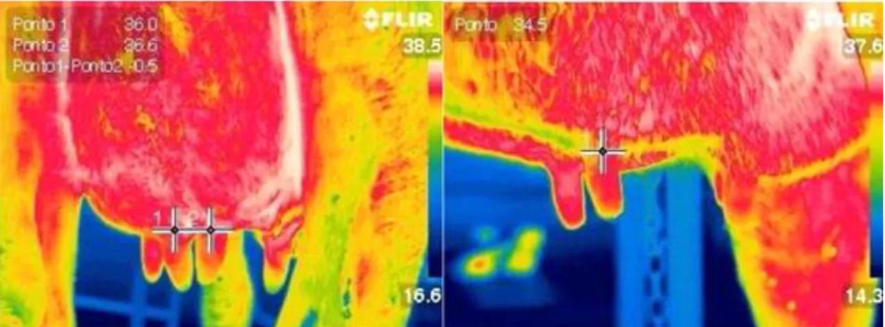
The thermal images were obtained using an infrared camera (FLIR T 440®), at 0.01ºC resolution, placed 1.5 m from the animal; the emission coefficient was adjusted at 0.97 Figure 1.
Dogiovani et al., Revista Brasileira de Higiene e Sanidade Animal (v.10, n.4) p. 685 – 692, out - dez (2016)
688 Images from the ocular globe, from
the front left quarter (FLQ), from the front right quarter (FRQ), from the rear left quarter (RLQ) and from the rear right quarter (RRQ) of the udder were recorded. The rectal temperature (RT) of each animal was measured using a digital thermometer. Subsequently, the CMT was performed and the scores ranging from 0 (the lowest) to 4 (the highest) were addressed according
to precipitation level and gel formation. The recorded data of the measured variables were presented through descriptive analysis.
The continuous quantitative data were compared through analysis of variance after the required assumptions were assessed in the Minitab 16.0 statistical package software to determine the Pearson’s linear correlation.
Results and Discussion
By taking all the data into consideration, regardless of the CMT result, it was possible seeing that the temperature (Tab. 1) was 31.62ºC ± 2.19, in FRQ; 30.85ºC ± 2.32, in FLQ; 32.08ºC ± 1.91, in RRQ; and 32.19ºC ± 2.01, in RLQ.
The mean temperature value was 38.32ºC ± 0.35, in RT; and 32.24ºC ± 1.33, inocular temperature (OT). Based on the variation coefficients, tone can state that the rectal temperatures were more homogeneous than the ocular ones, which were followed by the udder temperatures.
Table 1-Means, standard deviation, variation coefficient, maximum and minimum
temperatures in Celsius degrees, the front right quarter (1), front left quarter (2), rear right quarter (3), rear left quarter (4), rectal temperatures (5) and the ocular temperature (6) of 24 cows negative in the California Mastitis Test (7).
Parameter FRQ FLQ RRQ RLQ RT OT Mean 30.80 31.02 32.05 32.50 38.37 31.94 Standard Deviation 1.92 1.90 1.99 2.00 0.38 1.40 VariationCoefficient 6.24 6.11 6.21 6.16 1.00 4.38 Maximum 34.40 35.10 35.80 35.60 39.50 34.10 Minimum 27.30 28.20 28.60 28.10 37.80 28.60 Amplitude 7.10 6.90 7.20 7.50 1.70 5.50 1, FRQ, 2,FLQ, 3,RRQ, 4,RLQ, 5,RT, 6,OT, 7,negative CMT.
Dogiovani et al., Revista Brasileira de Higiene e Sanidade Animal (v.10, n.4) p. 685 – 692, out - dez (2016)
689 The analysis of negative CMT
animals has shown difference in the mean temperatures of the front quarters in comparison to those of the rear quarters (p=0.001). The mean and standard deviation of the temperature (ºC) obtained through the infrared thermography of the front quarters was 30.91±1.89; whereas it was 32.28±1.99, in the rear quarters. However, there is no difference between the right and left sides (p=0.399).
The mean temperature recorded through infrared thermography in the right side was 31.43± 2.03; and 31.76±2.07,in the left side. There was no interaction between position and side (p=0.763). The comparison of temperatures measured through the infrared thermography of the rear quarters of animals positive and negative in the CMT has not evidenced any difference (p=0.236); the mean temperatures recorded through the infrared thermography in the rear quarters of negative and positive CMT animals were 32.03±1.96 and 32.65±1.89, respectively. The comparison between 96 front quarters of positive CMT animals and front quarters of negative CMT animals has evidenced a difference between the mean temperatures recorded through infrared thermography in the front quarters of positive CMT animals (32.35±2.35) and those of negative CMT animals (31.00±2.20 (p=0.025)). The correlation coefficient values of the mean
udder surface temperature in negative animals, and the ocular and rectal temperatures were 0.577 and 0.475 (p= 0.000), respectively.
Unlike the present study, there was no difference in the temperature of the front and rear quarters. It may due to the fact that the authors have used controlled environments throughout the experiment and allowed the animals to rest for 30 minutes before the imaging tests (COLACK et al., 2008). Mastitis detection methods should take into account the daily variations under different environmental conditions depending on the changes in thetemperature of the udder skin; thus, reference baseline should be set (BERRY et al., 2003). It is worth considering the circadian oscillations in the body temperature of dairy cows, as it was already evidenced by authors such as (BITMAN et al., 1984).
According to Berry et al. (2003) the difference between the front and rear quarters may lie on the fact that the rear surface of the udder is in contact with the legs.
Studies involving sheep have also shown higher udder surface temperature in positive CMT animals (MARTINS et al., 2012). It corroborates the findings by Colak et al. (2008), who have found strong correlation (r=0.92) between udder surface temperature and CMT scores. The distribution of the 34 positive quarters
Dogiovani et al., Revista Brasileira de Higiene e Sanidade Animal (v.10, n.4) p. 685 – 692, out - dez (2016)
690 was 11 in FRQ and RLQ, and 6 in FLQ and
RRQ.
Thus, it resulted in 50% positive quarters between the front and rear quarters in CMT. These results contradict (BER et al., 2003), who only recorded rear quarter images, since they are more often affected by mastitis.
Although there is moderate correlation between the rectal temperature and the mean temperature in the udder, it stays within a normal range. It suggests the absence of systemic effect in positive CMT animals (COLACK et al., 2008).
Conclusion
The use of infrared thermography as a diagnostic tool to achieve early subclinical mastitis detection seems to be promising. However, a protocol to guide its use should be developed taking into
account the factors interfering in the udder temperatures, such as: the time the test is performed, environmental conditions, activity, and other management practices.
References
1. BALAS, C. Review of biomedical optical imaging - a powerful, non-invasive, nonionizing technology for improving in vivo diagnosis.
Measurement Science & Technology,
n.20, p104020, 2009.
2. BARBOSA, N.C.G.; DA SILVA, A.Z.R.; SOUZA, N.L.N.; CARVALHO, K.F.; NASCIMENTO, V.A.; DIAS, M. Relação entre a sujidade do úbere e a ocorrência de mastite subclínica.
Revista Brasileira de Higiene e Sanidade Animal, 2014 Setembro; 8 (5
Supl 1): 215-220.
http://dx.doi.org/10.5935/1981-2965.20140064029
3. BERRY, R.J.; KENNEDY, S.L.; SCOTT, S.L.; KYLE, B.L.; SCHAEFER, A.L. Daily variation in the udder surface temperature of dairy cows measured by infrared thermography.
Canadian Journal Animal Science,
v.83. p.687-693. 2003.
3. BITMAN, J.; LEFCOURT, A.M.; WOOD, D.L.; STROUD, B. Circadian and ultradian rhythms of lactating dairy cows. Journal of Dairy Science, v.67. p.1014-1023. 1984.
4. BLOOD, D.C.; RADOTITS, O. Clínica veterinária. 7th ed. Rio de Janeiro: Guanabara Koogan, 1991. 1263p.
5. COLACK, A.; POLAT, B.; OKUMUS, Z.; KAYA, M.; YANMAZ, L.E.; HAYRLI, A. Early detection of mastitis using infrared thermography in dairy cows. Journal Dairy Science, v.91. p.4244-4248. 2008.
6. COSTA, A.N.L., FEITOSA, J.V., MONTEZUMA JR, P.A., SOUZA, P.T., ARAÚJO, A.A. Rectal temperatures,
Dogiovani et al., Revista Brasileira de Higiene e Sanidade Animal (v.10, n.4) p. 685 – 692, out - dez (2016)
691 respiratory rates, production, and
reproduction performances of crossbred Girolando cows under heat stress in northeastern Brazil. International Journal of Biometeorol, n.59, v.11,
p.1647–1653, 2015.
7. FONSECA, L.F.L.; SANTOS, M.V. Qualidade do leite e controle de mastite. São Paulo: Lemos Editorial, 2000. 175p.
8. JUNIOR, G.A.F.; MANIERI, F.Z.; LOPES, N.S.S.; PILON, L.E.; ZAFALON, L.F. Ocorrência de mastite subclínica em ovelhas de diferentes raças em um mesmo sistema de produção.
Revista Brasileira de Higiene e Sanidade Animal, 2014 Setembro; 8 (5
Supl 1): 478-489.
http://dx.doi.org/10.5935/1981-2965.20140103068.
8. MARTINS, R.F.S.; PAIN. T.P.P.; CARDOSO. C.A.; DALLAGO, B.S.L.; MELO, C.B.; LOUVADINI, H.; MCMANUS, C. Mastitis detection in sheep by infrared thermography.
Research in Veterinary Science. n.94.
p.722-724. 2012.
9. MARTINS et al., Mastite subclínica em rebanhos leiteiros de propriedades rurais de Goiás. Revista Brasileira de
Higiene e Sanidade Animal, v.9, n.2,
206-214, abr–jun, 2015. DOI:
http://dx.doi.org/10.5935/1981-2965.20150019
10. MONTANHOLI, Y.R.,
NICHOLAS, E.O., KENDALL, C.S., SCHENKEL, F.S., MCBRIDE, B.W., MILLER, S. P. Application of infrared thermography as an indicator of heat and methane production and its use in the study of skin temperature in response to physiological events in dairy cattle (Bos taurus). Journal of Thermal Biology, v.33, p.468-475, 2008.
11. NÄÄS, I.A.; GARCIA, R.G.; CALDARA, F.R. Infrared thermal image for assessing animal health and welfare. Journal of Animal Behaviour
and Biometeorology, v.2, n.3, p.66-72,
2014.
12. NORBERG, E. Electrical conductivity of milking as a phenotypic and genetic indicator of bovine mastitis.
Livestock Production Science, v.96,
p.129-139, 2005.
13. PAULA, E.M.N.; DA COSTA FILHO, R.I.; NASCIMENTO, K.A.; GADÊLHA, D.F.B.G.; BARTOLI, R.B.M.; STELLA, A.E. Principais patógenos causadores de mastite subclínica em vacas na bacia leiteira do Sudoeste de Goiás. Revista Brasileira
de Higiene e Sanidade Animal, 2014
Setembro; 8 (5 Supl 1): 81-87.
http://dx.doi.org/10.5935/1981-2965.20140045010.
14. PILON, L.E; LUIZ ZAFALON, F; SANTANA, R.C.M.; JÚNIOR, G.A.F.; MANIERI, F.Z..; DOS SANTOS LOPES, N.S. Espécies de Staphylococcus coagulase-negativos isoladas da glândula mamária de ovelhas com mastite subclínica. Revista Brasileira de Higiene e Sanidade Animal, 2014 Setembro; 8 (5 Supl 1):
34-39 http://dx.doi.org/10.5935/1981-2965.20140038003.
15. RIBEIRO, M.; PETRINI, L.; AITA, M.; BALBINOTTI, M.; STUMPF JUNIOR, W.; GOMES, J.; SCHRAMM, R.; MARTINS, P.; BARBOSA, R. Relação Entre mastite clínica, subclínica infecciosa e não infecciosa em unidades de produção leiteiras na região sul do Rio Grande do Sul. Revista Brasileira de
Dogiovani et al., Revista Brasileira de Higiene e Sanidade Animal (v.10, n.4) p. 685 – 692, out - dez (2016)
692 16. SAKATANI, M.; TAKAHASHI,
M.; TAKENOUCHI, N. The efficiency of vaginal temperature measurement for detection of estrus in Japanese Black cows. Journal of Reprodroduction and
Development, v.22, n.62(2), p.201-207,
2016.
17. TIMMS, L. Mastitis diagnostic.
Western Dairy Digest, v.5, p.10-11,
2004.
18. VIGUIER, C.; ARORA, S.; SUSHRUT, A.; GILMARTIN, N.; WELBECK, K.; O’KENNEDY, R. Mastitis detection: current trends and future perspectives. Trends in Biotechnology, v.27, p.486-493, 2009.
19. WILLITS, S. Infrared thermography for screening and early detection of mastitis infection in working dairy herds.
Proceedings of Informations, v.42,