w w w . r b o . o r g . b r
Original
Article
Comparative
Study
of
the
Use
of
Intra-articular
and
Systemic
Meloxicam
to
Control
Experimentally
Induced
Osteoarthritis
in
Rabbit
Knees
夽
,
夽夽
Valéria
Trombini
Vidotto
a,b,c,∗,
Rodrigo
Tesser
da
Rocha
a,
Caroline
Lorraine
de
Paiva
d,
João
Ricardo
Nardotto
e,
Anderson
Farias
f,g,h,
Sandro
Alex
Stefanes
f,i,jaPostgraduateProgramonAnimalScience,UniãoPioneiradeIntegrac¸ãoSocial,Brasília,DF,Brazil
bDisciplineofDomesticAnimalAnatomy,VeterinaryMedicineCourse,FaculdadedeJaguariúna,Jaguariúna,SP,Brazil
cOrthopedicsandNeurologyService,VeterinaryHospital,FaculdadedeJaguariúna,Jaguariúna,SP,Brazil
dVeterinaryMedicineCourse,UniãoPioneiradeIntegrac¸ãoSocial,Brasília,DF,Brazil
eCentrodeDiagnósticoDiagnopet,Brasília,DF,Brazil
fPostgraduateProgramonVeterinaryMedicine,UniversidadeEstadualPaulistaJúliodeMesquitaFilho,SãoPaulo,SP,Brazil
gDisciplineofAnesthesiology,VeterinaryMedicineCourse,UniãoPioneiradeIntegrac¸ãoSocial,Brasília,DF,Brazil
hAnesthesiologyService,VeterinaryHospital,UniãoPioneiradeIntegrac¸ãoSocial,Brasília,DF,Brazil
iDisciplineofSurgery,VeterinaryMedicineCourse,UniãoPioneiradeIntegrac¸ãoSocial,Brasília,DF,Brazil
jOrthopedicsandNeurologyService,VeterinaryHospital,UniãoPioneiradeIntegrac¸ãoSocial,Brasília,DF,Brazil
a
r
t
i
c
l
e
i
n
f
o
Articlehistory:
Received9April2013
Accepted2May2013
Keywords:
Osteoarthritis
Anti-inflammatoryagents
Injections,intra-articular
Knee Rabbits
a
b
s
t
r
a
c
t
Objective:Thisstudyaimedtoevaluatemorphologicchanges,aswellaschondroprotective
andintra-articulareffectsofmeloxicamonjointrepairinrabbitsinducedbyexperimental
trochleoplasty,minimizingpossibleadversesideeffects.
Methods:Thirty-fiverabbitsweredividedintofourgroups:thecontrolgroup,whichdidnot
undergosurgery,andoperatedgroups,whichuseddifferentwaysofadministeringthe
anti-inflammatoryagent:systemic,0.2mg/kg;intra-articular,0.5mg/kg;positivegroupcontrol,
withoutmeloxicam.Eachoperatedgroupwasdividedaccordingtotheperiodsof7or30
daysevaluationaftersurgery.
Results:Regardingmacroscopicandhistologicalevaluationofcartilage,after30days,most
animalsshowedalmostcompletejointrepair,thepresenceoffewornoinflammatorycells;
whereaspartoftheanimalstreatedwithmeloxicampresentednecrosisinthetrochlear
ridgeandabsence ofinflammatorycellsafter7 days. Inpositive controlgroup,itwas
observedmoderateinflammationandconnectivetissueproliferation.Noneoftheanimals
intheoperatedgroupsshowedirregularities30daysaftersurgery.
Conclusion:Eitherintra-articularorsystemic,meloxicamrevealedtobefavorabletobeused
forjointrepairandcontrolofinflammatoryreaction.
©2013SociedadeBrasileiradeOrtopediaeTraumatologia.PublishedbyElsevierEditora
Ltda.Allrightsreserved.
夽
Pleasecitethisarticleas:VidottoVT,etal.Estudocomparativodousodemeloxicamporviaintra-articularesistêmicanocontroleda
osteoartriteexperimentalmenteinduzidaemjoelhodecoelhos.RevBrasOrtop.2013;48:524–531.
夽夽
PioneeringworkwasdoneintheUnionofSocialIntegration,Brasilia,DF,Brazil.
∗ Correspondingauthor.
E-mail:[email protected](V.T.Vidotto).
Estudo
comparativo
do
uso
de
meloxicam
por
via
intra-articular
e
sistêmica
no
controle
da
osteoartrite
experimentalmente
induzida
em
joelho
de
coelhos
Palavraschave:
Osteoartrite Anti-inflamatórios
Injec¸õesintra-articulares
Joelho Coelhos
r
e
s
u
m
o
Objetivo:Comoenfoquenoprocessodereparac¸ãodacartilagem,objetivou-seanalisarouso
domeloxicam,viaintra-articular,paraminimizarefeitosadversoscausadospelaaplicac¸ão
sistêmica.Avaliaram-sealterac¸õesmorfológicaseremodelamentodotecidocartilaginoso
emmodeloexperimental,emjoelhos.
Métodos: Usaram-se35 coelhos,divididosem quatrogrupos:grupocontrole(não
oper-ado),cincoanimais,egrupostratados,10animaiscada.Atécnicausadaparainduc¸ãode
osteartritefoitrocleoplastiaporabrasão. Grupostratadosforamsubdivididosdeacordo
comaviadeadministrac¸ãodamedicac¸ãoanti-inflamatória:sistêmica(0,2mg/kg),
intra-articular(0,5mg/kg)econtrolepositivo(semanti-inflamatório).Apósseteou30diasde
pós-operatório,acartilagemarticularfoiavaliadadeformamacroscópicaehistológica.
Resultados:Após30diasocorreureparac¸ãodacartilagemarticularem100%dosanimaisque
receberamamedicac¸ãosistêmicaede90%dosanimaisquereceberamviaintra-articular,
comapresenc¸adepoucasounenhumacélulainflamatória,enquantoquenogrupocomsete
diasdepós-operatórioobservou-seausênciadetecidocicatricialnosulcotroclearede
célu-lasinflamatórias.Nogrupocontroleoperado,semmedicac¸ão,observaram-seinflamac¸ão
moderadaeproliferac¸ãodetecidoconjuntivofibroso,apóssetedias.Emtodososgrupos
submetidosa30diasdepós-operatórioobservou-sediscretairregularidadenacartilagem
articular,ouausênciadela,macroemicroscopicamente.
Conclusão:Omeloxicamviaintrarticularmostrou-sefavorávelparausoemcoelhoseobteve
osmesmosresultadosdaadministrac¸ãosistêmicaquantoaremodelamentocartilaginoso
econtroledereac¸ãoinflamatória.Noentanto,sujeitoamenosefeitoscolateraisjádescritos
naviasistêmicaemaiorpraticidadeemcirurgias.
©2013SociedadeBrasileiradeOrtopediaeTraumatologia.PublicadoporElsevier
EditoraLtda.Todososdireitosreservados.
Introduction
Osteoarthrosis is the commonest aging process among
mammals.1 It is also known as degenerative joint
dis-ease (DJD) and is characterized by its non-infectious and
degenerative nature. It causes destruction of joint
carti-lage and leads to joint deformity due to disorders of
normalcelldifferentiation.2–4Althoughitisclassifiedas
non-inflammatory,acontinuouslow-gradeinflammatoryprocess
isassociatedwithDJDandthisleadstoosteoarthritis.4
Theetiologyofthedegenerativeprocessbeginswithaging,
buttheinflammatoryorinfectiousdiseasesthatdestroythe
cartilaginousstructure,ortraumainvolvingthecartilage,may
precipitateosteoarthrosis.2 Theprocess ischaracterizedby
progressiveerosionofthejointcartilageandleadsto
reduc-tion ofthejoint space, subchondralsclerosis,formation of
marginalosteophytes,subchondralcystsandsynovial
inflam-mation,which resultsin pain and reductionof functional
capacity.5
Theobjectivesoftherapyforosteoarthritisaretodiminish
thepainandmaintainorimprovejointfunction.Overthelast
fewyears,manystudieshaveinvestigatedthepotential
func-tionofanti-inflammatoryand chondroprotectiveagentsfor
repairingjointcartilage,controllinginflammatoryreactions
anddeceleratingthedegenerativeprocess.3
Non-steroidalanti-inflammatory drugs(NSAIDs) are the
agents mostused for alleviating pain over shortand long
periodsoftime.However,careneedstobetakeninviewofthe
possibleadverse effects, suchasgastrointestinal problems,
hepatotoxicityandnephrotoxicity.6–10
Withthefocusoncartilagerepair,theaimsherewereto
use the techniqueof trochleoplastyby means ofabrasion,
in orderto study themorphological changes and
cartilagi-noustissue remodelingthat wereinduced inexperimental
osteoarthritis inducedinrabbits, andtoanalyzethe useof
theNSAIDmeloxicamdirectlyonthetarget,intra-articularly,
which would provideanoptional routeforminimizing the
possibleadverseeffectscausedbysystemicadministration.
Material
and
Method
Thirty-fivehealthyNewZealandrabbits(Oryctolaguscuniculus)
ofbothsexes,weighingbetween1and2kgandofage90days,
wereused.Therabbitsweresubjectedtogeneralclinicaland
orthopedicexaminationsandlaboratorytests.Theprojectwas
approvedbytheEthicsCommitteeforAnimalUseofUnião
PioneiradeIntegrac¸ãoSocial(UPIS),underprotocolnumber
02/10.
Therabbitswererandomlydividedintofourgroups.Forthe
surgicalprocedure,itwasdecidedtostandardizeontheright
femorotibial-patellarjoint.
Controlgroup(CG):non-operated,withfiveanimals.
Treatedgroups,with10animalseach,subdivided
accord-ing to the administration route for the anti-inflammatory
Systemicgroup (SG): subcutaneousadministrationroute
forthe anti-inflammatorymedication, comprisingfive
ani-malswithapostoperativeperiodofsevendays(SG7)andfive
animalswith30days(SG30).
Intra-articulargroup(IAG):intra-articularadministration
routefortheanti-inflammatorymedication,comprisingfive
animalswithapostoperativeperiodofsevendays(IAG7)and
fiveanimalswith30days(IAG30).
Positive control group (CG+): without anti-inflammatory
medication, comprising five animals with a postoperative
periodofsevendays(CG+7)andfiveanimals with30 days
(CG+30).
Therabbitsreceivedanesthetic medicationconsistingof
ketamine(30mg/kg,intramuscularly)andxylazine(5mg/kg,
intramuscularly),together,andalsoanesthesiainthe
epidu-ral lumbosacral region, with application of 2% lidocaine
(0.3mL/kg).
Toexperimentallyinduceosteoarthrosis,thetechniqueof
trochleoplasty bymeans of abrasion was used. The
surgi-cal accesscomprised a lateral approach tothe knee joint,
as described by Fossum.4 The patella was dislocated to
enableexposureofthefemoraltrochlea.Thekneewasflexed
and, with the aid of a spherical milling device of 2mm
in diameter, coupled to a high-rotation microgrinder, the
trochleoplasty procedure was performed by deepening the
trochleargroovedowntothesubchondralbone,whichavoided
damagingthetrochlearbordersandtheadjacentjoint
carti-lage.
During the surgicalprocedure, after closing the capsule
and retinaculum, the animals in the intra-articular group
(IAG)receivedmeloxicam,inasingledoseof0.5mg/kg,
intra-articularly.
Theanimalsinthesystemicgroup(SG)receivedmeloxicam
atadoseof0.2mg/kg,subcutaneouslyevery24h,forthree
consecutivedays.
Alltheanimalsoperatedreceivedprophylacticantibiotic
therapy comprising anassociation of penicillins and
dihy-drostreptomycinsatadose of50,000UI/kg,intramuscularly
every48h(threeapplications).Theyalsoreceivedanalgesic
comprising tramadolhydrochloride ata dose of 4.0mg/kg,
subcutaneously every 12h, for three consecutive days, as
describedbyLichtenberger.11
Atthepreestablishedtimesof7and30daysafterthe
opera-tion,theanimalswereevaluatedtodescribedthemacroscopic
changes to the joint and to collect samples for
histologi-calanalysis.Theanimalswereanesthetizedusingketamine
(30mg/kg,intramuscularly)andxylazine(5mg/kg,
intramus-cularly)andweresacrificedbyapplyinganoverdoseof2.5%
sodiumthiopental and 19.1%potassiumchloride, in
accor-dancewiththerecommendedstandardsforuseofanimals
inscientificresearch.12
Thedistalepiphysesofthefemurwerecollectedandstored
inindividualflaskswith10%bufferedformaldehydesolution
atroomtemperature,forhistologicalevaluation.
Inthehistopathologicalanalysis,usingsectionsstainedby
meansofthehematoxylin–eosin(HE)andGomoritrichrome
(GT)methods,thebiologicalresponsewasdetermined asa
function of the cartilage repair process and inflammatory
changesinthejoint.Usingablindedanalysis,theresultswere
assessedaccordingtotheirhistologicalgrading,inscoretables
thathadbeenmodifiedfrompreviousstudiesconductedby
Oliveira13andSaricaogluetal.14
Toevaluatethenonparametricdatafromthehistological
analysisoncellmorphologyandjointinflammatoryreaction,
theMann–WhitneyRankSumtestwasusedtomake
compar-isonsbetweenthegroups.Allthecomparisonsweremadeat
thesignificancelevelof5%(p≤0.05).Forthis,theSigmaStatfor
Windowsstatisticalsoftware,version3.0.1,wasused.
For theother evaluations,on thedataobtainedthrough
macroscopicandhistologicalanalyses,descriptivemethods
wereused.
Results
Thetrochleargrooveofthenegativecontrolgroup(CG−)was
evaluatedasameansofmacroscopiccomparison.Nosurface
changeswereobserved(Fig.1A).
Sevendaysaftertheoperation,thefollowingmacroscopic
observationscouldbemadeinthegroupsevaluated:
• In four animals of the group CG+7 (4/5, 80%), areas of
irregularitywereobservedintherepairtissueandthe
red-dened borders atthetransition tothe adjacent cartilage
(Fig.2A).
• Thisfeaturewasalsoobservedinthreeanimalsofthegroup
SG7(3/5,60%)andinoneanimalofthegroupIAG7(1/5,20%).
• InthreeanimalsinthegroupIAG7(3/5,60%)andintwo
ani-malsofthegroupSG7(2/5,40%),theseareasofirregularity
intherepairtissuepresentedsmallareasofhyperemiaand
whitenedtissueattheextremitiesofthelesion(Fig.2B).
After 30 days, the following macroscopic observations
couldbemadeinthegroupsevaluated:
Fewerirregularitiesintherepairtissue,whichpresented
continuitywiththeadjacentnormalcartilageinfouranimals
ofthegroupCG+30(4/5,80%),inalltheanimalsinthegroup
SG30(5/5,100%)andinfouranimalsofthegroupIAG30(4/5,
80%)(Fig.3).
Duringthemicroscopicevaluationonthejointcartilage,it
waspossibletomakethefollowingobservations:
• IntheanimalsofthegroupCG−(5/5,100%),thetrochlear
groovepresentedacoveringofhyalinecartilaginoustissue,
withabsenceofinflammatorycells.
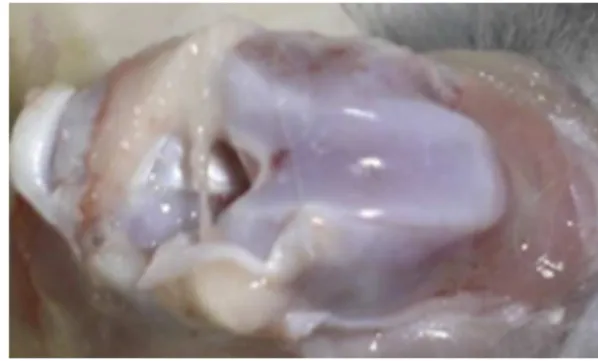
Figure1–Photographoftherightkneeofananimalin CG−,withoutabnormalities,withasmoothandshinyjoint
Figure2–(A)PhotographofthetrochleoplastyregionoftherightkneeofananimalinCG+7,withareasofirregularityin therepairtissueandreddenedborderatthetransitiontotheadjacentnormalcartilage(arrow).(B)Photographofthe trochleoplastyregionoftherightkneeofarabbitinIAG7,withareasofirregularityintherepairtissuebutwithoutareasof hyperemia(whitearrow),andpresentingwhitenedtissueattheextremitiesofthelesion(dashedarrow).
• Four animals ofthe group CG+7(4/5, 80%)and one
ani-malofthegroupSG7(1/5,20%)presentedmildtomoderate
inflammatoryreactionsintheareaofthetrochleargroove,
withintensedepositionoffibrousconnectivetissue,
sur-faceirregularity,congestionandedema,alongwithalow
neutrophilcount(Fig.4A).
• IntwoanimalsofthegroupSG7(2/5,40%)andthreeanimals
ofthegroupIAG7(3/5,60%),aminimalinflammatory
reac-tionwasobserved,withmildcongestionandedema,areas
ofintensehemorrhageandabsenceofhealingtissueinthe
regionofthetrochleargroove,wherethetrochleoplastywas
performed(Fig.4B).
Theother animals presented mild to moderate
inflam-matory reactions, with the presence of neutrophils and
macrophages,alongwithdepositionoffibrocartilaginous
tis-suestainedwithhematoxylinandeosin(HE)(Fig.4C),which
wasseenbetteronslidesstainedwithGomoritrichrome(GT)
(Fig.5D).
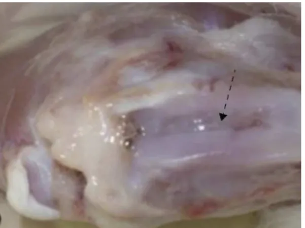
Figure3–Photographofthetrochleoplastyregionofan animalinSG30,withregularrepairtissuesurfacethatis continuouswiththeadjacentnormalcartilage(dotted arrow).
Alsoduringthemicroscopicevaluationonthejoint
carti-lage,inrelationtotheanimalsthatwereexamined30days
after the operation, it was possible tomake the following
observations:
• InfouranimalsofthegroupCG+30(4/5,80%),aminimal
inflammatoryreactionwasobserved,withmildcongestion
andedema,presenceofhyalinecartilageandlittle
fibrocar-tilage(Fig.5A).
• FouranimalsofthegroupsSG30andIAG30(4/5,80%)no
longerpresentedanyinflammatorycellsandonlypresented
hyalinecartilage,whichwasstainedusingHE(Fig.5B)and
GTandshowedthedisorganizationofthecollagen(Fig.5C).
In making statistical comparisons between the groups
sevendaysaftertheoperation,therewerenosignificant
dif-ferences(p≤0.05).However,incomparingtheanimalsafter30
days,itwasseenthattherewasagreaterinflammatory
reac-tionintheoperatedcontrolgroup(CG+30),inrelationtothe
groupthatreceivedsystemicmedication(Table1).
Inaddition,itwasobservedthattherewasalsoachangein
thesignificantinflammatoryreaction(p≤0.05),incomparing
therabbitsthatreceivedsystemicmeloxicamandwere
exam-inedsevendaysaftertheoperation(SG7)withthosethatwere
examinedafter30days(SG30).
Incomparingthecellmorphology,therewasasignificant
differencebetweentherabbitsthatreceivedsystemic
meloxi-camandwereexaminedsevendaysaftertheoperation(SG7),
andthosethatwereexaminedafter30days(SG30)(Table2).
Discussion
This study was characterized by being conducted using
an intra-articularroute ina rabbit modelforexperimental
osteoarthritis.Trochleoplastybymeansofabrasionwasused,
sincethisisaroutewithfewreportsinveterinarymedicine.
Inthehistopathologicalevaluationofthetrochleargroove,
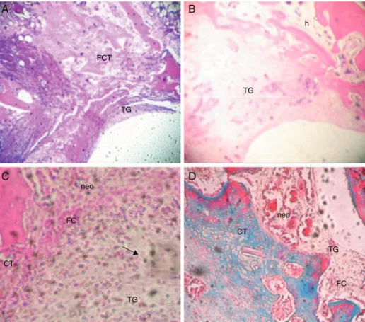
B
A
C
D
FCT
FC
CT
TG
TG
TG
CT
TG
FC h
neo neo
Figure4–Photomicrographofthetransitionareabetweenthetrochleagroove(TG)lesionandtheadjacentcartilaginous tissue(CT).(A)InarabbitofthegroupCG+7.Notetheintenseformationoffibrousconnectivetissue(FCT)withsubchondral lacunae.(B)InarabbitofthegroupIAG7.Notetheabsenceofformationofrepairtissueintheareaofthetrochleagroove (TG)lesion,andtheintensehemorrhaging(h).(C)InarabbitofthegroupSG7,withformationoffibrocartilage(FC)and slightinflammatoryreaction.Notethepresenceofmononuclearcells(arrow)andneovascularization(neo).(HE;40×).(D)In
arabbitofthegroupSG7,showingirregularity,withareasfilledwithfibrocartilage(FC).Noteareaofneovascularization (neo)(GT;40×).
A
B
C
c CT
CT
CT TG
TG TG
c
Figure5–Photomicrographofthetransitionareabetweenthetrochleargroove(TG)lesionandtheadjacentcartilaginous tissue(CT).(A)InarabbitofthegroupCG+30.Notethecontinuityofthetissue,withintenseformationofcartilaginous tissue(c).(B)InarabbitofthegroupIAG30.Notecontinuityofthetissue(HE;40×).(C)InarabbitofthegroupSG30,with
formationofcartilaginoustissue(CT).Notedisorganizationofthecollagen(c)(GT;40×).
jointsurfacewasreplacedbyfibroblast-richconnectivetissue
withadelicatestructure,withdepositionofimmaturetypeIII
collagenandnumerousbloodvessels,andwiththepresence
ofamildtomoderateinflammatoryreaction.Thisconfirmed
theobservationthatthejointcartilagehadlostits
homoge-nousnatureandwasbrokenandfragmented,withfibrillation.
Inthisregard,Silva15alsodescribedthepresenceofintensely
vascularizedtissue,withahighcellcontentanddense
con-nective tissue covering the area of the trochlear groove
lesion.
Corroborating Souza et al.,16 the macroscopic evaluated
showedthepresenceofirregularitiesandreddenedareason
the edgesofthelesion,whichconfirmedthatthejoint
tis-sue wasavascularandtheinflammatoryreactionmediated
bybloodvesselsbeganintheunderlyingtissue.
Thehistochemicalstainingofthematrixforproteoglycans
wasunequal,andthelineofseparationbetweenthecalcified
cartilageandtheradialzonehadbeeninvadedbycapillaries.5
Forthisreason,itneedstobeemphasizedthatthe
Table1–GradingoftheCellMorphologyFoundintheJointCartilageoftheGroups.
CG− CG+7 SG7 IAG7 CG+30 SG30 IAG30
0 5 2a 2 1 1# 2
0 5 3a 6 1 1# 1
0 5 6a 6 1 1# 1
0 5 5a 6 1 1# 5
0 2 6a 1 5 1# 1
0,normal;1,cartilageandsomefibrocartilage;2,fibrocartilage;3,somefibrocartilage,butmanynon-chondrocyticcells;4,onlynon-chondrocytic cells;5,fibroustissue;6,absenceofhealingtissue.GradingadaptedfromOliveira.13
a Statisticaldifferencebetweensubgroups(p≤0.05);gradingadaptedfromOliveira.13
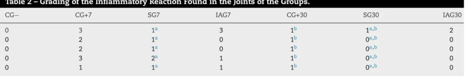
Table2–GradingoftheInflammatoryReactionFoundintheJointsoftheGroups.
CG− CG+7 SG7 IAG7 CG+30 SG30 IAG30
0 3 1a 3 1b 1a,b 2
0 2 1a 0 1b 0a,b 0
0 2 1a 0 1b 0a,b 0
0 3 2a 1 1b 0a,b 0
0 1 1a 1 1b 0a,b 0
0,noinflammation;1,minimalinflammation(slightcongestionandedema);2,mildinflammation(erosionofthejointsurface,congestion andedema;lowneutrophilcount);3,moderateinflammation(presenceofneutrophilsandmacrophages);4,severeinflammation(presenceof neutrophilsandmacrophages;fibrinexudate).GradingadaptedfromSaricaogluetal.14
a Statisticaldifferencebetweensubgroups(p≤0.05).GradingadaptedfromSaricaogluetal.14
b Statisticaldifferencebetweengroups(p≤0.05).
whichistheprimarysourcefordevelopingsuchresponses.
Thus,thepromotercellshadaccesstothelesionandenabled
formationoftissuecomposedoffibrocartilage.Thisfinding
reflectsinadequatetissuerepair,giventhatcollagensIand
IIare notusuallyexpressedincartilaginous tissue,as also
observedbyVelosaetal.17andRossi.18
Fibrocartilageor fibrouscartilage is atransitional tissue
andhasfunctionalandstructuralpropertiesthatarebetween
those ofdenseconnectivedisuse and hyalinecartilage. As
citedbyGhivizzanietal.19andOliveira,13although
fibrocarti-lageisresistanttotension,itischaracterizedbythepresence
ofcollagenI.Therefore, theformationoffibrocartilagethat
wasobservedwasundesired,becauseitalteredthestructural
andbiomechanicalpropertiesofthejoint.
Althoughgrowthofthistypeoftissuewasalsofoundin
someanimalsinthe groupsthatunderwent surgical
inter-vention(SG7andIAG7),whatdrewattentionmostwasthat
twoanimalsinthegroupSG7(2/5,40%)andinthreeanimals
inIAG7(3/5,60%),notonlyweretherenoinflammatorycells,
butalso,microscopically,thesiteonlypresentedareaswith
absenceofhealingtissueinthe area ofthetrochleoplasty,
without signs oftissuerepair. These were macroscopically
observedasirregularareas andwhitenedtissue.Thisleads
to the conclusion that, independent ofthe administration
route,thepresenceofanti-inflammatoryagentsblockedthe
metabolizationofarachidonicacid bytheCOX-2 routeand
impeded production of prostaglandins and consequently
theirinflammatorymetabolites,intherepairtissue,asalso
observedintheexperimentconductedbyMarchionnietal.20
Integrins, which form one of the main families of cell
surfacereceptors,participateinthemigrationofneutrophils
through bindingthese cells to vesselwalls, which enables
diapedesis. However, since integrins are inhibited by the
action of medications derived from oxicams, reduction of
polymorphonuclear cells takesplace. Thiswas seenin the
present investigation, which confirms what is said in the
literatureregardingmeloxicam,i.e.thatitisanon-steroidal
anti-inflammatory agent that is preferentially selective
for COX-2 and which demonstrates a capacity to inhibit
inflammationduringtheacutephase.20
These cells are responsible for absorption of the fibrin
of the coagulum. They synthesize growth factors that are
chemotaxicandmitogenictowardtheendothelialcellsthat
are present on the periphery of the lesion, and they
pro-motemigrationandformationofnewvessels.Forthisreason,
reductionofthesegrowthfactorsalsodiminishescellrepair,
giventhatthehealingprocessneedstofirstlygothroughthe
inflammationphase,sothatthenecrosedtissuecanbe
phago-cytizedandfibroblastscanberecruitedtostartthehealing
cascade,andthereaftergothroughthephasesofproliferation,
differentiationandtissuematuration.21,22
Inthesameanalysis,butnowontheanimalsexamined
30daysaftertheoperation,therewasnostatistically
signifi-cantdifferencebetweenthegroupsoperatedandthepositive
control(CG+30), inrelationtotissuerepair, giventhatina
largeproportionoftheanimalsinthegroupsCG+30,SG30and
IAG30,therewerelargequantitiesofhyalinecartilageandlittle
fibrocartilage,althoughtheystillpresenteddisorganizationof
thecollagen,asseenusingtrichromestaining.However,there
was asignificant differencebetweenthe medicatedgroups
andthepositivecontrolregardingtheinflammatoryreaction,
sincethecellularityobservedinthehistologicalanalysison
themedicatedanimalswasvisiblylower.
Itisknownthat jointcartilage isasparselycellular
tis-sue, and thatits biochemical characteristics mainly reflect
thecompositionoftheextracellularmatrix.Itisformedfrom
typeIIcollagenandproteoglycans,whichareresponsiblefor
isasmoothand shinytissue.5 Theseobservationscouldbe
madethroughmacroscopicandmicroscopicevaluationson
thelesion.Thesefindingsdemonstratethatlong-termuseof
anti-inflammatoryagentsenabledgoodcartilagerepair,
with-outanysignificantdifferencebetweenthecontrolgroupand
thegroupswithsystemicandintra-articularadministration,
butwithoutanycontinuationofaninflammatoryreaction.
With decreasing concentrations of inflammatory
prostaglandins,theincreaseinvascularpermeabilityandthe
tissueaggressionthatthesemediatorscauseareminimized
throughtheactionofthemedication.Thisactiontherefore
favors fibrogenesis and makes the extracellular matrix of
animalssubjectedtotheactionofmeloxicamricherincells
andmoreorganizedincollagenfibersatthelesionsite.20
Putting together thedata from the four groupsand the
observationtimes,analysisonthevariablesrelatingtoacute
andchronicinflammationandtorepairshowedthat
meloxi-camcontrolledtheacuteinflammatoryreaction(sevendays
aftertheoperation),independentoftheadministrationroute.
Thirtydaysaftertheoperation,thiscontroloverthe
inflam-matoryreactionwasmaintained,whichenabledsatisfactory
repairofthecartilaginoustissueofthefemorotibial-patellar
joint.
Itisknownthatmanypatientswhoundergojointsurgery
mayrequireprolongeduseofanti-inflammatoryagents.
How-ever,theiruseviaasystemicroute,evenatatherapeuticdose,
mayleadtoadversereactionssuchashepatotoxicityand,
par-ticularly,gastrointestinaldisorders,asdescribed byAlencar
etal.23
A single application of meloxicam intra-articularly
achievedaresultsimilartowhatwasfoundusingsystemic
administration. For this reason, it can be suggested that
intra-articularapplicationcanbeusedrationallyastreatment
intheimmediatepostoperativeperiod,forjointsurgery.
Conclusion
Fromevaluatingtheresultsobtainedfromtheexperimental
modelofthisstudy,bymeansofmacroscopicand
histopatho-logical examinations, it can be concluded that meloxicam
is effective for controlling the joint inflammatory process,
both in systemic and in intra-articular applications, and
it enables cartilage remodeling in an experimental model
using rabbits. Thus, this study contributes toward
advanc-ing knowledge and allows several new questions to be
asked,withnewproposalsforlocalanti-inflammatory
treat-ment.
Conflicts
of
Interest
Theauthorsdeclarenoconflictsofinterest.
r
e
f
e
r
e
n
c
e
s
1. PelletierJP,YaronM,HaraouiB,CohenP.Efficacyandsafetyof diacereininorteoarthritisoftheknee:adouble-blind, placebo-controlledtrial.TheDiacereinStudyGroup.Arthritis Rheum.2001;43(10):2339–48.
2.CamanhoGL.Tratamentodaosteoartrosedojoelho.RevBras Ortop.2001;36(5):135–40.
3.CaldeiraFMC,MuzziLAL,MuzziRAL.Artroseemcães. CadernoTécnicodeVeterináriaeZootecnia.2002;37(1): 53–83.
4.FossumTW.Cirurgiadepequenosanimais.SãoPaulo:Roca; 2005.
5.RezendeMA,GobbiRG.Tratamentomedicamentosoda osteoartrosedojoelho:drogasmodificadorasdadoenc¸a.Rev BrasOrtop.2009;44(1):14–9.
6.LeesP,LandonIMF,GiraudelJ,ToutainPL.Pharmacodynamics andpharmacokineticsofnonsteroidalanti-inflammatory drugsinspeciesofveterinaryinterest.JVetPharmacolTher. 2004;27(6):479–90.
7.ClarkTP.Theclinicalpharmacologyof
ciclooxigenase-2-selectiveanddualinhibitors.VetClinNorth AmSmallAnimPract.2006;36:1061–85.
8.FoxDB.Currenttreatmentstrategiesofcanineandfeline
osteoarthritis.In:NAVCproceedings(NorthAmerican
veterinaryconference)[serialontheInternet].2006.Available
at:http://www.ivis.org/proceedings/
navc/2006/SAE/319.asp?LA=1[cited01.10.2011];20:90–4[about
4p.].
9.JohnstonSA.Ostearthrits:jointanatomy,physiology,and pathobiology.VetClinNAm:SmallAnimPract.
2007;27:699–719.
10.FilhoMM,RahalSC.Ousodeanti-inflamatóriosinibidores Cox-2seletivosnaosteoartritecanina.Veterináriae Zootecnia.2008;15(3):407–15.
11.LichtenbergerM.Analgesiaintheferretandrabbit.In:56◦
CongressoInternazionaleMultisala,SCIVAC[periódicona
Internet].2007.Availableat:http://www.ivis.org[cited
03.10.2011];[aboutp.327–0].
12.CFMV.Eutanásia:resoluc¸ãodoCFMVinstituinormase procedimentosparaeutanásiaemanimais.Veterinária eZootecniaemMinasGeraisCRMV/MG.2002;17(75): 25.
13.OliveiraBJNA.Enxertoosteocondralalógeno,associadoà inoculac¸ãodecélulasmononuclearesdamedulaósseae proteínamorfogenéticaósseanoreparodosulcotroclearde coelhos.[dissertac¸ão].Uberlândia:UniversidadeFederalde UberlândiaFaculdadedeMedicinaVeterinária;2008.
14.SaricaogluF,DalD,AtillaP,ÌskitAB,TarhanO,AsanE,Aypar U.Effectofintraarticularinjectionoflornoxicamonthe articularcartilage&synoviuminrat.IndianJMedRes. 2008;127:362–5.
15.SilvaAA.Avaliac¸ãoclínicaderattusnorvegicusapósterapia antiinflamatóriacominibidorseletivoounãoparacox-2por extrapolac¸ãoalométrica.[tese].SantaMaria:Universidade FederaldeSantaMariaDepartamentodeMedicina Veterinária;2004.
16.SouzaR,RaiserA,GuimarãesL,RiosM,AraújoL,LeotteeA, HintzeC.Precursoresdeglicosaminoglicanosnareparac¸ão articularapóstraumaiatrogêniconojoelhodecães.RevClin Vet.1999;23(1):33–8.
17.VelosaAPP,OliveiraAM,CarrascoS,CapelozziVL,Teodoro WR,YoshinariNH.Meniscectomiaparcialcomomodelo experimentaldeosteoartriteemcoelhoseefeitoprotetordo difosfatodecloroquina.RevBrasReumatol.2007;47(6): 401–10.
18.RossiE.Envelhecimentodosistemaosteoarticular.Einstein. 2008;6(1):S7–12.
19.GhivizzaniSC,OliginoTJ,RobbinsPD,EvansCH.Cartilage injuryandrepair.PhysMedRehabilClinNAm.
2000;11(2):289–307.
21.LinTW,CardenasL,SoslowskyLJ.Biomechanicsoftendon injuryandrepair.JBiomech.2007;37(6):865–77.
22.IamagutiLS,BrandãoCVS.Usodemembranabiossintéticaa basedecelulosenaregenerac¸ãotecidualguiada.Semina: CiênciasAgrárias,Londrina.2007;28(4):701–8.