A R T I G O D E R E V I S Ã O
O S T E O B L A S T S A N D
B O N E F O R M A T I O N
Joana Caetano-Lopes,
*Helena Canhão,
*,**João Eurico Fonseca
*,**Abstract
Bone is constantly being remodelled in a dynamic process where osteoblasts are responsible for bone formation and osteoclasts for its resorption.
Osteoblasts are specialized mesenchymal cells that undergo a process of maturation where genes like core-binding factor α1 (Cbfa1) and osterix (Osx) play a very important role. Moreover, it was found recently that the Wnt/β-catenin pathway plays a part on osteoblast differentiation and proliferation. In fact, mutations on some of the proteins involved in this pathway, like the low-density lipoprotein re-ceptor related protein 5/6 (LRP5/6), lead to bone di-seases.
Osteoblasts have also a role in the regulation of bone resorption through receptor activator of nu-clear factor-κB (RANK) ligand (RANKL), that links to its receptor, RANK, on the surface of pre-osteo-clast cells, inducing their differentiation and fu-sion. On the other hand, osteoblasts secrete a so-luble decoy receptor (osteoprotegerin, OPG) that blocks RANK/RANKL interaction by binding to RANKL and, thus, prevents osteoclast differentia-tion and activadifferentia-tion. Therefore, the balance betwe-en RANKL and OPG determines the formation and activity of osteoclasts.
Another factor that influences bone mass is lep-tin, a hormone produced by adipocytes that have a dual effect. It can act through the central nervous system and diminish osteoblasts activity, or can have an osteogenic effect by binding directly to its receptors on the surface of osteoblast cells.
Keywords: Osteoblasts; Bone Formation;
Wnt/β--catenin Signalling; Leptin; Cbfa1.
Resumo
O osso está em constante remodelação num pro-cesso dinâmico, em que os osteoblastos são res-ponsáveis pela sua formação e os osteoclastos pela sua reabsorção.
Os osteoblastos derivam de células mesen-quimatosas que sofreram um processo de
ma-turação, no qual genes como o core-binding factor
α1 (Cbfa1) e osterix (Osx) assumem um papel de grande importância. Foi descoberto recentemente que a via de sinalização Wnt/β-catenina tem um papel relevante na diferenciação e proliferação do osteoblasto. Na realidade, mutações em deter-minadas proteínas envolvidas nesta via, como
o co-receptor low-density lipoprotein receptor
re-lated protein 5/6 (LRP5/6), são causa de doenças
ósseas.
Os osteoblastos desempenham também um pa-pel importante na regulação da reabsorção óssea
através do receptor activator of nuclear factor-κB
(RANK) ligand (RANKL) que interage com o seu re-ceptor RANK, expresso na superfície de pré-osteo-clastos, induzindo a diferenciação e fusão destas células. Por outro lado, os osteoblastos secretam um receptor solúvel, a osteoprotegerina (OPG), que bloqueia a interacção RANK/RANKL através da sua ligação ao RANKL, prevenindo assim a diferencia-ção e activadiferencia-ção do osteoclasto. Desta forma, o ba-lanço entre RANKL e OPG determina a formação e actividade do osteoclasto.
Outro factor que influencia a massa ós-sea é a leptina, uma hormona produzida pelos adipócitos, que tem um efeito duplo. Pode actuar através do sistema nervoso central e diminuir a actividade do osteoblasto, ou pode exercer um efeito osteogénico através da interacção com os receptores presentes na superfície do osteo-blasto.
Palavras-chave: Osteoblastos; Formação do Osso;
Sinalização Wnt/β-catenina; Leptina; Cbfa1.
*Unidade de Investigação em Reumatologia, Instituto de Medicina Molecular, Faculdade de Medicina da Universidade de Lisboa **Serviço de Reumatologia e Doenças Ósseas Metabólicas, Hospital de Santa Maria
O S T E O B L A S T S A N D B O N E F O R M AT I O N
Introduction
Bone must be stiff, slightly flexible and light in or-der to make loading possible and facilitate move-ment. In particular, bone has a specific characteris-tic that distinguishes it from other materials: this tissue can respond according to the location and extent of the damage, remove it and replace it with
new bone.1
Bone is composed by cells and an extracellular matrix which becomes mineralized by the depo-sition of calcium hydroxyapatite, giving the bone rigidity and strength. Bone has three distinct cell types: the osteoblasts, or bone-forming cells, the osteoclasts, or bone-resorbing cells, whose
functi-ons are intimately linked,2and the osteocytes,
which are osteoblasts entrapped within lacunae. In order to balance bone formation and resorption in healthy individuals, osteoblasts secrete factors that regulate the differentiation of osteoclasts and osteocytes secrete factors regulating the activity of
both osteoblasts3and osteoclasts.1Bone is
cons-tantly being resorbed by osteoclasts and then placed by osteoblasts in a process called bone
re-modelling.2Resorption is much faster than
forma-tion: an area of bone can be resorbed in 2-3 weeks
but it takes at least three months to rebuild it.4Bone
resorption is probably the first event that occurs
in response to a mechanical stress signal.5In fact,
resorption and formation are tightly coupled, so that after resorption a formation phase occurs and, normally, the amount of bone resorbed will be for-med in the succeeding phase (Figure 1). This coor-dination arises from the linkage between osteo-blasts and osteoclasts, which is mediated by the release of growth factors from bone during
re-sorption.6
On the other hand, osteoblasts, have, in addition to their classic role as bone forming cells, the abili-ty to regulate bone resorption through the expres-sion of receptor activator of nuclear factor-κB (RANK) ligand (RANKL), which links to its recep-tor, RANK, on the surface of pre-osteoclast cells, in-ducing their differentiation and causing bone resorption. On the contrary, the soluble decoy re-ceptor (osteoprotegerin, OPG), also produced by osteoblasts, is able to block RANK/RANKL interac-tion by binding to RANKL and, thus, prevent osteo-clast differentiation and activation, reducing bone resorption.6
In this article, we will review osteoblast biology and the processes that influence bone formation and resorption through this cell.
Osteoblasts
Osteoblasts have a very important role in creating and maintaining skeletal architecture; these cells are responsible for the deposition of bone matrix and for osteoclasts regulation. Osteoblasts are mo-nonuclear, not terminally differentiated,
speciali-zed cells.7When they are active, a large Golgi
ap-paratus and an abundant rough endoplasmic reti-culum is visible. In addition, osteoblasts form tight junctions with adjacent osteoblasts and have re-gions of the plasma membrane specialized in
ve-sicular trafficking and secretion.8As they
differen-tiate they acquire the ability to secrete bone
ma-trix.6Ultimately, some osteoblasts become trapped
in their own bone matrix giving rise to osteocytes
which, gradually, stop secreting osteoid.8
Osteo-cytes are the most abundant cells in bone; these cells communicate with each other and with the surrounding medium through extensions of their
plasma membrane.9,10Therefore, osteocytes are
thought to act as mechanosensors, instructing teoclasts where and when to resorb bone and
os-teoblasts where and when to form it.1,10
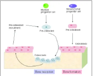
Figure 1. Bone is constantly being resorbed by osteoclasts
and formed by osteoblasts in a coupled process, mediated by the release of growth factors from bone during resorption and by molecules produced by osteoblasts, which regulate the differentiation of osteoclasts. (Osteoblasts and osteoclasts symbols were adapted from Rheugulation Database, knowledge base on rheumatoid arthritis, www.rheugulationdb.com)
J O A N A C A E TA N O-L O P E S E C O L.
Wnt pathway on osteoblastogenesis
Wnts are secreted glycoproteins that are found in
all animal species.11The human genome encodes
19 Wnt genes that bind to cell-surface receptors
and mediate a cascade of events that regulate a va-riety of cellular activities, including cell fate, deter-mination, proliferation, migration, polarity and gene expression. Wnts activate three distinct in-tracellular signalling cascades: the Wnt/β-catenin
pathway, the Wnt/Ca2+pathway and the
Wnt/pla-nar polarity pathway. The Wnt/β-catenin pathway is frequently referred to as the canonical pathway and it promotes cell fate determination, prolifera-tion and survival through the increase of β-catenin levels and alteration of gene expression by the transcription factor Lymphoid enhancer factor/T cell factor (Lef/Tcf ).12Activation of this signalling
pathway occurs with binding of Wnt to Frizzled (Fz), a transmembrane receptor, and low-density lipoprotein receptor related protein 5/6 (LRP5/6)
co-receptors11, 13(Figure 2). In the absence of a Wnt
ligand, the cytosolic level of β-catenin is kept low by its phosphorylation and degradation, thereby suppressing the expression of Wnt-responsive
ge-nes.12Inappropriate activation of Wnt signalling
contributes to cancer and reduced Wnt signalling
has been associated with osteoporosis.11,14
The first indication that Wnt signalling has an important role in bone formation came from stu-dies in humans where mutations that inactivate the Wnt co-receptor LRP5 were shown to cause os-teoporosis. Moreover, gain-of-function mutations in this receptor increased Wnt signalling and resul-ted in a high bone mass phenotype, both in
hu-mans and mice,15due to an elevated number of
ac-tive osteoblasts which seem to be protected from apoptosis. In fact, the mutation G171V on LRP5 is
a missense mutation14that decreases the
interac-tion between LRP5 and the Wnt antagonist Dick-kopf1 (Dkk1), thereby diminishing its inhibitory
effect on endogenous Wnt signalling.3In mice, null
mutations in LRP5 resulted in bone loss after birth, due to decrease in bone formation and osteoblast
proliferation.4Therefore, loss-of-function
mu-tations in LRP5 lead to low bone density whereas gain-of-function mutations cause high bone
mass.14It was also found that a series of genes
in-volved in the Wnt signalling pathway are induced during fracture repair in rodents, giving strength to the relation between Wnt signalling and LRP5 on bone.4
LRP5 is expressed at low levels in various tis-sues, shows little temporal changes, and constitu-tes a key factor in bone regulation. This effect is linked to the role of LRP5 on the Wnt signalling pathway. LRP5 interacts with proteins from the Wnt family to form a complex with Fz, leading to the activation of the canonical Wnt signalling
path-way.4In fact, β-catenin activity is essential for the
differentiation of mature osteoblasts and, conse-quently, for bone formation. However, lack of this molecule does not change the differentiation of osteoprogenitor cells into the early osteoblastic precursors but, instead, it blocks the expression of Osterix (Osx) and, as a consequence, these cells
ac-quire a chondrogenic phenotype.3The effect of
LRP5 on bone mass is mediated by the β-catenin
Wnt signalling pathway and, in vitro, it results in
Figure 2. Canonical Wnt signalling has multiple roles in
osteoblastogenesis, is essential for osteoblast lineage differentiation early in the development and, postnatally, is involved in osteoblast proliferation, survival and lifespan. Binding of Wnt to Frizzled and LRP5/6 receptors induce a signalling cascade that allows the accumulation of β-catenin in the citosol. β-catenin then enters the nucleus where it promotes the transcription of target genes. APC – Adenomatous Polyposis Coli; Dkk – Dickkopf; Dsh – Dishvelled protein; Fz – Frizzled receptor; GSK3 – Glycogen Synthase Kinase-3β; LRP5/6 – Low--density Lipoprotein Receptor Related Protein 5/6; sFRP – secreted frizzled-related protein;WIF-1 – Wnt inhibitory factor; Lef/Tcf – lymphoid enhancer factor/T cell factor. (Adapted from Krishnan et al.)13
O S T E O B L A S T S A N D B O N E F O R M AT I O N
the expression of an early osteoblast marker, alka-line phosphatase (ALP). Although this signalling pathway is involved in the regulation of osteoblas-togenesis and bone formation, a specific Wnt pro-tein that triggers its activation has still not been
identified.15 In fact, several Wnt genes, such as
Wnt1, Wnt4, Wnt5α, Wnt9α/14 and Wnt7b are ex-pressed in either osteoblast precursors or adjacent tissues during development, and Wnt3α and
Wnt10b are expressed in bone marrow,13but only
Wnt10b mutants express a postnatal decrease in
bone mass.3The expression of Wnt10b in
mesen-chymal progenitor cells, in vitro, induces the
ex-pression of the transcription factors core binding factor α1 (Cbfa1), Distal-less homeobox 5 (Dlx5) and Osx, stimulating osteoblastogenesis. These observations substantiate the fact that Wnt signal-ling influences the differentiation of the precursor cells by increasing the expression of key
osteoblas-togenic transcription factors15(Figure 3). On the
other hand, Wnt10b inhibits the transcription fac-tors CCAAT/enhancer-binding protein α (C/EBPα) and peroxisome proliferator-activated receptor γ
(PPARγ), blocking adipogenesis.15Thus, Wnts have
also a role on mesenchymal precursor cells linea-ge commitment and adipolinea-genesis is most likely
the default pathway for the cells that do not recei-ve proper inductirecei-ve signals to become osteoblasts, chondrocytes, myocytes or other mesodermal
cells.12As a matter of fact, a relationship between
osteoblast and adipocyte differentiation might ex-ist, where an increase in osteoblast differentiation is associated with a decrease in adipocyte differen-tiation.
Wnt signalling is tightly regulated by secreted antagonists. An example of this regulation is the teraction of Wnt with the receptor Fz which is in-hibited by members of the secreted frizzled-rela-ted protein family (sFRP) and Wnt inhibitory
fac-tor (WIF-1).12On the other hand, LRP5/6 activity is
antagonized by sclerostin (encoded by the SOST
gene) and members of the Dkk family.13Canonical
Wnts upregulate Dkk2 expression which, in turn, controls some of the processes required for termi-nal osteoblast differentiation, mostly by removing the cells from the cell cycle. In fact, Dkk2 is impor-tant in late stages of osteogenic differentiation, particularly for the formation of mineralized
ma-trix.16LRP5 and LRP6 are receptors for the Wnts
and for other types of molecules such as scleros-tin, which is produced by osteocytes, inhibiting the proliferation and maturation of osteoblasts and promoting their apoptosis. Mutations that reduce
SOST function, in humans, cause abnormal growth
of bone tissue, giving rise to a disease named scle-rosteosis. Also, the function of mature osteoblasts, including the ability to synthesize extracelular ma-trix proteins requires LRP5 as well as the signalling
protein activating-transcription factor 4 (ATF4).5
Wnt/β-catenin pathway acts by attenuating
os-teoclastogenesis13through transcriptional
regula-tion of OPG, since the expression of this molecule was found to be upregulated by canonical Wnt
sig-nalling in an in vitro screen for Wnt-regulated
ge-nes. OPG is also a direct target of the β-catenin-Tcf complex in osteoblasts, and Tcf1 is probably a
transcription factor required for OPG regulation.3
Genes expressed in osteoblasts
Osteoblasts derive from mesenchymal precursor
cells8,17which also originate chondrocytes,
myo-blasts, adipocytes and tendon cells, depending on the transcription factors that regulate the path-way.4,5,18Three of these transcription factors are
Cbfa1, Osx and ATF4, which have been identified
as controllers of the osteoblastic lineage.5,18In the
absence of Cbfa1 and Osx, no osteoblasts are for-med. Also, bone morphogenic proteins (BMPs),
Figure 3. Mesenchymal precursor cells give rise to
several cell types, among which are osteoblasts. ALP – alkaline phosphatase; ATF4 – activating transcription factor 4; cbfa1 – core binding factor α1; COL1 – collagen type Iα1; C/EBPα - CCAAT/
/enhancer-binding protein α; Dlx5 – distal-less homeobox 5; LRP5/6 – low – density lipoprotein receptor related protein 5/6; Osx – osterix; OPN – osteopontin; PPARγ – peroxisome proliferators-activated receptor γ; RANKL – receptor activator of nuclear factor-κB ligand. (Osteoblasts and osteoclasts symbols were adapted from Rheugulation Database, knowledge base on rheumatoid arthritis, www.rheugulationdb.com)
J O A N A C A E TA N O-L O P E S E C O L.
members of a family of secreted growth factors, provide important and specific signals that are
es-sential for full osteoblastogenic differentiation.5In
cell culture, osteoblasts resemble fibroblasts; the only morphological trait osteoblast specific is lo-cated outside the cell in the form of a mineralized extracelular matrix. Moreover, all the genes expres-sed in fibroblasts are also expresexpres-sed in osteoblasts. In fact, these cells have only two specific trans-cripts, one encoding Cbfa1 and other encoding os-teocalcin, an inhibitor of osteoclast function that is only expressed when these cells are completely differentiated.2
Cbfa1, also known as Runx2, Osf2 and AML3,4
has all the properties of a differentiation factor for the osteoblast lineage. During embryonic develop-ment, Cbfa1 is expressed just before osteoblast dif-ferentiation and only in mesenchymal cells com-mitted to become either chondrocytes or osteo-blasts. Subsequently, the expression of this
trans-cription factor becomes limited to osteoblasts2and
is required for the expression of
osteoblast-speci-fic proteins, such as osteocalcin.8Cbfa1-null mice
lack osteoblasts. However, they are able to develop
cartilage with late chondrocyte maturation.2,4Also,
through regulation of osteocalcin expression, Cbfa1 controls bone formation by differentiated osteoblasts. Binding sites for Cbfa1 are also present in the regulatory sequences of most genes that are
required for the synthesis of extracelular matrix.2
Osx is another factor involved on osteoblast diffe-rentiation and acts downstream of Cbfa1 to indu-ce differentiation of osteoblasts. Miindu-ce that are de-ficient in Osx develop a normal-shape skeleton composed only by cartilage, without osteoblasts or mineralized matrix. However, their cartilage is normal, containing fully differentiated chon-drocytes, which points to a specific role of Osx in
osteoblast differentiation.4
During differentiation, osteoblasts express a characteristic pattern of genes that distinguish them from other cell types. Collagen type Iα1 (COL1) is expressed from the beginning of osteo-blast differentiation and is the main structural component of bone matrix. Osteopontin (OPN), a non-collagenous matrix protein, and ALP are im-portant in stabilizing the matrix. Osteocalcin is another non-collagenous protein that is almost ex-clusively expressed in bone and is up-regulated in the late differentiation stage. This stage coincides with the onset of mineralization suggesting that osteocalcin may play a part in the regulation of
matrix mineralization.19
RANKL and OPG
RANKL is a member of the Tumour Necrosis Fac-tor (TNF) superfamily of cytokines. Also known as
TNFSF11, TRANCE, OPGL, ODF and CD252,20
RANKL was initially identified as a cytokine produ-ced by T cells and needed for their interaction with dendritic cells21and later it was found that this
pro-tein mediates the differentiation of T and B
lym-phocytes.22RANKL is produced as a
membrane-bound protein in osteoblasts, bone marrow
stro-mal cells, activated T lymphocytes23and smooth
muscle cells24and cleaved into a soluble form by a
metalloprotease.23The cell-bound form is the most
common and is expressed by many cell types. On the other hand, the expression of the secreted form is restricted to activated T cells25, 26and mast cells.27
Structurally, RANKL has an organization very simi-lar to that of other TNF family members, with a short intracellular domain and a long extracelular domain, where the first exons encode for the intra-cellular domain, and the extracelular domain is encoded by the middle and last exons. In fact, through analysis of the phylogeny of the TNF fa-mily, it was found that RANKL is closely related to CD40L and that these molecules appear to derive from an ancestor gene that diverged from other
TNF family members.20
TNF family members mediate several biological processes and RANKL is specially important in
bone, in the immune system22and in mammary
epithelium.28In bone, the expression of RANKL on
osteoblast cells allow the maturation, differentia-tion and activadifferentia-tion of osteoclasts by binding to its receptor, RANK (Figure 4), on osteoclast precur-sors, in the presence of macrophage-colony
sti-mulating factor (M-CSF).29,30Deletion of the
RAN-KL gene in mice resulted in severe osteopetrosis and a complete lack of osteoclasts, as a result of an inability of osteoblasts to support
osteoclastogene-sis.20Analysis of RANKL promoter revealed the
pre-sence of binding sites for two potent stimulators of this protein, vitamin D and glucocorticoids, as well as the binding site for Cbfa1.26,30
OPG, also known as TNFRS11B, OCIF and TR1, was first identified in 1997 as being a protein that
exhibits a protective effect on bone.31Although, it
is expressed ubiquitously and abundantly in many
O S T E O B L A S T S A N D B O N E F O R M AT I O N
TNF receptor superfamily, which consists of trans-membranar proteins that evoke signal transduc-tion, mediating several biological responses, such as cytotoxicity, apoptosis and cell survival,
prolife-ration and differentiation.31,32OPG was identified
only as a soluble protein,31,33closely related to
CD40,29which is able to bind to its respective
li-gand, thereby preventing the activation of cellular targets. In fact, the main function of OPG is to an-tagonize RANKL effects, by interrupting the sig-nalling between osteoblasts and osteoclast
proge-nitors28,34(Figure 4). Indeed, over expression of OPG
in mice resulted in osteopetrosis, due to inhibiti-on of osteoclast maturatiinhibiti-on, whereas
OPG-defi-cient mice exhibit osteoporosis.25
The human OPG gene is located on chromoso-me 8, in an area closely linked to other proteins in-volved in bone-forming activity, which raised the possibility that this region may enclose a cluster of genes involved in the regulation of bone
develop-ment and metabolism.31The promoter region of
the OPG gene has also several polymorphisms that
could result in altered binding of transcription
fac-tors, possibly affecting gene transcription,35and
result in changes in bone mineral density which, ultimately, might lead to bone metabolic disea-ses.34
RANKL and OPG are both produced by osteo-blasts and have an important role in regulating
os-teoclasts.5,6It has been proposed that several
fac-tors can regulate the RANKL/OPG ratio and, thus, regulate osteoclastogenesis. Among these factors
are vitamin D3,31IL-1α, IL-1β, TNFα, TNFβ, bone
morphogenic protein (BMP) 2, transforming growth factor β (TGFβ) and 17β-estradiol that increase OPG levels, whereas prostaglandin E2 (PGE2), parathyroid
hormone (PTH),29glucocorticoids and insulin-like
growth factor-1 (IGF-1) decrease them.36There is
also evidence pointing to a change of RANKL/OPG ratio in response to gene transcription activity due to polymorphisms enclosed in their promoter
regi-ons.35On the other hand, PTH, PGE
2, inflammatory
cytokines and vitamin D3stimulate RANKL, whereas
the expression of this molecule is attenuated by
es-trogen and TGFβ.37
In an in vitro study, RANKL mRNA levels were found to be higher in undifferentiated cells and decrease 5-fold during osteoblasts differentiation, whereas OPG mRNA levels were much lower in un-differentiated cells and increased 7-fold during dif-ferentiation. Moreover, these findings are in agree-ment with the fact that only undifferentiated cells can support osteoclastogenesis, while partially or
completely differentiated cells cannot.6
Accor-dingly, the amount of RANKL and OPG expressed by osteoblasts depends on their stage of differen-tiation: pre-osteoblast cells express high levels of RANKL and relatively low levels of OPG, thus sti-mulating osteoclast differentiation and function. On the other hand, more mature osteoblasts ex-press higher levels of OPG, in comparison to RAN-KL levels, inhibiting osteoclast differentiation and
function.5Hence, a high RANKL/OPG ratio in bone
microenvironment is the main molecular
mecha-nism that determines osteoclastogenesis.6,23
RAN-KL and OPG mRNA levels were shown to correlate with altered resorption in response to physiologi-cal stimuli, such as physiologi-calcium concentration and
hor-monal treatment.19
The role of Leptin
Leptin was first identified as an hormone secreted
Figure 4. Osteoblasts secrete two proteins, RANKL and
OPG, which are responsible for the communication between osteoclasts and their precursors.These two molecules have antagonistic effects on bone mass; while RANKL induce bone resorption, OPG blocks it. Leptin can act through the hypothalamus blocking osteoblast activity, or it can act directly on osteoblast receptors enhancing osteoblast function. IGF-1 – insulin-like growth factor 1; M-CSF – macrophage-colony stimulating factor; OB-R – leptin receptor; OPG – osteoprotegerin; PGE2–
prostaglandin E2; PTH – parathyroid hormone; RANK –
receptor activator of nuclear factor-κB; RANKL - RANK ligand;TGFβ – transforming growth factor β. (Osteoblasts and osteoclasts symbols were adapted from Rheugulation Database, knowledge base on rheumatoid arthritis, www.rheugulationdb.com)
J O A N A C A E TA N O-L O P E S E C O L.
by the white adipose tissue.23It acts by binding to
a hypothalamus receptor and regulates body weight through appetite suppression and
increa-sed energy expending.4Leptin is known to act in
the immune system, reproduction, development, hematopoiesis, angiogenesis and in the skeletal tissue.38
Two opposing mechanisms have been sugges-ted to explain the effect of leptin on bone metabo-lism. On one hand, leptin can act locally to promo-te the development of ospromo-teoprogenitor cells and stimulate osteoblasts to form new bone and, on the other hand, leptin can act through the central
nervous system decreasing osteoblast activity23
(Fi-gure 4). Mice genetically deficient in leptin, or lep-tin receptor, have an obese phenotype, with a high
bone mass.2,38However, when leptin is injected
in-tracerebroventricularly, there is a decrease in bone-forming activity and a return to the bone mass seen in a wild-type mice, suggesting that leptin acts through a central pathway after binding to its
re-ceptor in the hypothalamic nuclei.38,39In addition,
increased serum levels of leptin decrease bone mass suggesting that serum levels of this hormo-ne might control bohormo-ne mass through a hormo-neuronal
pathway.40It is still unclear which are the
mecha-nisms relating leptin, central nervous system and bone. However, it is known that in the hypothala-mic nuclei leptin receptors are co expressed in neu-rons expressing the appetite-suppressing neuro-peptides proopiomelanocortin (POMC) and cocai-ne-and amphetamine-regulated transcript (CART) and in neurons expressing the appetite-inducing neuropeptide Y (NPY) and agouti-related peptide (AgRP). Interestingly, intracerebroventricular in-jection of leptin results in reduced expression of NPY and AgRP and increased expression of POMC. Thus, this provides preliminary evidence that the melanocortin pathway might be involved in leptin signalling, at least for its appetite regulator role.41
When leptin acts directly on osteoblast and chondrocyte surface receptors it can have an os-teogenic effect.38,39There are six isoforms of the
lep-tin receptor (OB-R), created by different splicing
variants of the gene ob, but only four of them
(OB--Ra, OB-Rb, OB-Rc and OB-Rf ) are expressed on osteoblasts. From these, just OB-Ra and OB-Rb are able to mediate leptin-induced signalling
trans-duction into the cell.42It was also found that
pri-mary human osteoblasts transcribe, translate and secrete leptin and that leptin expression fluctuates during its differentiation. In fact, in mesenchymal
cells, leptin expression is present and in prolifera-ting osteoblasts is suppressed, while it reappears in late stage osteoblasts. Accordingly, it can be ar-gued that leptin increases bone formation by enhancing human osteoblast proliferation,
colla-gen synthesis and mineralization.43Moreover,
lep-tin serves as an osteoblastic antiapoptotic agent by reducing the mRNA levels of Bax-α/Bcl-2, which facilitate the transition of mature osteoblasts to os-teocytes.39,43
Due to the fact that RANKL and OPG are invol-ved in the interaction between osteoblasts and os-teoclasts, the influence of leptin upon these
mole-cules was investigated. In an in vitro study, it was
found that leptin decreases RANKL mRNA
expres-sion but had no effect on OPG mRNA levels.23This
observation raised the hypothesis that leptin can affect bone remodelling through the RANKL/OPG pathway. This effect may depend on leptin serum levels which, in humans, are influenced by food
intake, periods of growth and reproduction.23.
Conclusions
Cbfa1 and Osx genes are critical in osteoblast dif-ferentiation. In addition, the Wnt/β-catenin path-way plays a role not only on osteoblast differenti-ation but also on its proliferdifferenti-ation. In fact, muta-tions in some of the proteins involved in this path-way, like LRP5/6 lead to bone diseases. Osteoblasts are also influenced by leptin in a dual way: inhibi-tory, through the central nervous system, or sti-mulatory through the direct binding on surface re-ceptors. On the other hand, osteoblasts are res-ponsible for the RANKL/OPG balance which is the major determinant of osteoclast differentiation.
Although most of the current therapeutic opti-ons for osteoporosis antagonize bone resorption, the increasing knowledge on osteoblast regulation is creating new hypothetical targets that can trans-form the future of osteoporosis management.
Correspondence to: João Eurico Fonseca
Unidade de Investigação em Reumatologia, Instituto de Medicina Molecular, Faculdade de Medicina da Universidade de Lisboa, Edifício Egas Moniz Av. Professor Egas Moniz, 1649-028 Lisboa, Portugal E-mail: [email protected]
References
O S T E O B L A S T S A N D B O N E F O R M AT I O N
structural basis of bone strength and fragility. N Engl J Med 2006; 354: 2250-2261.
2. Ducy P, Schinke T, Karsenty G. The osteoblast: a sophistica-ted fibroblast under central surveillance. Science 2000; 289: 1501-1504.
3. Hartmann C. A Wnt canon orchestrating osteoblastogenesis. Trends Cell Biol 2006; 16: 151-158.
4. Harada S, Rodan GA. Control of osteoblast function and re-gulation of bone mass. Nature 2003; 423: 349-355.
5. Krane SM. Identifying genes that regulate bone remodeling as potential therapeutic targets. J Exp Med 2005; 201: 841--843.
6. Gori F, Hofbauer LC, Dunstan CR, Spelsberg TC, Khosla S, Riggs BL. The expression of osteoprotegerin and RANK li-gand and the support of osteoclast formation by stromal-os-teoblast lineage cells is developmentally regulated. Endocri-nology 2000; 141: 4768-4776.
7. Canhão H, Fonseca JE, Queiroz MV. Epidemiologia da osteo-porose. Mecanismos de remodelação óssea e factores pro-tectores do osso. Acta Reumatológica Portuguesa 2005; 30: 225-240.
8. Mackie EJ. Osteoblasts: novel roles in orchestration of skele-tal architecture. Int J Biochem Cell Biol 2003; 35: 1301-1305. 9. Knothe Tate ML, Adamson JR, Tami AE, Bauer TW. The
oste-ocyte. Int J Biochem Cell Biol 2004; 36: 1-8.
10. Manolagas SC. Birth and death of bone cells: basic regula-tory mechanisms and implications for the pathogenesis and treatment of osteoporosis. Endocr Rev 2000; 21: 115-137. 11. Cadigan KM, Liu YI. Wnt signaling: complexity at the
surfa-ce. J Cell Sci 2006; 119: 395-402.
12. Westendorf JJ, Kahler RA, Schroeder TM. Wnt signaling in osteoblasts and bone diseases. Gene 2004; 341: 19-39. 13. Krishnan V, Bryant HU, Macdougald OA. Regulation of bone
mass by Wnt signaling. J Clin Invest 2006; 116: 1202-1209. 14. He X, Semenov M, Tamai K, Zeng X. LDL receptor-related
proteins 5 and 6 in Wnt/beta-catenin signaling: arrows point the way. Development 2004; 131: 1663-1677.
15. Bennett CN, Longo KA, Wright WS, et al. Regulation of oste-oblastogenesis and bone mass by Wnt10b. Proc Natl Acad Sci U S A 2005; 102: 3324-3329.
16. Li X, Liu P, Liu W, et al. Dkk2 has a role in terminal osteoblast differentiation and mineralized matrix formation. Nat Genet 2005; 37: 945-952.
17. Karsenty G. The complexities of skeletal biology. Nature 2003; 423: 316-318.
18. Stains JP, Civitelli R. Genomic approaches to identifying transcriptional regulators of osteoblast differentiation. Ge-nome Biol 2003; 4: 222.
19. Thomas GP, Baker SU, Eisman JA, Gardiner EM. Changing RANKL/OPG mRNA expression in differentiating murine primary osteoblasts. J Endocrinol 2001; 170: 451-460. 20. Kodaira K, Kodaira K, Mizuno A, et al. Cloning and
characte-rization of the gene encoding mouse osteoclast differentiati-on factor. Gene 1999; 230: 121-127.
21. Anderson DM, Maraskovsky E, Billingsley WL, et al. A homo-logue of the TNF receptor and its ligand enhance T-cell growth and dendritic-cell function. Nature 1997; 390: 175-179. 22. Lam J, Nelson CA, Ross FP, Teitelbaum SL, Fremont DH. Crystal structure of the TRANCE/RANKL cytokine reveals determinants of receptor-ligand specificity. J Clin Invest 2001; 108: 971-979.
23. Lamghari M, Tavares L, Camboa N, Barbosa MA. Leptin ef-fect on RANKL and OPG expression in MC3T3-E1 osteob-lasts. J Cell Biochem 2006; 98: 1123-1129.
24. Kim HH, Shin HS, Kwak HJ, et al. RANKL regulates endothelial cell survival through the phosphatidylinositol 3’-kinase/Akt signal transduction pathway. Faseb J 2003;17:2163-2165. 25. Kartsogiannis V, Zhou H, Horwood NJ, et al. Localization of
RANKL (receptor activator of NF kappa B ligand) mRNA and protein in skeletal and extraskeletal tissues. Bone 1999; 25: 525-534.
26. Hofbauer LC, Heufelder AE. Role of receptor activator of nu-clear factor-kappaB ligand and osteoprotegerin in bone cell biology. J Mol Med 2001; 79: 243-253.
27. Ali AS, Lax AS, Liljestrom M, et al. Mast cells in atherosclero-sis as a source of the cytokine RANKL. Clin Chem Lab Med 2006; 44: 672-674.
28. Theoleyre S, Wittrant Y, Tat SK, Fortun Y, Redini F, Heymann D. The molecular triad OPG/RANK/RANKL: involvement in the orchestration of pathophysiological bone remodeling. Cytokine Growth Factor Rev 2004; 15: 457-475.
29. Khosla S. Minireview: the OPG/RANKL/RANK system. En-docrinology 2001; 142: 5050-5055.
30. Atkins GJ, Kostakis P, Pan B, et al. RANKL expression is rela-ted to the differentiation state of human osteoblasts. J Bone Miner Res 2003; 18: 1088-1098.
31. Simonet WS, Lacey DL, Dunstan CR, et al. Osteoprotegerin: a novel secreted protein involved in the regulation of bone density. Cell 1997; 89: 309-319.
32. Yasuda H, Shima N, Nakagawa N, et al. Identity of osteoclas-togenesis inhibitory factor (OCIF) and osteoprotegerin (OPG): a mechanism by which OPG/OCIF inhibits osteo-clastogenesis in vitro. Endocrinology 1998; 139: 1329-1337. 33. Yamaguchi K, Kinosaki M, Goto M, et al. Characterization of
structural domains of human osteoclastogenesis inhibitory factor. J Biol Chem 1998; 273: 5117-5123.
34. Yamada Y, Ando F, Niino N, Shimokata H. Association of polymorphisms of the osteoprotegerin gene with bone mi-neral density in Japanese women but not men. Mol Genet Metab 2003; 80: 344-349.
35. Arko B, Prezelj J, Komel R, Kocijancic A, Hudler P, Marc J. Se-quence variations in the osteoprotegerin gene promoter in patients with postmenopausal osteoporosis. J Clin Endocri-nol Metab 2002; 87: 4080-4084.
36. Walsh MC, Choi Y. Biology of the TRANCE axis. Cytokine Growth Factor Rev 2003; 14: 251-263.
37. Wada T, Nakashima T, Hiroshi N, Penninger JM. RANKL-RANK signaling in osteoclastogenesis and bone disease. Trends Mol Med 2006; 12: 17-25.
38. Cock TA, Auwerx J. Leptin: cutting the fat off the bone. Lan-cet 2003; 362: 1572-1574.
39. Reseland JE, Gordeladze JO. Role of leptin in bone growth: central player or peripheral supporter? FEBS Lett 2002; 528: 40-42.
40. Elefteriou F, Takeda S, Ebihara K, et al. Serum leptin level is a regulator of bone mass. Proc Natl Acad Sci U S A 2004; 101: 3258-3263.
41. Plum L, Ma X, Hampel B, et al. Enhanced PIP3 signaling in POMC neurons causes KATP channel activation and leads to diet-sensitive obesity. J Clin Invest 2006; 116: 1886-1901. 42. Lee YJ, Park JH, Ju SK, You KH, Ko JS, Kim HM. Leptin
recep-tor isoform expression in rat osteoblasts and their functional analysis. FEBS Lett 2002; 528: 43-47.
43. Gordeladze JO, Drevon CA, Syversen U, Reseland JE. Leptin stimulates human osteoblastic cell proliferation, de novo collagen synthesis, and mineralization: Impact on differen-tiation markers, apoptosis, and osteoclastic signaling. J Cell Biochem 2002; 85: 825-836.