Authors
Julia Izadora da Silva Martins1
Isabela Maria Bertoglio1
Amanda Carolina Damasceno Zanuto Guerra2
Mariana Espiga Maioli2
Vinicius Daher Alvares Delfino2
1Universidade Estadual de Londrina,
Programa de Clínica Médica, Londri-na, PR, Brasil.
2Universidade Estadual de Londrina,
Londrina, PR, Brasil.
Submitted on: 04/02/2018. Approved on: 07/13/2018.
Correspondence to:
Julia Izadora da Silva Martins. E-mail: [email protected]
associated with purpura fulminans: a case report
Achados histológicos renais em paciente com injúria renal aguda
associada à purpura fulminans: relato de caso
Introdução: Purpura Fulminans (PF) é uma doença trombótica de rápida pro-gressão, com infarto hemorrágico da pele e coagulação intravascular disseminada (CIVD). É potencialmente causadora de injúria renal aguda (IRA). Porém, não há descrição na literatura médica dos achados histológicos renais causados por PF. Relato de caso: Mulher, 20 anos, previamente hígida, hospitalizada por odinofagia, febre, mialgia generalizada e anúria, evoluiu com aparecimento de placas purpúricas em face e membros. Necessitou de hemodiálise (HD) já na admissão. Exames laboratoriais mostra-vam anemia, leucocitose, plaquetopenia e elevação de desidrogenase lática. As lesões purpúricas tornaram-se bolho-sas com rompimento e progressão para necrose, se aprofundaram, atingindo derme, subcutâneo e musculatura, até a exposição óssea. Não houve melhora com antibioticoterapia inicial voltada para tratamento de meningococemia. Suspeitou-se, então, de microangiopa-tia trombótica (MAT) e PF. A paciente permaneceu em HD diária e necessitou também de plasmaférese, após mel-hora sustentada da plaquetopenia, foi submetida à biópsia renal, que não foi compatível com MAT, possivelmente caracterizando PF. Houve recuperação completa da função renal e as sequelas cutâneas foram tratadas com enxerto. Conclusão: Em casos nos quais os fenô-menos trombóticos e hemorrágicos se sobrepõem, a obtenção da biópsia renal se torna difícil. Neste caso, a biópsia permitiu excluir IRA causada por MAT e mostrar, pela primeira vez, achados compatíveis com PF.
R
ESUMOPalavras-chave: Púrpura Fulminante; Microangiopatias Trombóticas; Lesão Renal Aguda; Biópsia.
Introduction: Purpura fulminans (PF) is a rapid progressive thrombotic disease in which hemorrhagic infarction of the skin and disseminated intravascular coa-gulation (DIC) occurs. It can potentially cause acute kidney injury (AKI). Howe-ver, there is no description in the medical literature of renal histological findings of PF. Case report: A 20-year-old female patient, previously healthy, was admitted to the emergency department (ED) with odynophagia, fever, generalized myal-gia and anuria, which evolved with the appearance of purpuric plaques on the face and limbs. She required dialysis on admission. Laboratorial tests showed ane-mia, leukocytosis, thrombocytopenia, and elevation of lactic dehydrogenase (LDH). The purpuric lesions became bullous with ruptures and then necrotic and erosive, reaching the dermis, subcutaneous tissue and musculature, until bone exposure. There was no improvement with initial antibiotic therapy aimed at the treatment of meningococcemia. Thrombotic mi-croangiopathy (TMA) and PF were then suspected. The patient remained in daily dialysis, requiring plasmapheresis. After sustained improvement of the thrombo-cytopenia, she underwent renal biopsy, which was not compatible with TMA, characterizing possible PF. A complete recovery of the renal function was achie-ved and cutaneous sequels were treated with grafts. Conclusion: When thrombo-tic and hemorrhagic phenomena overlap, obtaining a renal biopsy can be difficult. However, in the presented case, the biopsy allowed the exclusion of AKI caused by TMA, presenting for the first time, histo-logical findings compatible with PF.
A
BSTRACTINTRODUCTION
Purpura fulminans (PF) is a rapid progressive thrombotic disease in which hemorrhagic infarction of the skin and disseminated intravascular coagula-tion (DIC) occurs. The condicoagula-tion can also evolve into multiple organ failure or venous thrombosis of large vessels.1 PF lesions can be clinically
distingui-shed from simple skin hemorrhage for usually being well demarcated, hardened, and with an erythema-tous circumferential area. With time, the lesions in-terconnect and evolve into tissue necrosis.2
There are three categories of PF: neonatal PF, associated with hereditary deficiency of anticoagu-lants; acute infectious PF or sepsis-associated PF, which causes DIC; and, idiopathic PF, subdivided into post-infectious PF (commonly associated with
Varicella and Streptococcus infections) and PF of unknown etiology.1-5 Acute infectious PF is
consi-dered a synonymous for severe meningococcemia, since 10-20% of acute meningococcemia cases re-sult in PF. However, there are records of bactere-mia by Staphylococcus aureus and Streptococcus pneumoniae with PF as a complication;6,7 PF was
also reported in some viral infections, such as dengue.8
Thrombotic microangiopathy (TMA) usually results from microangiopathic hemolytic anemia (MAHA), thrombocytopenia, and the presence of schistocytes in peripheral blood. Histologically, it is characterized by damaged and edematous endothelial cells, without arterioles and capillaries, and clusters of platelets and hyaline thrombi that cause partial or complete micro-vascular occlusions.9 In the kidneys, edematous
endo-capillary cells (endotheliosis), fibrin thrombi, platelet clusters, fibrosis of the intima and a membranoprolife -rative pattern can be found. The diseases associated wi-th TMA include wi-thrombotic wi-thrombocytopenic purpura (TTP), hemolytic uremic syndrome (HUS), malignant hypertension, antiphospholipid syndrome, preeclamp-sia/HELLP syndrome, HIV infection, and others.9 Regardless of the etiology, TMA is a hematological emergency that requires immediate treatment,10 and
it can be classified into primary and secondary TMA. Primary TMA occurs spontaneously, without an asso-ciated cause; the secondary form occurs in gestation, autoimmune diseases, use of certain medications and malignant disease, as examples.11
HUS and TTP are the prototypes of MAHA.9 HUS is characterized by MAHA, thrombocytopenia and re-nal dysfunction. In most cases, the etiologic agent is E. coli producing Shiga toxin. However, in a minority of
cases, atypical HUS (aHUS) may occur - a rare genetic disorder characterized by complement-mediated TMA resulting from mutations affecting the regulation of the alternative complement pathway.12 In TTP, hemolytic anemia and thrombocytopenia can be found and fever may occur; renal impairment and neurological manifes-tations are variable. TTP is associated with deficiency or dysfunction of the ADAMTS13. TTP may be conge-nital (mutations in ADAMTS13) or acquired (autoanti-bodies). Like the aHUS, infections and other stressors can trigger TTP, but in aHUS the ADAMTS13 levels are normal.13 PF can also trigger hemolytic anemia or other forms of anemia.14,15 leading to the combination of tissue extravasation, external losses, and microan-giopathic hemolysis, which are the characteristics of the disease.15
All these alterations are potentially the cause of acute kidney injury (AKI); however, in view of a clinical situa-tion in which the thromboembolic and hemorrhagic risks are present, it may be difficult to obtain renal biopsy to define the cause of injury. There is no description in the reviewed medical literature of the renal histological fin -dings of PF.
CASE REPORT
limbs. The patient was anuric and required dialysis on admission. Laboratory tests showed anemia (Hb 10.7 g/dL), leukocytosis (44,990/µL), thrombocytopenia (24,000/µL), impaired renal function (Cr 4.15 mg/ dL), hyponatremia and hyperkalemia, and elevation of LDH to 3,579 U/L.
The initial diagnostic hypotheses on admission we-re meningococcemia and staphylococci. Blood cultuwe-res were collected and antibiotic therapy (ceftriaxone and vancomycin) was started. Despite the antibiotics, the patient continued with worsening of the clinical condi-tion, hypotension, tachycardia, and tachypnea, and pur-puric lesions became bullous (Figure 1). The presence of schistocytes and elevated serum LDH levels indica-ted a possible TMA. The patient did not present neu-rological alterations, thus excluding the diagnosis of thrombotic TTP. Considering the hypothesis of aHUS, we started investigating the possible causes of TMA. All blood cultures and rheumatologic tests were nega-tive. The serology performed was also negative, except IgM positive for dengue, collected on the sixth, twelve and twenty-eight days of medical history, all positive. The result for arboviruses (dengue 1, 2, 3 and 4, chi-kungunya and zika) was negative. However, this was collected on the sixth day of the onset of the disease,
and ideally, the arbovirus survey sample is collected on the first day of symptoms and is acceptable up to the fifth day of the disease.
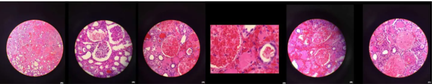
The patient completed the antibiotic regimen and afterwards, she was maintained only in supportive tre-atment, performing daily hemodialysis due to anuria and frequent need for transfusion of red blood cells due to anemia. On the fifteenth day of hospitalization, after sustained improvement in thrombocytopenia, she underwent renal laparoscopic biopsy, which iden-tified extensive areas of ischemic and hemorrhagic infarction, interstitial hemorrhage, and medium-sized vessels with fibrin thrombi (Figure 2). Skin lesions were also analyzed by biopsy, which identified in-flammatory infiltrate with extensive necrosis in adi-pose tissue, absence of signs of vasculitis in muscles, and absence of dermis and local epidermis surrounded by areas of epidermal repair. These lesions evolved with rupture of blisters and necrotic areas that pro-gressively deepened, reaching the dermis, subcuta-neous tissue, musculature, and tibial bone exposure (Figure 1). There was a need for frequent debridement of the necrotic areas in lower limbs, performed by the plastic surgery team of the burn treatment center of the service. As there was incessant progression of the
Figure 1. Evolution of skin lesions: petechiae and purpuric plaques (A), blisters (B), necrosis (C), bone exposure (D), and skin grafting results (E).
lesions in legs and feet soft tissues, it was considered that the underlying disease that was causing the ne-crosis was be active. All available markers were nega-tive for vasculitis and there was no purulent secretion or positive culture that would justify the clinical evo-lution. On the 32nd day of hospitalization, plasmaphe-resis was chosen as an alternative measure since there was association with TMA, and initially, aHUS was one of the diagnostic hypotheses. Around the 40th day of hospitalization, the patient started to show a signi-ficant diuresis and the necrotic areas in the legs sta-bilized. Hemodialysis was suspended and the plastic surgery team started skin grafting with good response (Figure 1). The patient was discharged after 68 days of hospitalization and the diagnosis of TMA and idio-pathic PF associated with AKI was established.
CONCLUSION
This study presents a report of a patient with severe acute onset and rapid evolution of purpuric lesions complicated by AKI requiring hemodialysis. Although PF is considered a thrombotic disease, wi-th indication of full anticoagulation wi-therapy, in wi-this case, the use anticoagulant was not possible due to its association with hemolytic and thrombocytope-nic conditions, making management more difficult. Initially, it was believed to be a self-limiting disea-se, since the investigation of infectious causes was negative, including a false positive test for dengue. However, the active disease, with risk of lower limb amputation due to progressive necrosis in bone, caused concern. Plasmapheresis was indicated as an alternative measure, in order to stop the aggressive progression of the cutaneous disease. In addition, the association with TMA and the hypothesis of aHUS that had been initially considered corrobora-ted this indication. There was a clinical response to treatment, stabilizing the areas of necrosis, allowing the skin grafting. The renal biopsy, which is very di-fficult to obtain in these cases, was important and allowed the differentiation of the possible causes of AKI, with histological findings compatible wi-th TMA, such as microwi-thrombus in wi-the lumen of the glomerular capillaries, arterioles and arteries, myointimal proliferation, leading to glomerular is-chemia and tuft retraction.16 The presence of these
hemorrhagic findings in the interstitium led us to consider, therefore, the diagnosis of AKI caused by PF. Further studies are needed to corroborate these findings, although obtaining a renal biopsy in these cases is still of great technical difficulty.
REFERENCES
1. Chalmers E, Cooper P, Forman K, Grimley C, Khair K, Min-ford A, et al. Purpura fulminans: recognition, diagnosis and management. Arch Dis Child [Internet] 2011 Jan [cited 2018 Jan 18];96:1066-71. Available from: http://adc.bmj.com/con-tent/96/11/1066. DOI: 10.1136/1066 adc.2010.199919 2. Smith OP, White B. Infectious purpura fulminans: diagnosis
and treatment. Br J Haematol [Internet] 2001 Dec [cited 2018 Jan 18];104:202-7. Available from: http://dx.doi.org/10.1046/ j.1365-2141.1999.01186.x
3. Darmstadt GL. Acute Infectious Purpura Fulminans: Patho-genesis and Medical Management. Pediatr Dermatol [Internet] 2002 Jan [cited 2018 Jan 18];15:169-83. Available from: http:// dx.doi.org/10.1046/j.1525-1470.1998.1998015169.x 4. Adcock DM, Brozna J, Marlar RA. Proposed Classification and
Pathologic Mechanisms of Purpura Fulminans and Skin Necro-sis. Semin Thromb Hemost [Internet] 1990 [cited 2018 Jan 18];16:333-40. Available from: https://www.thieme-connect. com/DOI/DOI?2007-1002686. DOI: 10.1055/s-2007-1002686
5. Honarpisheh H, Camp R, Lazova R. Staphylococcal Pur-pura Fulminans: Report of a Case. Am J Dermatopathol [In-ternet] 2015 Aug [cited 2018 Jan 18];37:643-6. Available from: http://journals.lww.com/amjdermatopathology/Ab- stract/2015/08000/Staphylococcal_Purpura_Fulminans___Re-port_of_a.10.aspx DOI: 10.1097/DAD.0000000000000175 6. Chambers HF. Staphylococcal Purpura Fulminans: A
Toxin-Mediated Disease? Clin Infect Dis [Internet] 2005 Apr [cit-ed 2018 Jan 18];40:948-50. Available from: http://dx.doi. org/10.1086/428584
7. Kravitz GR, Dries DJ, Peterson ML, Schlievert PM. Purpura Fulminans Due to Staphylococcus aureus. Clin Infect Dis [In-ternet] 2005 Apr [cited 2018 Jan 18];40:941-7. Available from: http://dx.doi.org/10.1086/428573
8. Karunatilaka DH, De Silva JRS, Ranatunga PK, Gunasekara TMR, Faizal MAM, Malavige GN. Idiopathic purpura fulmi-nans in dengue hemorrhagic fever. Indian J Med Sci [Internet] 2007 Aug [cited 2018 Jan 18];61:471-3. Available from: http:// www.bioline.org.br/request?ms07076
9. Leung N, Textor SC. Vascular Injury to the Kidney. In: Kasper D, Fauci A, Hauser S, Longo D, Jameson JL, Loscalzo J, eds. Harrison’s Principles of Internal Medicine, 19e [Internet]. New York, NY: McGraw-Hill Education; 2015 [cited 2018 Jan 18]. Chapter 341. Available from: https://accessmedicine.mhmedi-cal.com/Content.aspx?bookId=1130§ionId=79747076 10. Arnold DM, Patriquin CJ, Nazy I. Thrombotic
microangio-pathies: a general approach to diagnosis and management. CMAJ [Internet] 2017 Jan [cited 2018 Jan 18];189:E153-E159. Available from: http://www.cmaj.ca/content/189/4/E153. DOI: 10.1503/cmaj.160142
12. Shen YM. Clinical evaluation of thrombotic microangio-pathy: identification of patients with suspected atypical hemolytic uremic syndrome. Thromb J [Internet] 2016 Oct [cited 2018 Jan 18];14:19. Available from: https://www. ncbi.nlm.nih.gov/pmc/articles/PMC5056489/ DOI: 10.1186/ s12959-016-0114-0
13. Karpman D, Loos S, Tati R, Arvidsson I. Haemolytic uraemic syndrome. J Intern Med [Internet] 2016 Oct [cited 2018 Jan 18];281:123-48. Available from: http://dx.doi.org/10.1111/ joim.12546
14. Hollingsworth JH, Mohler DN. Microangiopathic Hemolytic Anemia Caused by Purpura Fulminans. Ann Intern Med
[Internet] 1968 Jun [cited 2018 Jan 18];68:1310-4. Available from: http://annals.org/aim/article-abstract/682452/microan-giopathic-hemolytic-anemia-caused-purpura-fulminans. DOI: 10.7326/0003-4819-68-6-1310
15. Lalitha AV, Aruna D, Prakash A, Swamy HMN, Rao SDS. Spectrum of Purpura Fulminans. Indian J Pediatr [Internet] 2009 Jan [cited 2018 Jan 18];76:87-9. Available from: http:// doi.org/10.1007/s12098-009-0034-0