S C I E N C E A D V A N C E S
|
R E S E A R C H A R T I C L E
C H E M I S T R YA 1000-year-old mystery solved: Unlocking
the molecular structure for the medieval blue
from Chrozophora tinctoria, also known as folium
P. Nabais1, J. Oliveira2*, F. Pina1, N. Teixeira2, V. de Freitas2*, N. F. Brás3, A. Clemente4,
M. Rangel5, A. M. S. Silva6, M. J. Melo1*
The molecular structure of the medieval watercolor known as folium has finally been solved in the 21st century. The interdisciplinary approach taken was the key to producing extracts that had been prepared following medieval instructions, and shows the blue/purple chromophore as the major dye in Chrozophora tinctoria fruits (shell). A
multi-analytical characterization of its structure was made using HPLC-DAD-MS, GC-MS, NMR (1H, 13C, COSY, HSQC,
HMBC, INADEQUATE), and computational studies. The results demonstrate that the blue compound corresponds to 6′-hydroxy-4,4′-dimethoxy-1,1′-dimethyl-5′-{[3,4,5-trihydroxy-6-(hydroxymethyl)tetrahydro-2H-pyran-2-yl] oxy}-[3,3′-bipyridine]-2,2′,5,6(1H,1′H)-tetraone, a hermidin derivative, which we named chrozophoridin. Experimental data and computational modeling studies show that this mono-glycosylated dimer is represented by two stable isomers (atropisomers). This is an indispensable piece of knowledge for the characterization of this medieval dye in works of art such as medieval manuscript illuminations and for testing its stability and contributes to the preservation of our cultural heritage.
INTRODUCTION
Chrozophora tinctoria in medieval and 19th century written sources
The use of the plant Chrozophora tinctoria (L.) A.Juss. to produce colors for illuminated manuscripts is extensively described in medieval written sources (1–4). What distinguishes C. tinctoria from other medieval natural sources to dye or produce paints is that, until now, the blue color structure remained elusive (5–7), despite the efforts by many groups in the last decades of the 20th century and into the 21st century (8–11). To tackle this mystery, our interdisciplinary group assembled a team of chemists who have expertise in natural products identification; conservation scientists, working in the re-production of medieval colors; and a biologist with a great deal of botanical and field knowledge of the Portuguese flora, who oversees plant sourcing. This interdisciplinary approach proved essential to solving the complex structure of the blue dye.
On the basis of the detailed descriptions that were selected from three medieval treatises, we planned the field expeditions and the sampling methods for collecting plant materials. Fruits were collected during July, August, and September 2017 and 2018 (unripe and ripe) in southern Portugal (Granja/Mourão). C. tinctoria extracts were prepared following the treatises’ instructions, and the main colorant was isolated, purified, and characterized through a multi-analytical
approach: high-performance liquid chromatography–high-resolution mass spectrometry–diode array detector (HPLC-HRMS-DAD), gas chromatography–mass spectrometry (GC-MS), nuclear magnetic res-onance (NMR) [1H, 13C, correlation spectroscopy (COSY),
hetero-nuclear single-quantum coherence (HSQC), heterohetero-nuclear multiple- bond correlation (HMBC), INADEQUATE], and electron paramagnetic resonance (EPR) (12, 13). On the basis of the experimental results, the molecular structure for the blue color compound has finally been solved and will be discussed in this paper. Theoretical calculations supported this assignment, and the predicted ultraviolet-visible (UV-VIS) absorption spectra overlapped well the experimental spectra. Other families of chromophores, present in minor amounts, were also detected and characterized by HPLC-HRMS.
C. tinctoria and its uses were well known in antiquity and medieval
times. However, the practice of creating the blue color fell out of use and it was lost in the 19th century. Its medicinal properties were first described by Dioscorides (De Materia Medica, 1st century) and were also mentioned in medieval pharmacopoeia texts, and studies focusing on its anti-inflammatory properties have been published recently (14–16). The dyeing properties of this species and their applications are a fascinating subject that will be revisited, as it is relevant to this research.
In medieval times, the blue and purple solutions extracted from
C. tinctoria were stored, after adsorption onto cloth and drying, as
watercolors (clothlets), and were applied as paint by cutting a piece of cloth and extracting its color with the appropriate binding medium. Complete descriptions of the plant, when to collect it, and how it was processed are found in important medieval treatises such as
The book on how to make all the colour paints for illuminating books
(15th century), Montpellier liber diversarum arcium (14th century), and Theophilus on divers arts (12th century), hereafter referred to as
Book of all color paints (1), Montpellier (2), and Theophilus (3). In
these treatises, only the fruits were collected (Fig. 1). The paint thus obtained was named folium or tornasol (turnsole); this latter desig-nation is common to the blue/purple watercolors obtained from
1REQUIMTE–Laboratório Associado para a Química Verde, Faculdade de Ciências e Tecnologia, Universidade NOVA de Lisboa, Campus Caparica, 2829-516 Monte de Caparica, Portugal. 2REQUIMTE–Laboratório Associado para a Química Verde, Faculdade de Ciências, Universidade do Porto, Rua do Campo Alegre, s/n, 4169-007 Porto, Portugal. 3REQUIMTE–UCIBIO, Faculdade de Ciências, Universidade do Porto, Rua do Campo Alegre, s/n, 4169-007 Porto, Portugal. 4cE3c–Centre for Ecology, Evolution and Environmental Changes, Faculdade de Ciências, Universidade de Lisboa, Campo Grande, 1749-016 Lisboa, Portugal. 5REQUIMTE–Laboratório Associado para a Química Verde, Instituto de Ciências Biomédicas de Abel Salazar, Universidade do Porto. 6REQUIMTE–Laboratório Associado para a Química Verde, Departamento de Química, and QOPNA, University of Aveiro, 3810-193 Aveiro, Portugal. *Corresponding author. Email: [email protected] (M.J.M.); [email protected] (J.O.); [email protected] (V.d.F.)
Copyright © 2020 The Authors, some rights reserved; exclusive licensee American Association for the Advancement of Science. No claim to original U.S. Government Works. Distributed under a Creative Commons Attribution NonCommercial License 4.0 (CC BY-NC). on October 16, 2020 http://advances.sciencemag.org/ Downloaded from
lichens (e.g., Roccella tinctoria and Lasallia pustulata), and conse-quently, C. tinctoria dyes remained forever interlinked to these lichens. Clarke, in his critical edition of texts written in the 14th and 15th centuries, goes further and suggests that the term tornasol was used generically to designate any water-based color stored in cloth (17). This had been already posited by Wallert (9) in his proposal to identify folium in manuscript illuminations based on molecular flu-orescence. Fortunately, we were able to select some medieval reci-pes to produce C. tinctoria blues, because they are so detailed and offer accurate descriptions of the fruit and instructions to not break the fruits and free the seeds, thus pointing to the use of C. tinctoria (and not of lichens). We also have 19th century documental evidence that this medieval knowledge was preserved, until nearly the turn of that century, in the region of Grand-Gallargues (now, Gallargues- le- Montueux), in France. In 1842, Joly (18) published a clear, con-cise, and complete text on this subject, including references to earlier works such as Nissolle’s (19) publication that offers a precise description of the plant, accompanied with an equally accurate illustration. Joly’s text is remarkable in several aspects; first, he visits Grand-Gallargues to interview a priest by the name of Hugues and to gather information directly from the makers; precisely during the period when the plant should be collected (August/September), “vers la fin de septembre 1838, et dans les derniers jours du mois d’août
1839.” According to Joly, in this region, this activity has been
docu-mented since 1600 (when records are available), but even so, ac-cording to Father Hugues, it is one of the most mysterious of crafts (18, 20):
“peu d’industries sont aussi mystérieuses: ceux qui l’exploitent
n’en connaissent point la destination; ceux qui en profitent n’en connaissent point la préparation, et ceux qui l’ont décrite n’ont débité que des mensonges, parce qu’ils ne transcrivaient que de fausses indications” [few industries are so mysterious: those who exploit
it do not know its final applications; those who profit by it do not know how to prepare it, and those who have described it have only told lies, because they have only transcribed false directions] (18).
Joly describes the process using the entire plant to produce the dark blue color and the innovations that were introduced in the me-dieval process, and concludes that, at that time, the clothlets were mainly sold to dye cheese rinds red in the Netherlands (21): “mais il
paraît que l’usage en est borné à donner aux croûtes du fromage de Hollande cette teinte rouge qui les distingue, (...) Il suffit de tremper les formages dans un baquet d’eau bleuie par les chiffons, et de les en retirer presque aussitôt pour les faire sécher” [but it seems that the
use is limited to giving the rind of Dutch cheese that red hue that distinguishes it, (...) All you have to do is soak the cheese in a bucket of water dyed blue by the clothlets (rags), and to remove them
almost immediately to left them dry] (18). Joly proposes that not only other species could be tested for dyeing more efficiently, such as other Chrozophora species, but also Mercurialis perennis (dog’s mercury) and Mercurialis tomentosa. This is extraordinary, as now from this current project, we know that these dyes share a common molecular structure (Fig. 1). Joly also tested and concluded that the precursors for the blue may be found in all the parts of the plant and are more abundant in fruits. He concluded, “Sous l’influence
de la vie, il existe dans ces organes à l’état incolore; après la mort du végétal, et sous l’influence de l’oxygène atmosphérique et d’une prompte dessiccation, il peut devenir bleu” [Under the influence
of life, it exists in these organisms in a colorless state; after the death of the plant, and under the influence of atmospheric oxygen and a rapid desiccation, it becomes blue] (18), which is precisely the way the appearance of the blue color in M. perennis is described in more recent studies performed by Swan and Lorentz et al. (22–25). These authors have characterized the molecular structure for the blue chromophore as will be described below.
Medicinal properties in Mercurialis species and molecular structures for its blue colors
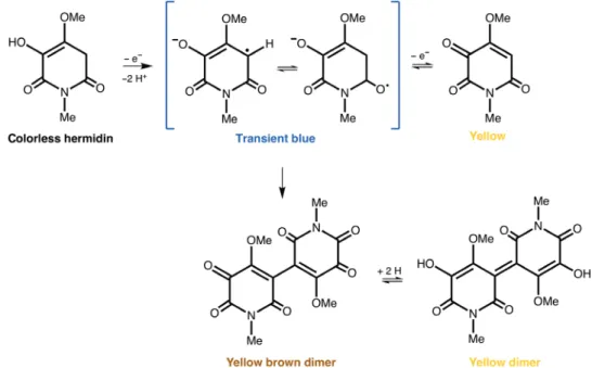
In 1985, Swan proposed the first molecular structure for the “blue chromogen” isolated from M. perennis (dog’s mercury), a perennial herb used in remedies for medicinal purposes (22, 23). Swan synthesized 2,3,6-trihydroxy-4-methoxy-1-methyl-pyridinium (fig. S1), a colorless compound in solution that he could oxidize to a blue transient color. In his own words: “To summarize, it is suggested that the chromogen present in the colorless aqueous extract of Mercurialis perennis (...) is compound hermidin (2,3,6- trihydroxy-4-methoxy-1-methyl-pyridinium) and that on oxidation by air or K3[Fe(CN)6] the blue transient color is due to a semiquinone-
like (17), and that at the moment when the solution turns yellow it contains compounds (16), (22), and (23).” (fig. S1).
On drying, M. perennis plants take on a dark blue color. As Swan could only obtain a temporary blue in solution, this made him doubt that this chromogen could be the only source of the blue color. Later, in 2010 and 2014 (23, 24), the instability of the blue structure in solution was studied in detail by Lorenz et al. (Fig. 2). That research allowed this group to confirm the molecular structures proposed by Swan as the origin of the blue color in M. perennis and to suggest interconversion mechanisms of the colorless hermidin into the blue hermidin quinone and the latter into yellow dimeric reduced forms, as described in detail in the Supplementary Materials and depicted in Fig. 2 (23, 24).
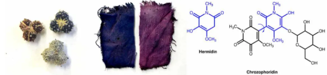
In 1984, Forrester had already reported that aqueous solutions of hermidin readily give rise to a transient blue radical-anion on Fig. 1. The molecule of this study, chrozophoridin. Left: Close-up of C. tinctoria fruits (collected in Alentejo, Portugal) and clothlets prepared with the juice of the fruits
following the instructions in the Book of all color paints. Light green fruits were used in this study shortly after collection. Right: Molecular structures of the blue colorants, hermidin (from M. perennis), and chrozophoridin (from C. tinctoria). Photo credit: Paula Nabais, Universidade NOVA de Lisboa.
on October 16, 2020
http://advances.sciencemag.org/
exposure to air, the identity of which has been established by elec-tron spin resonance spectroscopy (25).
Plant description
C. tinctoria (L.) A.Juss. (Euphorbiaceae) is an annual herb native to
the Mediterranean region, north Africa, and central and southwestern Asia. This species can be found on dry and disturbed lands, ruderal habitats, fallows, and along the edges of cultivated fields, mostly in limestone. The plants are 10 to 40 cm tall, gray-green, and tomentous (densely covered with stellate hairs). Stems are erect and branched and leaves are alternate, rhombic to ovate, cuneate at the base, and with sinuate leaf margin (26).
C. tinctoria is monoecious, bearing male and female reproductive
organs in different flowers but on the same plant. Flowers are grouped into spike-like racemes, with male flowers at the top and female flowers at the base, usually solitary. Male flowers are yellow and inconspicuous. Female flowers exhibit a spherical ovary and petals are not present. Flowering occurs from May to September and fruit maturation is between July and October. Mature fruits are dark green, subspheric, and slightly lobed capsules, covered with white scales and 5 to 8 mm in diameter (Fig. 1 and fig. S2). During dehiscence, each of the three single-seeded loculus opens into two valves, releasing three gray to light brown, rough-textured obovate seeds approximately 4 mm in size (26).
Molecular structures for colorants in C. tinctoria
In contrast to the successful discovery of the molecular structure for the blue in M. perennis (dog‘s mercury), the works published for
C. tinctoria have been inconclusive or have suggested incorrect
struc-tures. In 1997, Guineau (8) proposed orcinol for the blue color based on time-of-flight secondary ion MS data, which supported the previous suggestions of the presence of lichen colorants in C. tinctoria that were reviewed by Wallert (9). As we will show in this paper, orcinol- based structures are not present in the extracts of the fruits of C. tinctoria
(27). More recently, the dye was investigated by Aceto’s group and, although they did not proposed any structure for the blue dye, they reported that “the mass spectrum is dominated by a peak at 266 m/z” (10). Anthocyanins have also been proposed, but never identified (9).
In the past 2 years, we have endeavored to unlock the chemical structure behind the blue/purple color in C. tinctoria. This is an in-dispensable piece of knowledge for the preservation of our European cultural heritage, in works of art such as medieval manuscript illu-minations where this dye could have been used.
RESULTS
Characterization of the blue dye extracted from the fruits (shell) of C. tinctoria
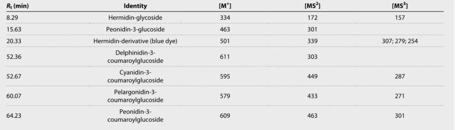
The blue extract of C. tinctoria, obtained from the fruits as reported in Materials and Methods, was analyzed using HPLC-DAD and LC-MS (Table 1 and fig. S3). The extract essentially showed the presence of one chromatographic peak characterized by a maximum absorption wavelength, max, at circa 540 nm, indicative of a purple/
bluish color, and an ion mass mass/charge ratio (m/z) at 501, in positive ion mode. The following fragments were also detected, at
m/z 339, compatible with the loss of a hexose moiety (shown to be
an O-glucose linkage), and at m/z 307, 279, and 254, in the MS3 spectrum, consistent with the loss of methoxyl and methyl groups. In addition, an ion mass at m/z 334 with a MS2 spectrum at m/z 172 (loss of 162 unified atomic mass unit) and a MS3 spectrum with two
fragments at m/z 157 and 115 were also detected in the extract. The ion mass m/z 172 and the respective fragments at m/z 157 and 115 were described in the literature by Lorenz et al. (23) and correspond to hermidin pre sent in M. perennis. In this case, the ion mass at m/z 334 should correspond to the glycosylated form of hermidin (Fig. 1). We did not observe a peak at 266 m/z, contrary to what was observed by Aceto et al. (10) that reported that in their extracts of C. tinctoria “the mass spectrum is dominated by a peak at 266 m/z.”
Fig. 2. Causes of color in hermidin extracted from M. perennis. Conversion of colorless hermidin into the blue hermidin quinone and formation of dimeric structures
as proposed by Lorenz et al. (23, 24).
on October 16, 2020
http://advances.sciencemag.org/
Adding to this, several anthocyanins were also identified as minor compounds in the fruits’ extract, peonidin-3-O-glucoside, and the coumaroylated derivatives of delphinidin, cyanidin, pelargonidin, and peonidin (Table 1 and figs. S3 and S4). These anthocyanins were tentatively identified by mass spectra and the chromatographic re-tention time compared to standards.
On the basis of its exact mass (501.1371), the elemental composition of the blue dye should correspond to C20H24O13N2 (500.41 g mol−1).
On a first attempt, and due to the presence of a hermidin-glucoside in the extract, a hermidin-like structure was considered for it. Con-trary to what was reported for the transient blue radical-anion of cyanohermidin in M. perennis (fig. S1), the blue extract from C. tinctoria was stable for several days, pointing to the absence of a radical species. Despite this indication, EPR experiments were performed and con-firmed that no radical species were present in the aqueous solution of the purified compound.
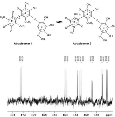
To elucidate the structure of the blue dye present in C. tinctoria fruits, the purified compound was analyzed using one-dimensional (1D; 1H and 13C) and 2D (COSY, HSQC, HMBC and INADEQUATE)
NMR in DMSO-d6/D2O (9:1). The assignment of the proton and
carbon chemical shifts (Table 2) of the purified compound showed the presence of two atropisomers (Fig. 3). The proposed structures correspond to two rotamers (atropisomer 1 and atropisomer 2), which were perceived in the 13C experiment, with all the carbon signals appearing in duplicate and with the same intensity (Fig. 3).
The anomeric protons H-1′ from the two isomers were assigned to the two doublets (J = 7.7 and 7.5 Hz, respectively) present at 4.32 and 4.20 parts per million (ppm), respectively. Protons H-2′, H-3′, H-4′, H-5′, H-6′a, and H-6′b of glucose of both isomers were at-tributed to the signals present at 3.13, 3.10, 2.90, 3.16, 3.45, and 3.63 ppm, respectively, by their correlations with each other in the COSY spectrum. Because of the superimposition of signals involving two similar compounds, it was not possible to accurately attribute the chemical shifts and coupling constants of all the protons for both isomers. The O-CH3 protons present in carbon C-4 and C-10 were
assigned to the singlets present at 3.75/3.77 and 3.71/3.78 ppm, re-spectively, and the protons N-CH3 present in rings A and B of both
isomers appeared at 3.12 ppm as a singlet.
The assignment of the carbon resonances was made possible using 2D NMR techniques (gHSQC, gHMBC, and INADEQUATE).
Car-bons C-1′, C-2′, C-3′, C-4′, C-5′, and C-6′ from the glucose moiety of atropisomers 1 and 2 were assigned to 108.3/108.7, 74.1/74.1, 77.4/77.5, 75.0/75.0, 76.9/76.9, and 61.3/61.5 ppm through their direct correlations with the respective protons. Carbons O-CH3 (rings
A and B) and N-CH3 (rings A and B) from isomers 1/2 were
as-signed to 59.2/59.4, 60.8/60.1, 27.4/27.5, and 27.3/27.5 ppm (Table 2). Carbons C-5 from both atropisomers were assigned to 122.0 and 122.3 ppm, by the long-distance correlation (HMBC) with the ano-meric protons H-1′. Carbons C-4 and C-10 from both atropisomers were attributed to 161.1/161.0 and 156.0/156.1 ppm, respectively, by their long-range correlations with methyl groups. Carbons C-2, C-6, C-8, and C-12 from atropisomers 1 and 2 were attributed to 164.0/163.7, 159.0/158.8, 161.7/161.6, and 157.0/156.9 ppm, respec-tively, and by their long-distance correlation (HMBC spectrum) with the protons present at the N-methyl group.
We could not make an unequivocal assignment of carbons C-3 and C-9 through NMR, because these two carbons do not present any proton in their vicinity (Fig. 3). Moreover, the chemical shift of carbon C-11 could only be determined using a 2D 13C-13C
INADEQUATE experiment, where the correlation between this carbon with carbon C-12 was observed. The appearance of carbon C-11 at high chemical shifts concurs with a carbonyl group in that position.
The results are consistent with the structure of the blue compound corresponding to 6′-hydroxy-4,4′-dimethoxy-1,1′-dimethyl-5′- {[3,4,5-trihydroxy-6-(hydroxymethyl)tetrahydro-2H-pyran-2-yl] oxy}-[3,3′-bipyridine]-2,2′,5,6(1H,1′H)-tetraone, a hermidin-mono- glycosylated dimer that we would hereafter designate as chrozophoridin (Fig. 3). This is a notable result, considering that previously, in
M. perennis, the dimeric forms of hermidin were associated with
yellow and yellow-brown solutions, whereas the blue color was based on a monomeric radical transient species. To get further insight into the proposed structure and as a means to predict its blue color, computational calculations were performed.
Computational calculations
By exploring the conformational space of chrozophoridin, 10 en-ergetically favored isomers (local minima) were found with E < 1.0 kcal mol−1 (table S1 reports their relative internal energy and
both 1 and 2 values after optimization in an aqueous environment). Table 1. API-LC-ESI-MS/MS data for the major and minor compounds extracted from C. tinctoria fruits. Molecular ion and respective fragments MS2 and MS3 obtained by atmospheric pressure ionization (API)–LC–electrospray ionization (ESI)–MS/MS (positive ion mode) found in the blue extract of C. tinctoria fruits.
Rt (min) Identity [M+] [MS2] [MS3]
8.29 Hermidin-glycoside 334 172 157
15.63 Peonidin-3-glucoside 463 301
20.33 Hermidin-derivative (blue dye) 501 339 307; 279; 254
52.36 coumaroylglucosideDelphinidin-3- 611 303 52.67 coumaroylglucosideCyanidin-3- 595 449 287 60.07 coumaroylglucosidePelargonidin-3- 579 433 271 64.23 coumaroylglucosidePeonidin-3- 609 463 301 on October 16, 2020 http://advances.sciencemag.org/ Downloaded from
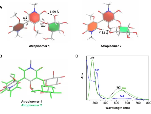
The various isomers of chrozophoridin can be assembled in two groups based on their dihedral values (group I composed of isomers 1, 3, 4, 5, 8, 9, and 10 that have similar dihedral values: 1 = −61.1 ± 1.5° and 2 = 83.0 ± 0.9°, and group II composed by isomers 2, 6, and 7 that have similar dihedral values: 1 = 60.6 ± 0.0° and 2 = −80.9 ± 0.3°). Each group can be represented by the most thermodynamically fa-vored atropisomers 1 and 2. A mixture of both atropisomers is ex-pected because of their very small energy difference (0.004 kcal mol−1, which corresponds to 50.2% and 49.8% quantities of atropisomers 1 and 2, respectively, according to the Maxwell-Boltzmann distribution, and using a temperature of 298 K). The analysis of the molecular structures of both atropisomers in solution revealed that the elec-tronic delocalization provided by the perpendicular orientation of both aromatic rings may be essential to their formation. However, atropisomer 2 has the B ring and the glucose group close to each other to make one hydrogen bond (as seen in Fig. 4). This stereo-chemical effect may cause the slight energy difference between the two isomers.
Time-dependent density functional theory (TD-DFT) calculations were performed to obtain the UV-VIS absorption spectra for the
two atropisomers. max was evaluated using various density
func-tionals (see table S4). It would appear that in comparison with ex-periments, the Boese-Martin for kinetics (BMK) density functional was the best-performing functional for both atropisomers (Fig. 4 compares their predicted UV-VIS spectra with an experimental spectrum at pH 7). Theoretical and experimental spectra overlap quite well. The small discrepancy might be the result of errors associated with the TD-DFT determination of max values (28) and from the
use of an implicit solvent model approach instead of an explicit description of the solvent molecules. The latter may affect the intra-molecular interactions and stability of the atropisomers (29). The spectral shift (max) to experimental data obtained in water (pH 7)
is −23.8 and 5.0 nm for atropisomers 1 and 2, respectively (Fig. 4). Again, the hypsochromic shift observed in the maximum absorption wavelength for atropisomer 1 in relation to atropisomer 2 could be associated to the lower proximity between the B ring and the glucose unit. The max of absorption, in the visible, is essentially attributed
to the electronic transition from the highest occupied molecular orbital (HOMO) to the lowest unoccupied molecular orbital (LUMO). In both conformers, the HOMO is spread through both A and B rings, while the LUMO is mainly distributed on B ring (fig. S5). The transition from HOMO to LUMO should have an intramolecular charge transfer character. Slight MO distribution differences are re-lated with the marginally higher energy gap between HOMO and LUMO in isomer 1 in relation to isomer 2 (fig. S5). With respect to the stability of the blue color, it is interesting to note that the bond between the two aromatic rings may be viewed almost as a single bond (length of 1.464 Å in both conformers).
Overall, the UV-VIS spectra predicted for chrozophoridin cor-roborates that this dimer is the cause of the blue/purple colors (Fig. 4). Potentially, the blue color is the result of the abovementioned charge transfer.
Atropisomer 1 Atropisomer 2
Fig. 3. Results of NMR (13C). Structure for the blue dye present in C. tinctoria fruits (shell), chrozophoridin, and 13C zoomed spectra (bottom).
Table 2. Essential NMR data for the identification of chrozophoridin.
1H and 13C chemical shifts of the blue dye present in C. tinctoria fruits, determined in DMSO-d6:D2O (9:1). Position 1Η (ppm); J (Hz) 13C (ppm)* A ring 1-N-CH3 3.12; s 27.4/27.5 2 C═O – 164.0/163.7 3 – 88.5/88.4 4 – 156.0/156.1 4-OCH3 3.71; 3.78; s 59.2/59.4 5 – 122.0/122.3 6 – 159.0/158.8 B ring 7-N-CH3 3.12; s 27.3/27.5 8 C═O – 161.7/161.6 9 – 128.4/127.7 10 – 161.1/161.0 10-OCH3 3.75; 3.77; s 60.8/61.0 11 C═O – 172.6/172.3 12 C=O – 157.0/156.9 Glucose moiety 1′ 4.32; d, 7.7/4.20; d, 7.5 108.3/108.7 2′ 3.13;† 74.1/74.1 3′ 3.10;† 77.4/77.5 4′ 2.90;† 75.0/75.0 5′ 3.16;† 76.9/76.9 6a′ 3.45; 61.3/61.5 6b′ 3.63; 61.3/61.5
*The carbon signals are duplicated, which indicates that at least two isomers are present. †Unresolved (superposition).
on October 16, 2020
http://advances.sciencemag.org/
DISCUSSION
Knowledge was lost on how to extract the blue color from C. tinctoria, but it was recovered through research on medieval written sources on the art of painting, in particular, the art of illuminating books (30, 31). Following medieval instructions, only the fruits were col-lected between July and September, and care was taken to not grind the seeds that are found inside. The extracts thus obtained presented a blue dye as a major chromophore (fig. S3). Last, in the 21st century, an interdisciplinary approach led to the discovery of a molecular structure for the blue colorant extracted from the fruits of C. tinctoria, namely, a hermidin-based mono-glycosylated dimer (Fig. 3), named chrozophoridin. Experimental data and computational quantum me-chanics studies show that this dimer is represented by two isomers, atropisomers 1 and 2 (Fig. 4). Together with the blue hermidin-based dimer, several anthocyanins were identified for the first time (Table 1 and figs. S3 and S4) and are present as minor compounds. Chrozo-phoridin was compared to the blue found in a species from the same family, M. perennis (dog’s mercury), an old medicinal plant known mostly through ethnomedicine (22). The blue chromophore in both species includes the hermidin ring (Fig. 1), but in contrast to the transient radical species in M. perennis, the blue in C. tinctoria has a stable glycosylated dimeric structure and is therefore water soluble. To summarize, this molecular structure is key to identifying folium in works of art and to studying the structural, electronic, and reactive properties of this complex dye (32, 33). This will pave the way for evaluating conservation conditions and the determination and plan-ning of the best preservation strategies.
In conclusion, chrozophoridin was used in ancient times to make a beautiful blue dye for painting, and it is neither an anthocyanin— found in many blue flowers and fruits—nor indigo, the most stable natural blue dye. It turns out to be in a class of its own. Thus, we believe that this will be not our final word on this amazing plant and its story and that further discoveries will follow soon.
MATERIALS AND METHODS
The fruits of C. tinctoria were collected between July and September of 2016, 2017, and 2018, in Alentejo (Granja/Mourão), Portugal (by A.C., F.P., M.J.M., and P.N.). The coordinates were provided by (34). Fresh fruits (160 g) were extracted with 4 liters of methanol:water (70:30, v/v) during 2 hours under stirring, giving rise to a deep blue–colored solution. Methanol was evaporated under vacuum, and the obtained crude blue extract was analyzed by HPLC-DAD and LC-MS.
For the characterization of the blue dye, the extract was further purified. In a first step, by applying the full extract to a reversed- phase C18 silica gel in a Büchner funnel under vacuum (G3 porosity) washed with deionized water, it was possible to observe the elution of yellow compounds and then the blue fraction was recovered slowly with water. This blue fraction was concentrated in a rotoevaporator and then was purified, twice, by low-pressure column chromatography using the same C18-RP gel as stationary phase (250 mm × 16 mm inside diameter) and deionized water as a mobile phase. The purified blue fraction was lyophilized, and the blue powder obtained was analyzed by NMR, HPLC-DAD, HPLC-DAD-MS, GC-MS, and EPR.
A
B C
Fig. 4. Analysis of the thermodynamically favored atropoisomers of chrozophoridin, in solution. Molecules are depicted as sticks and colored by atom type. (A) A
ring, B ring, and glucose ring are colored in red, orange, and green, respectively. Both 1 and 2 dihedrals and hydrogen bonds are also indicated. (B) Superposition of the A ring of the two molecules. Colored by element (atropisomer 1) and green (atropisomer 2). (C) UV-VIS spectra (water, pH 7, blue) for chrozophoridin compared with the predicted spectra [Boese-Martin for kinetics (BMK) functional] for atropisomers 1 (black) and 2 (green).
on October 16, 2020
http://advances.sciencemag.org/
The initial geometry of chrozophoridin was built with the GaussView software (35–37). Geometry optimizations were carried out, and the most energetically favored isomers (local minima with E < 1.0 kcal mol−1) were further optimized in solvent. TD-DFT
single-point calculations were applied to determine the UV-VIS ab-sorption spectra for the two most energetically stable atropisomers of chrozophoridin. All calculations were performed by the Gaussian 09 package (38). For comprehensive details, please see the Supple-mentary Materials.
SUPPLEMENTARY MATERIALS
Supplementary Materials for this article is available at http://advances.sciencemag.org/cgi/ content/full/6/16/eaaz7772/DC1
REFERENCES AND NOTES
1. M. J. Melo, R. Castro, P. Nabais, T. Vitorino, The book on how to make all the colour paints for illuminating books: Unravelling a Portuguese Hebrew illuminators’ manual. Herit. Sci.
6, 44 (2018).
2. M. Clarke, Mediaeval Painters’ Materials and Techniques: The Montpellier Liber Diversarum
Arcium (Archetype Publications, 2011), pp. 110–111.
3. J. G. Hawthorne, C. S. Smith, Theophilus on Divers Arts: The Foremost Medieval Treatise on
Painting, Glassmaking and Metalwork (Dover Publications, 1979), pp. 38–40.
4. F. Brunello, De Arte Illuminandi e Altri Trattati Sulla Tecnica Della Miniatura Medievale (Neri Pozza Editore, 1992), pp. 63–67.
5. M. J. Melo, P. Nabais, R. Araújo, T. Vitorino, The conservation of medieval manuscript illuminations: A chemical perspective. Phys. Sci. Rev. 4, 20180017 (2019).
6. I. Degano, Liquid chromatography: Current applications in heritage science and recent developments. Phys. Sci. Rev. 4, 20180009 (2018).
7. F. Pozzi, M. Leona, Surface-enhanced Raman spectroscopy in art and archaeology.
J. Raman Spectrosc. 47, 67–77 (2015).
8. B. Guineau, Le folium des enlumineurs, une couleur aujourd'hui disparue: Ce que nous rapportent les textes sur l'origine et la fabrication de cette couleur, son procédé d'emmagasinage sur un morceau d'étoffe et son emploi dans l'enluminure médiévale. Identification de folium dans des peintures du IXe s., du Xe s. et du début du XIe s.
Archéologie Médiévale 26, 23–44 (1996).
9. A. Wallert, Chrozophora tinctoria Juss. Problems in identifying an illumination colorant.
Restaurator 11, 141–155 (1990).
10. M. Aceto, A. Arrais, F. Marsano, A. Agostino, G. Fenoglio, A. Idone, M. Gulmini, A diagnostic study on folium and orchil dyes with non-invasive and micro-destructive methods. Spectrochim. Acta A Mol. Biomol. Spectrosc. 142, 159–168 (2015). 11. C. Krekel, thesis, Ludwig-Maximilians-Universität München, Munich, Germany
(1996).
12. J. Oliveira, J. Azevedo, A. M. S. Silva, N. Teixeira, L. Cruz, N. Mateus, V. de Freitas, Pyranoanthocyanin dimers: A new family of turquoise blue anthocyanin-derived pigments found in Port wine. J. Agric. Food Chem. 58, 5154–5159 (2010). 13. J. Oliveira, C. Santos-Buelga, A. M. S. Silva, V. de Freitas, N. Mateus, Chromatic
and structural features of blue anthocyanin-derived pigments present in Port wine.
Anal. Chim. Acta 563, 2–9 (2006).
14. A. Menghini, Ed., De Materia Medica, Il Dioscoride di Napoli (Aboca Edizioni, 2016), vol. 1, pp. 413.
15. O. Meung, La Pharmacie des Moines: Traité de la Vertu des Plantes (Macer floridus) (Éditions Paleo, 2011), pp. 127.
16. H. M. Abdallah, F. M. Almowallad, A. Esmat, I. A. Shehata, E. A. Abdel-Sattar, Anti-inflammatory activity of flavonoids from Chrozophora tinctoria. Phytochem. Lett. 13, 74–80 (2015).
17. M. Clarke, The crafte of lymmyng and The maner of steynyng: Middle English recipes for
painters, stainers, scribes, and illuminators (Early English Text Society, Oxford, 2016),
pp. 44: ‘turnesole, tornesole , tournesole, tursole; n. any color of direct dye extracted from a flower or berry that is stored by being absorbed into a clothlet, to be subsequently released by immersion in a medium for e.g. an illuminator's color, typically purple or blue (not a botanical name, i.e. not referring to the turnsole plants Chrozophora tinctoria (Juss.) or Helotropium spp., although the former plant itself may be used as the color source)’.
18. M. N. Joly, Recherches sur la Fabrication du Tournesol en Drapeaux, et sur le Principe Colorant du Chrozophora tinctoria (Ad. de jussieu; Croton tinctorium, Linné) Employé à cette Fabrication. Ann. Chim. Phys. 111–126 (1842).
19. M. Nissolle, Description du Ricinoïdes, ex quâ paratur Tournesol Gallorum, Inst. Rei Herb. App. 565, in Histoire de l’Academie Royale des Sciences (Imprimerie Royale Paris, 1712), pp. 332–338.
20. We were not yet able to have access to the publication of Father Hugues. 21. On the other hand, a “tournesol en pains” was imported from the Netherlands, which
was prepared with diverse species of lichens (Roccella tinctoria, used in the past, had been substituted by Lecanora tartarea - cudbear).
22. G. A. Swan, Isolation, structure, and synthesis of hermidin, a chromogen from Mercurialis
perennis L. J. Chem. Soc. Perkin Trans. 1, 1757–1766 (1985).
23. P. Lorenz, M. Hradecky, M. Berger, J. Bertrams, U. Meyer, F. C. Stintzing, Lipophilic constituents from aerial and root parts of Mercurialis perennis L. Phytochem. Anal. 3, 234–245 (2010).
24. P. Lorenz, J. Conrad, S. Duckstein, D. R. Kammerer, F. C. Stintzing, Chemistry of hermidin: Insights from extraction experiments with the main alkaloid of Mercurialis perennis L. tracked by GC/MS and LC/MSn. Helv. Chim. Acta 97, 1606–1623 (2014).
25. A. R. Forrester, Autoxidation of hermidin: An ESR study. Experientia 40, 688–689 (1984).
26. C. Benedí, in Flora Iberica 8, S. Castroviejo, C. Aedo, M. Laínz, F. Muñoz Garmendia, G. Nieto Feliner, C. Paiva, J. Benedí, Eds. (Real Jardín Botánico, CSIC, 1997), pp. 196–199. 27. M. J. Melo, P. Nabais, M. Guimarães, R. Araújo, R. Castro, M. C. Oliveira, I. Whitworth,
Organic dyes in illuminated manuscripts: A unique cultural and historic record.
Philos. Trans. A Math. Phys. Eng. Sci. 374, 20160050 (2016).
28. P. Trouillas, F. Di Meo, J. Gierschner, M. Linares, J. C. Sancho-García, M. Otyepka, Optical properties of wine pigments: Theoretical guidelines with new methodological perspectives. Tetrahedron 71, 3079–3088 (2015).
29. P. Trouillas, J. C. Sancho-García, V. De Freitas, J. Gierschner, M. Otyepka, O. Dangles, Stabilizing and modulating color by copigmentation: Insights from theory and experiment. Chem. Rev. 116, 4937–4982 (2016).
30. N. Eastaugh, V. Walsh, T. Chaplin, R. Siddall, Pigment Compendium: A Dictionary of
Historical Pigments (Elsevier Butterworth-Heinemann, 2004), p. 372.
31. D. Cardon, Natural Dyes: Sources, Tradition, Technology and Science (Archetype Publications, 2007).
32. C. Miliani, L. Monico, M. J. Melo, S. Fantacci, E. M. Angelin, A. Romani, K. Janssens, Photochemistry of artists’ dyes and pigments: Towards better understanding and prevention of color change in works of art. Angew. Chem. Int. Ed. 57, 7324–7334 (2018). 33. P. Lorenz, M. Bunse, S. Sauer, J. Conrad, F. C. Stintzing, D. R. Kammerer, Conversion
of plant secondary metabolites upon fermentation of Mercurialis perennis L. Extracts with two lactobacteria strains. Fermentation 5, 42 (2019).
34. M. Porto, A. J. Pereira, C. Tauleigne Gomes, J. L. Vitorino, Distribution of
Chrozophora tinctoria (L.) Raf. Flora-On: Flora de Portugal Interactiva, Sociedade
Portuguesa de Botânica (2016); http://www.flora-on.pt/#wChrozophora+tinctoria [accessed 7 January 2016].
35. F. Di Meo, J. C. Sancho-García, O. Dangles, P. Trouillas, Highlights on anthocyanin pigmentation and copigmentation: A matter of flavonoid -stacking complexation to be described by DFT-D. J. Chem. Theory Comput. 8, 2034–2043 (2012). 36. E. H. Anouar, J. Gierschner, J.-L. Duroux, P. Trouillas, UV/Visible spectra of natural
polyphenols: A time-dependent density functional theory study. Food Chem. 131, 79–89 (2012).
37. J. Oliveira, P. Araújo, A. Fernandes, N. F. Brás, N. Mateus, F. Pina, V. Freitas, Influence of the structural features of amino-based pyranoanthocyanins on their acid-base equilibria in aqueous solutions. Dyes, Pigm. 141, 479–486 (2017).
38. M. J. Frisch, G. W. Trucks, H. B. Schlegel, G. E. Scuseria, M. A. Robb, J. R. Cheeseman, G. Scalmani, V. Barone, G. A. Petersson, H. Nakatsuji, X. Li, M. Caricato, A. Marenich, J. Bloino, B. G. Janesko, R. Gomperts, B. Mennucci, H. P. Hratchian, J. V. Ortiz, A. F. Izmaylov, J. L. Sonnenberg, D. Williams-Young, F. Ding, F. Lipparini, F. Egidi, J. Goings, B. Peng, A. Petrone, T. Henderson, D. Ranasinghe, V. G. Zakrzewski, J. Gao, N. Rega, G. Zheng, W. Liang, M. Hada, M. Ehara, K. Toyota, R. Fukuda, J. Hasegawa, M. Ishida, T. Nakajima, Y. Honda, O. Kitao, H. Nakai, T. Vreven, K. Throssell, J. A. Montgomery Jr., J. E. Peralta, F. Ogliaro, M. Bearpark, J. J. Heyd, E. Brothers, K. N. Kudin, V. N. Staroverov, T. Keith, R. Kobayashi, J. Normand, K. Raghavachari, A. Rendell, J. C. Burant, S. S. Iyengar, J. Tomasi, M. Cossi, J. M. Millam, M. Klene, C. Adamo, R. Cammi, J. W. Ochterski, R. L. Martin, K. Morokuma, Ö. Farkas, J. B. Foresman, D. J. Fox, Gaussian 09 (Gaussian Inc., 2009).
Acknowledgments: We thank Z. Azevedo for the LC-ESI-MS analysis, M. Andrade for the NMR
analysis, M. C. Oliveira for the first HRMS spectra and anthocyanins’ identification, and E. Eis for providing the plant M. perennis. Funding: This work was supported by the Associate Laboratory for Green Chemistry-LAQV, which is financed by national funds from FCT/MCTES (UID/QUI/50006/2019), and by project STEMMA—From singing to writing—survey on material production and routes of Galician-Portuguese Lyric, PTDC/LLT-EGL/30984/2017. J.O. and N.F.B. thank the FCT for their IF grant (IF/00225/2015 and IF/01355/2014, respectively). N.T. thanks contract REQUIMTE/EEC2018/PTDC/QUI-OUT/29925/2017, within the project “Polyphenols in
on October 16, 2020
http://advances.sciencemag.org/
QUI-OUT/29925/2017, and P.N. thanks Ph.D. grant within the CORES Ph.D. programme (PD/ BD/105895/2014). Author contributions: Experimental work: P.N., J.O., F.P., N.T., A.C., M.R., A.M.S.S., and M.J.M.; computational work: N.F.B.; project development and funding: V.d.F., F.P., and M.J.M. Competing interests: The authors declare that they have no competing interests.
Data and materials availability: All data needed to evaluate the conclusions in the paper are
present in the paper and/or the Supplementary Materials. Additional data related to this paper may be requested from the authors.
Accepted 22 January 2020 Published 17 April 2020 10.1126/sciadv.aaz7772
Citation: P. Nabais, J. Oliveira, F. Pina, N. Teixeira, V. de Freitas, N. F. Brás, A. Clemente, M. Rangel, A. M. S. Silva, M. J. Melo, A 1000-year-old mystery solved: Unlocking the molecular structure for the medieval blue from Chrozophora tinctoria, also known as folium. Sci. Adv. 6, eaaz7772 (2020).
on October 16, 2020
http://advances.sciencemag.org/
originally published online April 17, 2020 DOI: 10.1126/sciadv.aaz7772
(16), eaaz7772.
6
Sci Adv
ARTICLE TOOLS http://advances.sciencemag.org/content/6/16/eaaz7772
MATERIALS
SUPPLEMENTARY http://advances.sciencemag.org/content/suppl/2020/04/13/6.16.eaaz7772.DC1
REFERENCES
http://advances.sciencemag.org/content/6/16/eaaz7772#BIBL
This article cites 22 articles, 0 of which you can access for free
PERMISSIONS http://www.sciencemag.org/help/reprints-and-permissions
Terms of Service
Use of this article is subject to the
is a registered trademark of AAAS.
Science Advances
York Avenue NW, Washington, DC 20005. The title
(ISSN 2375-2548) is published by the American Association for the Advancement of Science, 1200 New
Science Advances
License 4.0 (CC BY-NC).
Science. No claim to original U.S. Government Works. Distributed under a Creative Commons Attribution NonCommercial Copyright © 2020 The Authors, some rights reserved; exclusive licensee American Association for the Advancement of
on October 16, 2020
http://advances.sciencemag.org/