S Y M P O S I U M : A D V A N C E D H I P A R T H R O S C O P Y
Surgical Technique
Endoscopic Gluteus Maximus Tendon Release for External Snapping Hip Syndrome
Giancarlo C. Polesello MD, PhD, Marcelo C. Queiroz MD, Benjamin G. Domb MD, Nelson K. Ono MD, PhD,
Emerson K. Honda MD, PhD
Published online: 20 October 2012
ÓThe Association of Bone and Joint Surgeons12012
Abstract
Background While many authors have recommended
surgery for patients with persistent symptoms of external snapping hip, it is unclear which one best relieves symptoms. Concerns with iliotibial band (ITB)-modifying techniques include altering the shape of the lateral thigh and overload of the contralateral abduction mechanism. We describe a new endoscopic technique that decreases the tension of the ITB
complex by releasing the femoral insertion of the gluteus maximus tendon (GMT).
Description of Technique Via an endoscopic approach, we
tenotomize the GMT near its insertion at the linea aspera through two trochanteric portals, developing a space beneath the ITB.
Methods We reviewed eight patients (nine hips) with
external snapping hip nonresponsive to nonoperative treatment treated by endoscopic GMT release. There were seven women (one bilateral) and one man, with a mean±SD age of 35±13.1 years (range, 18–55 years). Mean symptom duration was 36±20.3 months (range, 16–84 months). Minimum followup was 22 months (mean, 32±9.3 months; range, 22–45 months).
Results Snapping and pain resolved in seven patients after
the initial procedure. We performed one revision procedure with complete relief of symptoms. All eight patients returned to their previous level of activity. Average modified Harris hip score was 61 points (range, 45–70 points) preoperatively and 78 points (range, 62–93 points) at latest followup. We observed no neurovascular complications.
Conclusions Our small series suggests endoscopic release
of the GMT resolves pain and snapping symptoms in most patients.
Level of Evidence Level IV, therapeutic study. See
Instructions for Authors for a complete description of levels of evidence.
Introduction
Snapping hip syndrome, or coxa saltans, is a diagnosis that describes an audible and/or palpable snap about the hip, sometimes accompanied by pain [13]. The most common form of snapping hip, the external type, is caused by
One of the authors (BGD) certifies that he or she, or a member of his or her immediate family, has received or may receive payments or benefits, during the study period, an amount of $10,000 to $100,000 from Arthrex Inc (Naples, FL, USA) and an amount less than $10,000 from MAKO Surgical Corp (Ft Lauderdale, FL, USA). Each other author certifies that he or she, or a member of his or her immediate family, has no funding or commercial associations (eg, consultancies, stock ownership, equity interest, patent/licensing arrangements, etc) that might pose a conflict of interest in connection with the submitted article.
All ICMJE Conflict of Interest Forms for authors andClinical Orthopaedics and Related Researcheditors and board members are on file with the publication and can be viewed on request. Each author certifies that his or her institution approved the human protocol for this investigation, that all investigations were conducted in conformity with ethical principles of research, and that informed consent for participation in the study was obtained.
This work was performed at FCMSCSP (Sa˜o Paulo, Brazil) and the American Hip Institute and Hinsdale Orthopedics (Chicago, IL, USA).
G. C. Polesello (&), M. C. Queiroz, N. K. Ono, E. K. Honda
The Hip Group, Department of Orthopedic Surgery and Traumatology, Faculdade de Cieˆncias Me´dicas da Santa Casa de Sa˜o Paulo (FCMSCSP), Rua Dr. Cesario Mota Jr, 112, CEP 01221-020 Sa˜o Paulo, SP, Brazil
e-mail: giancarlopolesello@hotmail.com
B. G. Domb
American Hip Institute and Hinsdale Orthopedics, Chicago, IL, USA
Clin Orthop Relat Res (2013) 471:2471–2476
DOI 10.1007/s11999-012-2636-5
and Related Research
®
structures external to the hip. Possible structures involved include the proximal hamstring tendon snapping over the ischial tuberosity or, more commonly, snapping of the iliotibial band (ITB), fascia lata, gluteus maximus (GM), or a combination of these over the greater trochanter (GT). Trochanter deformities, big offsets, or bone spurs may also have a role [1, 7, 11,12]. The latter situation refers to a conflict in the relationship between the posterior ITB or the anterior aspect of the GM muscle as it travels over the GT [10,12]. Subluxation of the ITB over the GT may occur with hip extension, and in this situation, the ITB moves from an anterior to a posterior position relative to the GT. In addition to this, during hip extension, the distal border of the GM tendon (GMT) may snap over the GT [1,8].
Many authors [2, 9, 13, 14] have described open pro-cedures to treat patients whose symptoms are not relieved by nonoperative measures. These open operative treat-ments include resection of a posterior portion of the ITB [7], elliptical resection [14] or step-cut procedure [13] of the ITB over the GT, and Z-plasty of the ITB [2,9]. The procedures attempt to decrease the tension of the ITB complex as it passes over the GT [10]. Only Ilizaliturri et al. [6] have described an endoscopic procedure to relieve symptoms. The principle of their procedure is to create a diamond-shaped ITB defect over the GT, thereby decreasing tension. However, concerns with techniques that create a defect at the ITB or different kinds of Z-plasty are that any surgical modifications of the ITB may cause a deformity of the shape of the lateral thigh [4] and an overload to the contralateral abduction mechanism [4] and may not act at the cause of the ITB tension.
Based on this, we describe a novel technique for endo-scopic release of the GMT and report the preliminary clinical results of this procedure.
Description of Technique
The indication for this endoscopic technique was a painful snapping not relieved by at least 3 months of nonoperative treatment (physical therapy and local corticosteroid bursal injection). The contraindications were (1) the presence of intraarticular disease, (2) patients with previous hip sur-gery, and (3) the presence of bony deformities.
The preoperative evaluation included physical exami-nation and AP pelvis radiographs. The snap was identified by palpation of the trochanteric region with flexion-extension of the hip, accompanied by pain. The snapping was evaluated by asking the patient to repro-duce it voluntarily; then it was reprorepro-duced with passive flexion-extension of the hip with the patient in the lateral decubitus position. Palpation of the trochanteric region caused pain.
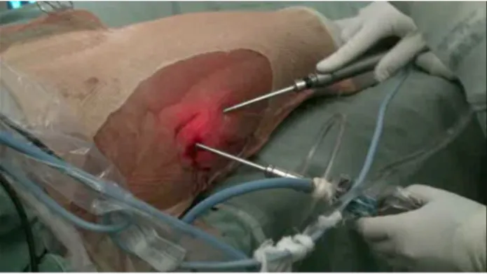
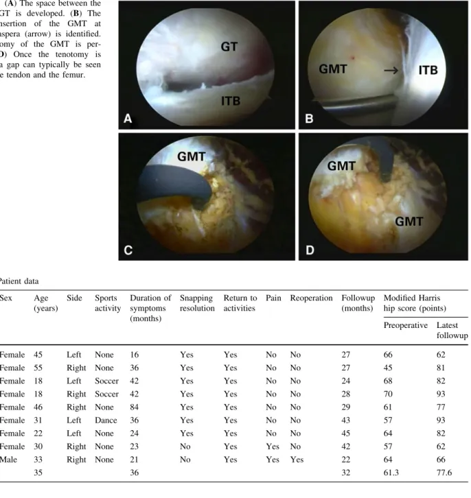
Two authors (GCP, BGD) performed all surgeries. All procedures were performed with general anesthesia fol-lowing the technique described below. The patient was placed in a supine position on a standard operating table. Posterior and anterior preparing and draping of the surgical site and leg were carried out. The leg was draped so that the hip could be moved through a full ROM during the oper-ation. With the surgical site prepared, the GT was outlined and two portal sites were marked on the skin: the superior trochanteric portal (STP) was placed at the superior tip of the GT and the inferior trochanteric portal (ITP) was placed approximately 10 cm below the tip of the GT, in line with the axis of the femur (Fig.1). We started by injecting 100 mL normal saline directly over the prominence of the GT and down to the ITB to develop the potential space just beneath the ITB (Fig.2A). A needle was inserted distally at a 45° angle into the STP and the guidewire and the 4.5-cm arthroscopic cannula were subsequently advanced. The 70°4-mm arthroscope was then introduced and a water inflow portal with an arthroscopy pump at low pressure was initiated to develop the space between the trochanteric bursa, vastus lateralis, and ITB. Next, a needle was intro-duced at the site of the ITP in a proximally oriented 45° angle, localized in the subcutaneous tissue under direct endoscopic view. The skin incision was then made and a shaver blade was introduced through the portal and the ITB. The cannula was placed through the ITB and no additional release was performed. The space down to the ITB was further developed, from inside to outside, with the shaver until the ITB could be easily identified. The femoral insertion of the GMT was identified (Fig.2B) and com-pletely transected using a radiofrequency device close to the linea aspera (Fig.2C). Once the tenotomy was com-plete, a gap could be seen between the tendon and the femur (Fig.2D). A reproduction of the snapping sound was tested at different times during the procedure (seven of nine hips in our study produced snapping under anesthesia and
this finding corresponded well to the reproduction of snapping during passive motion with the patient in the supine position) so that adequate GM resection was obtained. When the procedure was complete, fluid was aspirated from the tissue and the skin portals were sutured closed. The average surgical time was 62 minutes (range, 31–97 minutes). We had no abnormal vascular bleeding and no nerve lesions related to the surgical procedure.
All patients were released on the same day of the sur-gery, at which time full weightbearing and full ROM of the hip were allowed. During the postoperative period, patients were encouraged to squat so that the GMT could be stretched. Crutches were used for a week. Supervised physiotherapy was performed for 3 months and initiated
with GMT stretching exercises (squats and assisted adduc-tion with hip extension, 15 repetiadduc-tions, three times a day) followed by active ROM and finally strengthening. Return to full activities was allowed after at least 3 months or when snapping, pain, and swelling had subsided.
Patients and Methods
Between October 2008 and September 2010, we treated eight patients (nine hips) with external snapping hip using the procedure described above. There were seven women (one bilateral) and one man, with a mean±SD age of 35±13.1 years (range, 18–55 years) (Table1). Two Fig. 2A–D (A) The space between the
ITB and GT is developed. (B) The femoral insertion of the GMT at the linea aspera (arrow) is identified. (C) Tenotomy of the GMT is per-formed. (D) Once the tenotomy is complete, a gap can typically be seen between the tendon and the femur.
Table 1. Patient data
Patient Sex Age (years)
Side Sports activity
Duration of symptoms (months)
Snapping resolution
Return to activities
Pain Reoperation Followup (months)
Modified Harris hip score (points)
Preoperative Latest followup
1 Female 45 Left None 16 Yes Yes No No 27 66 62
2 Female 55 Right None 36 Yes Yes No No 27 45 81
3 Female 18 Left Soccer 42 Yes Yes No No 24 68 82
3 Female 18 Right Soccer 42 Yes Yes No No 28 70 93
4 Female 46 Right None 84 Yes Yes No No 29 61 77
5 Female 31 Left Dance 36 Yes Yes No No 43 57 93
6 Female 22 Left None 24 Yes Yes No No 45 64 82
7 Female 30 Right None 23 No Yes Yes No 42 57 62
8 Male 33 Right None 21 No Yes Yes Yes 22 64 66
patients participated in amateur sports activity (soccer and dance). The mean duration of symptoms before endoscopic GMT release was 36±20.3 months (range, 16–84 months). Minimum followup was 22 months (mean±SD, 32± 9.3 months; range, 22–45 months). No patients were lost to followup. This study was approved by our institutional review board, and all patients signed an informed consent.
Patients were evaluated preoperatively and at 1, 3, 6, and 12 months postoperatively and yearly thereafter. At each visit, the presence of the snapping was investigated and a modified Harris hip score (HHS) [3] was obtained. Only a clinical examination was performed and no imaging examinations were obtained.
Pre- and postoperative HHSs were compared using Wilcoxon tests. SPSS1
10.0 (SPSS Inc, Chicago, IL, USA) was used for analyses.
Results
Seven of nine hips had their pain and snapping resolved (Table1). One revision procedure was performed in a 33-year-old male patient whose snapping and pain remained. In the revision procedure, a GMT and tensor fasciae latae (TFL) release was performed; at latest followup (22 months), the patient remains free of symptoms and snapping. The second failure was in a 30-year-old female patient who had her lateral snapping and pain resolved but remained with a mild ischium snapping and pain. Eight of eight patients returned to their previous level of activity. No patient complained of weakness at most recent followup. The mean modified HHS increased (p=0.011) from 61.3±7.5 points (range, 45–70 points) preoperatively to 77.6±11.9 points (range, 62–93 points) at latest followup.
Discussion
External snapping hip may be caused by the ITB or the GM muscle snapping over the GT. Many surgical techniques have been described and most of them aim at decreasing the tension of the ITB complex with Z-plasty or other forms of resection [2,9,13,14]. The main reason for the increase in tension of the ITB complex is still unknown, as are the biomechanical consequences of its resection. The anatomic and functional relation between the GMT and the ITB [5] stimulated us to formulate the hypothesis that a GMT tenotomy could decrease the tension to the ITB. We therefore describe a novel technique for endoscopic release of the GMT and report the preliminary clinical results of this procedure.
Our study has limitations. First, we had a small sample size. This is due to the low frequency of this condition and
to the fact that most patients with symptomatic external snapping hip improve with nonoperative treatment [14]. Since our primary objective was to describe the new technique, this does not jeopardize our observations. Sec-ond, ours was not a randomized study comparing the new technique to the one described by Ilizaliturri et al. [6], so we cannot say whether our technique is equivalent or superior to another. However, seven of our nine patients were completely relieved of snapping after recovering from the procedure. Third, we did not measure the loss of extension strength, but no patient had weakness when asked directly. Despite the fact that loss of extension strength was not a complaint in this group of patients, it could be more relevant in a group of high-level athletes. In a low-demand population, with weak hip abductors and extensors, releasing the GMT may hypothetically impair hip function. On the other hand, releasing the ITB can cause an overload to the contralateral abduction mecha-nism, as the ITB is a static and dynamic stabilizer of the hip and must be preserved. Fourth, the shape of the buttock was not analyzed with an objective parameter, only with our subjective observation. Further studies are needed to better understand the external snapping hip, ITB-TFL-GMT complex, and long-term clinical results of an endo-scopic GMT release.
Results from previously described techniques are vari-able. Zoltan et al. [14] performed an elliptical resection of the ITB over the trochanter in five patients, and one patient required a second intervention due to persistent symptoms. All patients had complete resolution of the pain and snapping. Brignall and Stainsby [2] performed an ITB Z-plasty in six patients, and again, one patient required a second intervention. A second Z-plasty was performed, and all patients had complete resolution of the symptoms. Provencher et al. [9] also performed ITB Z-plasty in eight patients. All patients had complete resolution of the snap-ping, and all but one returned to full activities. This patient had persistent pain but no snapping. White et al. [13] performed a step-cut procedure of the ITB over the tro-chanter in 16 patients, and 14 had improvement of the pain and snapping. Ilizaliturri et al. [6] were the only ones that performed an arthroscopic procedure, which involved a diamond-shaped resection of the ITB in 11 patients. Ten patients had complete resolution of the pain and snapping, and one patient had improvement but not resolution of the symptoms.
any deformity over the trochanter as it does not require fat removal. In addition to this, resection of part of the ITB and the accommodation of the remaining parts over the anterior and posterior aspects of the GT can cause further defor-mity. This novel procedure is technically easier than the one described by Ilizaliturri et al. [6] as it only requires a simple tenotomy to the GMT. On the other hand, ITB preservation hinders instrument maneuverability. The mean postoperative modified HHS with this new technique was 77. As ours was a small case series, the two patients who still had pain and snapping impaired the results. Excluding them, the mean modified HHS would have been 81. In our series, all patients returned to their previous level of function and there were no cases of nerve injury. The sciatic nerve is posterior to the femur diaphysis at this location, and the GMT is attached to the linea aspera, so the sciatic nerve is far from the release region. The first perforating artery is below the GMT and may be damaged only if the tenotomy is performed too deep.
When compared to previously described open tech-niques [2, 9, 13, 14] and to the arthroscopic technique described by Ilizaliturri et al. [6], the complete resolution of symptoms in seven of nine hips is satisfactory. The two hips that failed to have their snapping/pain resolved had a visible GM contracture and hypotrophy before surgery. This might imply these snapping hips had a different bio-mechanical etiology and highlights the importance of the physical examination of these patients and special attention to the shape of the buttock and the presence of posterior muscle contractures. Maybe a combined release (ITB and GM) would be indicated in such patients. New criteria need to be created to better assess these patients, and with these criteria, a correct surgical indication could be obtained.
Our findings suggest an external snapping hip may be treated by releasing the GMT instead of the ITB. The ITB is tensioned anteriorly by the TFL and posteriorly by the GM, both working synergistically in normal conditions [5]. Therefore, the ITB, GM, and TFL work as a single com-plex [5]. The current literature reports the ITB is the main
cause of an external snapping hip, but the ITB is closely related to the GMT, and by relaxing the GMT, the ITB may relax as well [5]. Despite the good results [12], an ITB release acts at the effect not at the cause of the ITB tension. By previous observations, the authors realized the GMT is closely related to the ITB and may be a posterior tensor of the fascia lata (Fig.3). Therefore, releasing it could decrease the ITB tension and end the snapping. The pro-cedure described here may even be a more physiologic operation to decrease ITB tension. Some of the clinical failures (persistent snapping and/or pain) of other ITB release techniques may be due to a tight GM; therefore, this technique can also be used as a salvage procedure for an ITB release failure.
In light of our preliminary results, the described endo-scopic technique to release the GMT for the treatment of the external snapping hip syndrome is reproducible and its results are comparable to those reported after releasing the ITB. We believe these results justify a future randomized trial comparing the endoscopic ITB and GMT release or a combination of both.
Acknowledgments The authors thank Sheila Ingham MD, PhD (Federal University of Sa˜o Paulo, Brazil) for valuable advice, reviewing of the paper, and help with the English version.
References
1. Brignall CG, Brown RM, Stainsby GD. Fibrosis of the gluteus maximus as a cause of snapping hip: a case report.J Bone Joint Surg Am. 1993;75:909–910.
2. Brignall CG, Stainsby GD. The snapping hip: treatment by Z-plasty.J Bone Joint Surg Br. 1991;73:253–254.
3. Byrd JW, Jones KS. Prospective analysis of hip arthroscopy with 2-year follow-up.Arthroscopy. 2000;16:578–587.
4. Evans P. The postural function of the iliotibial tract.Ann R Coll Surg Engl. 1979;61:271–280.
5. Falvey EC, Clark RA, Franklyn-Miller A, Bryant AL, Briggs C, McCrory PR. Iliotibial band syndrome: an examination of the evidence behind a number of treatment options.Scand J Med Sci Sports. 2010;20:580–587.
6. Ilizaliturri VM Jr, Martinez-Escalante FA, Chaidez PA, Cama-cho-Galindo J. Endoscopic iliotibial band release for external snapping hip syndrome.Arthroscopy. 2006;22:505–510. 7. Larsen E, Johansen J. Snapping hip.Acta Orthop Scand. 1986;
57:168–170.
8. Nam KW, Yoo JJ, Koo KH, Yoon KS, Kim HJ. A modified Z-plasty technique for severe tightness of the gluteus maximus.
Scand J Med Sci Sports. 2011;21:85–89.
9. Provencher MT, Hofmeister EP, Muldoon MP. The surgical treatment of external coxa saltans (the snapping hip) by Z-plasty of the iliotibial band.Am J Sports Med. 2004;32:470–476. 10. Schaberg JE, Harper MC, Allen WC. The snapping hip
syn-drome.Am J Sports Med. 1984;12:361–365.
11. Teitz CC, Garrett WE Jr, Miniaci A, Lee MH, Mann RA. Tendon problems in athletic individuals.Instr Course Lect. 1997;46:569–582. 12. Wahl CJ, Warren RF, Adler RS, Hannafin JA, Hansen B. Internal coxa saltans (snapping hip) as a result of overtraining: a report of 3 cases in professional athletes with a review of causes and the role of ultrasound in early diagnosis and management. Am J Sports Med. 2004;32:1302–1309.
13. White RA, Hughes MS, Burd T, Hamann J, Allen WC. A new operative approach in the correction of external coxa saltans: the snapping hip.Am J Sports Med. 2004;32:1504–1508.

