RESUMO.- [Avaliação clínica e parasitológica do fluazu-ron e da ivermectina pour-on no tratamento da demo-diciose canina.] O objetivo do presente estudo foi avaliar a eficácia do fluazuron e da ivermectina pour-on em diferen -tes protocolos terapêuticos no tratamento da demodiciose,
através da quantificação de ácaros por raspados cutâneos e exames histológicos, além da avaliação dos cães. Foram avaliados 18 cães com raspados cutâneos positivos para o ácaro Demodex canis, divididos em três grupos. Todos os animais foram tratados a cada 14 dias, totalizando seis tra -tamentos em cada cão (Dias 0, 14, 28, 42, 56 e 70). No gru -po 1 foi utilizado fluazuron 2,5% pour-on na dosagem de 20mg/kg; no grupo 2 foi empregado fluazuron 2,5% pour--on na dosagem de 20mg/kg associado a ivermectina 0,5% pour-on, na dosagem de 0,6mg/kg e, no grupo 3, somente ivermectina 0,5% pour-on 0,6mg/kg. Raspados cutâneos e acompanhamento clínico das lesões foram realizados a cada 14 dias por 84 dias e realizado exame histopatológico
Clinical and parasitological evaluation of pour-on fluazuron
and ivermectin for treating canine demodicosis
1Clarissa P. Souza2*, Regina H.R. Ramadinha3 and Fabio B. Scott2
ABSTRACT.- Souza C.P., Ramadinha R.H.R. & Scott F.B. 2014. Clinical and parasitologi-cal evaluation of pour-on fluazuron and ivermectin for treating canine demodicosis.
Pesquisa Veterinária Brasileira 34(11):1094-1100. Departamento de Parasitologia Animal, Instituto de Veterinária, Universidade Federal Rural do Rio de Janeiro, BR-465 Km 7, Sero -pédica, RJ 23890-000, Brazil. E-mail: [email protected]
The objective of the study was to evaluate the efficacy of pour-on formulations of flu -azuron and ivermectin in different therapeutic protocols for treatment of demodicosis by means of quantifying mites with skin scraping, histological and clinical evaluation in dogs. Eighteen dogs with skin scrapings positive for Demodex canis were evaluated, divided into three groups. All the animals were treated every 14 days, completing 6 treatments for each animal (days 0, 14, 28, 42, 56 and 70). In group 1, pour-on 2.5% fluazuron was used at the dose of 20mg/kg; in the group 2 pour-on 2.5% fluazuron at a dose of 20 mg/kg in asso -ciation with pour-on 0.5% ivermectin at the dose of 0.6mg/kg; and in group 3, pour-on 0.5% ivermectin alone was used, at the dose of 0.6mg/kg. The treatment was evaluated and monitored through skin scrapings and clinical follow-up of the lesions every 14 days for 84 days, and through histopathological examination at the end of each treatment protocol. The success rate was defined as the percentage of dogs in each group that had negative skin scrapings after the treatment: this was 16.67% for group 1, and 50% for groups 2 and 3. The reduction in mite counts reached effectiveness of 67.66%, 88.99% and 84.29% for groups 1, 2 and 3 respectively. The Wilcoxon test showed that there was a significant diffe -rence between the number of mites before and after treatment in groups 2 and 3. The his -topathological examination revealed that only group 1 showed no significant difference in the intensity of infestation between days 0 and 84. Clinically, there was no significant diffe -rence between the evaluation before and after treatment in the three groups. Pour-on 2.5% fluazuron and pour-on 0.5% ivermectin were not effective for treating canine demodicosis, either in association or as single therapy, when applied every 14 days for a period of 70 days. Quantification of mites using skin scrapings and histological evaluation proved to be ineffective, either one as sole therapeutic evaluation parameters, for canine demodicosis. INDEX TERMS: Demodex canis, control, fluazuron, ivermectin, pour-on.
1 Received on January 8, 2014.
Accepted for publication on August 12, 2014.
2 Departamento de Parasitologia Animal, Instituto de Veterinária (IV),
Universidade Federal Rural do Rio de Janeiro (UFRRJ), Campus de Sero
-pédica, BR-465 Km 7, Sero-pédica, RJ 23890-000, Brazil. *Corresponding author: [email protected]
3 Departamento de Medicina e Cirurgia Veterinárias, IV-UFRRJ, BR-465
ao final de cada protocolo terapêutico. A taxa de sucesso foi definida pela porcentagem de cães em cada grupo com raspados negativos ao final do tratamento, que foi 16,67% para o grupo 1 e 50% para os grupos 2 e 3. A redução na contagem no número de ácaros alcançou eficácia de até 67,66%; 88,99% e 84,29%, nos grupos 1, 2 e 3, respecti -vamente. O teste de Wilcoxon mostrou que houve diferen -ça significativa entre a quantidade de ácaros antes e após o tratamento nos grupos 2 e 3. No exame histopatológico apenas o grupo 1 não apresentou diferença significativa na intensidade da infestação entre os dias 0 e 84. Clinicamen -te não houve diferença significativa entre as avaliações an -tes e após o tratamento dos três grupos. O fluazuron 2,5% pour-on e a ivermectina 0,5% pour-on associados ou como terapia única, não foram eficazes no tratamento da demodi -ciose canina, quando aplicados a cada 14 dias em um perí -odo de 70 dias. A quantificação de ácaros através do exame parasitológico em raspado cutâneo e em exame histopato -lógico demonstrou-se ineficaz como parâmetro isolado de avaliação pós-terapêutica para demodiciose canina. TERMOS DE INDEXAÇÃO: Demodiciose canina, Demodex canis, controle, fluazuron, ivermectina, pour-on.
INTRODUCTION
Demodectic mange or canine demodicosis is an inflamma -tory parasitic skin disease caused by excessive proliferation of the mite Demodex canis, which lives in hair follicles and sebaceous glands. These parasites are transmitted from mother to offspring, which also inherit a specific cell-me -diated condition of immunodeficiency (Heine et al. 2005, Delayte et al. 2006).
It has now been shown through molecular techniques that all dogs, even without lesions, house a small popula -tion of mites on their skin. Their immune systems tolerate the presence of these parasites and keep them under con -trol. But at some point, the number of mites can increase, thereby developing a pathogenic role in relation to the host (Ravera et al. 2013).
Routinely, infestation is diagnosed through viewing the mite D. canis under an optical microscope in parasitological examinations of hair plucking or skin scrapings, or histopa -thological examination (Larsson 1989).
Several drugs have been used for treating canine de -modicosis. Amitraz and macrocyclic lactones are the most common drugs and they are used in different therapeutic schemes. In general, they show varied levels of effective -ness and they may generate side effects that make it impos -sible to complete the therapy. Another negative point is that some protocols are not very practicable, which decreases the pet owners’ compliance. For this reason, it is necessary to search for alternative therapies, due to the possibility of failure or impracticality of the currently recommended tre -atments (Mueller et al. 2012).
Ivermectin was the first macrocyclic lactone to be ma -rketed and has been regularly used for treating various infestations in small animals (Lynn 2003). In the 1990s a pour-on 0.5% formulation became available for the treat -ment of ecto and endoparasites in cattle, which highlighted
the practicality of topical therapy (Paradis & Pagé 1998). A recent advance in the effort to control ectoparasites on animals came through the development of insect growth inhibitors. Fluazuron is in this class and it is considered to be a good drug for controlling ticks on ruminants, due to its high specificity, low mammalian toxicity, effectiveness at low concentrations and long-lasting residual effect against reinfestation (Kryger et al. 2005). The action and safety of this antiparasitic drug has now also been tested against fle -as and ticks on dogs, with promising results (Vieira 2009, 2012, Oliveira et al. 2012).
The objective of the present study was to evaluate the efficacy of pour-on formulations of fluazuron and ivermec -tin in different therapeutic protocols for trea-ting demodi -cosis by means of quantifying mites using skin scraping, histological and clinical evaluations on dogs.
MATERIALS AND METHODS
The study was approved by the Ethics Committee on Animal Expe
-rimentation of the Universidade Federal Rural do Rio de Janeiro (CEUA-FAPUR/UFRRJ). Eighteen dogs of both sexes and various ages and breeds that presented clinical and laboratory diagnoses of localized and generalized demodicosis were evaluated. These dogs were treated at the Dermatology service of the Federal Rural University of Rio de Janeiro Veterinary Hospital, over a period of 20 months.
The initial diagnosis was made through skin scrapings of two areas with lesions, each of 1cm2, and these were delineated on
the dog’s skin using a marking pen. As close as possible to this site, still within the lesional area, a biopsy was performed under local anesthesia using a punch of 8 mm of diameter on order to collect skin material on day 0. The fragment collected was fixed in a solution of 10% formalin and was sent for routine histology processing and hematoxylin-eosin staining.
Dogs that presented one to four small, circumscribed areas of alopecia were considered cases of localized demodicosis. All the animals that presented at least five small damaged areas (<100 cm2 of the body) or were affected over an entire region of the body
(>100 cm2) or had an entire limb compromised (Mueller et al.
2009) were deemed to have the generalized form of infestation. The dogs were divided into localized and generalized demo
-dicosis cases and therefore randomly placed into one of the three groups, with six animals each group.
The group 1 received a pour-on application of 20mg/kg of 2.5% fluazuron (Acatak®; Novartis Animal Health) arthropod gro -wth inhibitor formulation, as used for cattle. The drug was admi
-nistered with the aid of a disposable hypodermic syringe at a dose of 20mg/kg, along the back of the dogs.
The animals of the group 2 were treated with fluazuron at 20mg/kg and also with pour-on 0.5% ivermectin (Ivomec®; Me -rial Animal Health) at a dose of 0.6mg/kg. Both drugs were admi
-nistered at the same time in parallel on opposite sides along the back of each dog, using a disposable hypodermic syringe for each product.
The treatment for the group 3 consisted of pour-on 0.5% iver
-mectin at 0.6mg/kg.
All the dogs were treated every 14 days for 70 days, thus com
-pleting a total of six treatments for each animal (days 0, 14, 28, 42, 56 and 70). The total treatment time was established in ac
Possible side effects relating to the treatment were evaluated. The animals were weighed to calculate the volume that should be administered, and at every new administration, the weight was measured again and the volume adjusted.
For evaluation and follow-up treatment, two skin scrapings of 1cm2 each were repeated on all the dogs every 14 days, for 84
days, always in the same areas of the skin. The different stages of D. canis were identified, counted and compared on each evalua
-tion day, as recommended by Mueller et al. (2012).
On day 84 another sample of skin was collected from each animal at the same area where biopsy was performed at the be
-ginning of the protocol. The intensity of infestation seen in the histopathological examination, both before and after treatment, was determined using the score recommended by Caswell et al. (1995), where (0) indicates no mites seen in the examination, (1) rare fragments of mites (slight infestation), (2) 2 to 4 mites per slide (mild infestation), (3) several mites, but three or less per follicle (moderate infestation) and (4) more than three mites per hair follicle (severe infestation).
At the initial and final consultations (days 0 and 84), the main skin lesions of the dogs were examined and there were evalua
-ted: alopecia, dyschromia (erythema and/or hyperpigmentation), crusts, seborrhea, papules and pustules, and swelling. The inten
-sity of these alterations was classified as: (0) absent, (1) slight, (2) intermediate or (3) severe (Heine et al. 2005, Delayte et al. 2006). These parameters were used for clinical evaluation of the effecti
-veness of the proposed therapeutic protocols.
Dogs with secondary bacterial infection diagnosed through cytological examination using skin imprints, which were stained by means of fast panoptic (Instant Prov), were treated with sys
-temic antibiotic: cephalexin at a dose of 30mg/kg every 12 hours. Antibiotics were used until bacteria were no longer found in cyto
-logy, or any lesions suggestive of pyoderma.
The effectiveness of therapeutic protocols based on reduction of the number of mites in relation to the number prior to treat
-ment was determined by means of the geometric mean, calculated using the following formula:
(Geometric mean before treatment - geometric mean after treatment) x100 Geometric mean before treatment
The success rate was defined as the percentage of dogs in each group that presented negative skin scraping tests (Fourie et al. 2007).
The results obtained from the skin scraping tests, clinical eva
-luation scores and histological examinations were statistically compared before and after treatment using the Wilcoxon test for differences between ordered pairs (Sampaio 2002).
All the animals were monthly monitored for twelve months following the withdrawal of the treatment protocols, which is the time period suggested in the literature for monitoring recurrence of clinical disease in dogs (Gortel 2006, Mueller et al. 2012).
RESULTS
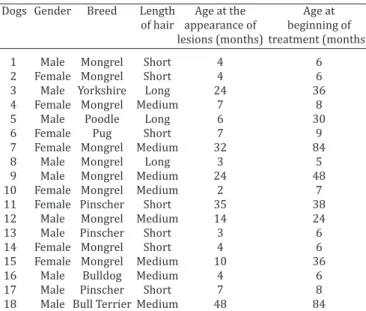
The Table 1 shows the ages of the dogs studied at the time of appearance of the lesions and at the beginning of the tre -atment; data about sex, breed and length of fur of the dogs were also included.
Results from therapy using pour-on 2.5% fluazuron
The dogs included in this treatment protocol were iden -tified by the numbers 1 to 6 (Group1). The evaluations per -formed at the beginning and during the treatment, through
the numbers of mites in the skin scraping tests, are descri -bed in Table 2. The reduction in the count of the number of mites reached levels that ranged from 38.90 to 67.66% in the six evaluations after the therapy began. The success rate at the end of treatment was 16.67% (1/6) (Table 2). There was no significant difference between the numbers of mites before and after the therapy.
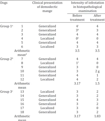
Through histological examination, it was found that the -re was no significant dec-rease in the intensity of infestation from the beginning (day 0) to after the treatment (day 84) (Table 3).
Clinical evaluation of the lesions showed that the tre -atment failed to resolve the skin alterations caused by the mite Demodex canis. There was no significant difference be -tween the clinical observations made before treatment and at the end of the treatment protocol (Table 4).
Results from therapy using pour-on 2.5% fluazuron and pour-on 0.5% ivermectin
The dogs included in this treatment protocol were iden -tified by the numbers 7 to 12 (Group 2).. The reduction in the number of mites reached levels that ranged from 67 to 88.99% in the six evaluations conducted after the thera -py protocol began. The success rate at the end of the tre -atment time was 50%. There was a significant difference in the numbers of mites from before to after the therapy (Table 2).
Through histological examination, it was found that the -re was no significant dec-rease in the intensity of infestation from before (day 0) to after the treatment (day +84) (Table 3).
Clinical evaluation of the lesions showed that the treat -ment was partially effective for controlling the skin altera -tions caused by D. canis. Dogs 7, 9 and 10 (3/6) presented clinical failure, dogs 11 and 12 (2/6) showed clinical im -provement and dog 8 (1/6) presented clinical cure. There was no significant difference between the clinical observa
-Table 1. Characteristics and history of 18 dogs with demodicosis used in evaluating different treatment
protocols for infestation control
Dogs Gender Breed Length Age at the Age at
of hair appearance of beginning of
lesions (months) treatment (months)
1 Male Mongrel Short 4 6
2 Female Mongrel Short 4 6
3 Male Yorkshire Long 24 36
4 Female Mongrel Medium 7 8
5 Male Poodle Long 6 30
6 Female Pug Short 7 9
7 Female Mongrel Medium 32 84
8 Male Mongrel Long 3 5
9 Male Mongrel Medium 24 48
10 Female Mongrel Medium 2 7
11 Female Pinscher Short 35 38
12 Male Mongrel Medium 14 24
13 Male Pinscher Short 3 6
14 Female Mongrel Short 4 6
15 Female Mongrel Medium 10 36
16 Male Bulldog Medium 4 6
17 Male Pinscher Short 7 8
tions made before treatment and at the end of the treat -ment protocol (Table 4).
Results from therapy using pour-on 0.5% ivermectin
The dogs included in this treatment protocol were iden -tified by the numbers 13 to 18 (Group 3). The reduction in the number of mites reached levels that ranged from 28.24 to 84.29% in the six evaluations conducted after the treat -ment began. The success rate at the end of treat-ment was 50%. There was a significant difference in the numbers of mites from before to after the therapy (Table 2). Through histological examination, it was found that there was no significant decrease in the intensity of infestation from be -fore (day 0) to after the treatment (day +84) (Table 3).
Clinical evaluation of the lesions showed that the tre -atment was partially effective for controlling the skin alte -rations caused by D. canis. Dogs 14 and 16 (2/6) presen -ted clinical failure, dogs 15 and 18 (2/6) showed clinical improvement and dogs 13 and 17 (2/6) presented clinical cure. There was no significant difference between the clini -cal observations made before treatment and at the end of the treatment protocol (Table 4).
Dogs 1, 2, 3, 5 (group 1), 10, 11 (group 2), 14, 15, 16 and 18 (group 3) (10/18, 55,6%) received systemic antibiotic therapy until remission of pyoderma and, for some ani -mals, this treatment continued throughout the trial period. In dogs 1, 2, 3, 5, 6 (group 1), 7, 10, 12 (group 2), 14, 16
and 18 (group 3) (11/18, 61,1%) mites could be seen in the skin scraping, and dogs 4, 9 and 15 (3/18, 16,67%) still presented clinical alterations on the skin on the last day of evaluation. These dogs were then started on treatment with oral ivermectin at a daily dose of 0.6 mg/kg until com -plete remission of lesions and until they presented negative skin scrapings, which took up to four months of treatment for some animals.
The dogs were monitored for 12 months after the treat -ment protocol ended, through periodic reviews or telepho -ne contacts, or even through photos sent by the ow-ners.
Dogs 8, 11 (group 2), 13 and 17 (group 3) (4/18, 22,2%), which did not receive any drugs other than the ini -tially instituted protocol, did not show any clinical signs of recurrence after 12 months.
None of the dogs presented any side effects relating to the treatment during the evaluation period of this study.
DISCUSSION
Fluazuron is an insect growth inhibitor that belongs to the group of chitin synthesis inhibitors, also called benzoyl -phenylureas. It does not have any direct action that causes the death of insects and mites but, rather, it interferes with the molting and hatching processes of the parasites, there -by interrupting their life cycle. Fluazuron has been used es -pecially for controlling Rhipicephalus (Boophilus) microplus (Graf 1993). Vieira (2009) and Oliveira et al. (2012) also
Table 2. Quantity of Demodex canis mites found in skin scraping tests on the
dogs evaluated by means of three therapeutic protocols over the 70 days of
treatment
Groups Dogs Measurement Quantity of Demodex canis (different stages)
method Day 0 Day 14 Day 28 Day 42 Day 56 Day 70 Day 84
Group 1a 1 429 193 184 214 124 96 78
2 55 16 22 16 14 13 13
3 25 10 8 31 61 11 13
4 2 1 0 1 4 2 0
5 20 16 15 15 17 11 10
6 88 79 39 29 15 22 18
Geometric meand 35.71 18.42 16.33 18.94 21.82 13.71 11.55
% Reduction - 48.42 52.56 46.96 38.90 61.61 67.66
% Success rate 0 0 16.67 0 0 0 16.67
Group 2b 7 8 6 10 6 2 1 1
8 3 0 0 0 0 0 0
9 2 3 4 4 3 0 0
10 54 8 12 7 6 5 4
11 72 10 2 1 0 0 0
12 12 1 3 4 2 1 1
Geometric mean 11.44 3.36 3.77 2.96 2.04 1.31 1.26
% Reduction - 70.63 67 74.13 79 88.55 88.99
% Success rate 0 16.67 16.67 16.67 33.33 50 50
Group 3c 13 2 1 0 0 0 0 0
14 8 6 6 7 11 12 2
15 14 13 3 2 0 0 0
16 3 2 6 5 5 2 1
17 12 25 3 1 1 0 0
18 365 103 68 45 38 32 22
Geometric mean 11.97 8.59 5.29 3.83 3.58 3.03 1.88
% Reduction - 28.24 55.81 68 70.10 74.69 84.29
% Success rate 0 0 16.67 16.67 33.33 50 50
a dogs treated with pour-on 2.5% fluazuron; b dogs treated with pour-on 2.5% fluazuron and
demonstrated the potential of pour-on fluazuron against different stages of R. sanguineus, at diverse concentrations.
Because the mite D. canis belongs to the same taxono -mic class as ticks (Arachnida), it was considered possible that fluazuron might have some efficacy against these pa -rasites.
The results obtained through pour-on application of fluazuron confirm what was previously found through tes -ting lufenuron, which is another compound in the same group of chitin synthesis inhibitors. Lufenuron was also used for treating canine demodicosis and was considered to be ineffective (Schwassmann et al. 1997). This drug was developed to control fleas on cats and dogs, and it is admi -nistered as pills. Schwassmann et al. (1997) suspected that the failure of oral lufenuron against mites was associated with small penetration of the drug into the skin. There is also the possibility that the arthropods may have different mechanisms of chitin synthesis, since it has been proven that lufenuron provides excellent results in environmental control of different stages of fleas’ life cycle, and fluazuron used orally also has shown good efficacy in controlling lar -vae and nymphs of R. sanguineus (Vieira 2012). Thus, it is believed that even if administered orally, fluazuron would not show good efficacy against Demodex canis.
In recent decades, ivermectin has been extensively eva -luated for treating canine demodicosis in various protocols, especially orally, and it is one of the most widely used drugs in the world (Mueller et al. 2012). Efficacy of up to 89.7% has been reported when administered orally every day, but with treatment protocols of duration of up to 150 days (Ristic et al. 1995, Delayte et al. 2006). The present study evaluated the therapeutic potential of ivermectin applied using a pour-on formulation separately and in combination
Table 3. Clinical presentation of demodicosis and quantity of Demodex canis in histopathological examinations on the
dogs used in the three therapeutic protocols, before treatment and after 84 days of evaluation
Dogs Clinical presentation Intensity of infestation
of demodectic in histopathological
mange examination
Before After
treatment treatment
Group 1a 1 Generalized 4i 4
2 Generalized 3h 3
3 Generalized 4 4
4 Localized 0e 0
5 Generalized 4 4
6 Localized 3 3
Arithmetic 3.5 3.5
meand
Group 2b 7 Generalized 4 4
8 Localized 1f 0
9 Generalized 3 0
10 Generalized 3 2 g
11 Generalized 4 1
12 Localized 4 2
Arithmetic 3.17 1.5
mean
Group 3c 13 Localized 3 2
14 Generalized 3 2
15 Generalized 4 3
16 Generalized 3 2
17 Localized 3 0
18 Generalized 3 2
Arithmetic 3.17 1.83
mean
a dogs treated with pour-on 2.5% fluazuron; b dogs treated with pour-on
2.5% fluazuron and pour-on 0.5% ivermectin; c dogs treated with pour
--on 0.5% ivermectin, d p>0.05; e 0 = no mites; f 1 = slight infestation; g 2 = mild infestation; h = moderate infestation; i 4 = severe infestation.
Table 4. Clinical signs observed in the dog’s skin in the three treatment protocols evaluated, before (day 0) and at the end (day 84) of treatment
Animals Clinical signs
Alopecia Dyschromia Crusts Seborrhea Papule / Pustule Edema
Day 0 Day 84 Day 0 Day 84 Day 0 Day 84 Day 0 Day 84 Day 0 Day 84 Day 0 Day 84
Group 1a 1 2f 2 3g 3 2 3 1e 1 0d 0 0 0
2 3 3 2 2 1 0 1 1 0 0 0 0
3 1 2 3 3 3 3 0 0 0 0 0 0
4 1 1 0 0 1 1 0 0 0 0 0 0
5 2 3 3 3 2 3 1 1 0 0 3 3
6 1 1 1 1 0 0 0 0 1 1 0 0
p value 0.1797 0 0.5930 0 0 0
Group 2b 7 2 2 3 3 1 0 1 2 0 0 0 0
8 1 0 0 0 0 0 0 0 0 0 0 0 9 1 1 1 0 0 0 1 1 0 0 0 0
10 1 1 1 1 0 0 3 2 0 0 0 0
11 2 0 3 0 1 0 1 0 0 0 0 0
12 2 1 2 1 2 0 0 0 0 0 0 0
p value 0.1088 0.1088 0.1088 0.5930 0 0
Group 3c 13 2 0 0 0 0 0 1 0 0 0 0 0
14 2 3 2 2 0 2 0 0 1 0 0 0
15 3 1 2 0 2 0 2 0 0 0 0 0
16 2 2 2 2 0 0 1 0 3 3 0 0
17 1 0 1 0 1 0 0 0 0 0 0 0
18 3 2 2 1 3 1 0 0 0 0 3 0
p value 0.1380 0.1088 0.4652 0.1088 0.3173 0.3173
a dogs treated with pour-on 2.5% fluazuron; b dogs treated with pour-on 2.5% fluazuron and pour-on 0.5% iver
with fluazuron, also in a pour-on formulation, applied at the dose of 0.6mg/kg that is usually used in applications via other routes, but at intervals of 14 days for a period of 84 days.
The topical formulation of ivermectin has already been compared with the oral product among goats, and this de -monstrated that percutaneous administration promotes prolonged persistence of the drug in plasma. However, the bioavailability is significantly lower than is seen with oral administration (Scott, Kinabo & McKellar 1990). In the present evaluation, no satisfactory results supporting the hypothesis that it might be possible to administer pour-on ivermectin over a longer treatment interval than could be done through oral administration were obtained, over the treatment period that had been established.
The pour-on formulation of ivermectin was previously tested at a dose of 1.5mg/kg applied three times a week, but only among dogs with chronic and generalized demo -dicosis and with a history of unsuccessful treatment with amitraz. A reduction in the severity of clinical signs and up to 75% in the number of mites in skin scrapings was obser -ved, but the effectiveness according to the recurrence rate after 12 months of monitoring was 8% (Paradis & Pagé 1998). The disease is considered to be chronic and genera -lized when it persists for at least six months with involve -ment of no less than 50% of the dog’s body or involve-ment of the four limbs. This form has been reported as being di -fficult to treat and the outcome is often frustrating (Paradis & Pagé 1998, Fourie et al. 2007). Dogs 3, 5, 7, 9, 12, 15 and 18 (7/18) had been developing clinical signs of infestation for at least six months prior to being enrolled into the study and they presented the generalized form of demodicosis. With the exception of number 12, in which the disease was presented in the localized form, these animals were consi -dered to be chronic cases. None of these dogs showed com -plete clinical remission and/or absence of parasites in the diagnostic examinations at 84 days of evaluation.
There is a need for further studies on the pharmacoki -netics in dogs that are administered different formulations of ivermectin. The pour-on formulation used at the conven -tional dose every 15 days for treating canine scabies (Pa -radis et al. 1997) has demonstrated excellent results. This report makes us believe that there is a reasonable level of systemic absorption and dispersion in dogs’ skin.
In each of the three groups, there were two animals with the localized form and four animals with the generalized form of canine demodicosis. Several literature reports have commented on the difficulty of treating generalized mange, in comparison with localized mange. It has even been cited that 10% of the localized cases may have spontaneous re -mission of clinical signs. Nonetheless, this does not occur routinely in clinical practice and there are no scientific data to support this finding (Mueller et al. 2009). In the present study, different therapeutic responses were observed, re -gardless of the clinical presentation of the demodicosis. Even among the six dogs with localized mange (numbers 4, 6, 8, 12, 13 and 17), two of them showed clinical failure at the end of the treatment (Dogs 4 and 6).
The duration of treatment required for evaluating the
therapeutic protocols in the present study needed to be determined. This was established in accordance with the recommendation from Schwassmann et al. (1997), who stated that after two to three months of therapy, it was pos -sible to observe whether the medication had been effective or not. On the other hand, there are comments in the lite -rature indicating that, for clinical remission of the lesions and absence of parasites in the skin scrapings, the treat -ment may be very lengthy. Moreover, premature termina -tion may be the major cause of treatment failure (Gortel 2006). Differences in the duration of treatment protocols are reported by some authors, such as Delayte et al. (2006), who obtained the first negative skin scraping result after 90 days of treatment and parasitological clearance after 130 days. Ristic et al. (1995) obtained these same results at 45 and 70 days, respectively. It is possible that, with con -tinuity of the treatment, or if the drugs were applied more often, better clinical and parasitological results might have been observed among the animals studied.
The results found in this study allowed us to observe that although groups 2 and 3 showed significant reductions in the numbers of mites between day 0 and day 84, this was not translated into effective reduction of the general cli -nical signs among these animals. In other cases, the dogs may present clinically normal results after treatment, but still demonstrate mites in skin scraping tests (Mueller et al. 2012). Thus, studies evaluating therapeutic protocols against demodicosis should not use observation of the pre -sence or ab-sence of Demodex mites solely, or clinical appea -rance alone. Both parameters (clinical and parasitological) must be considered in interpreting the results and in deter -mining whether therapeutic success was achieved.
CONCLUSIONS
Pour-on 2.5% fluazuron and pour-on 0.5% ivermectin, either in association or as single therapies, were not effec -tive for treating canine demodicosis when applied every 14 days over a period of 70 days.
Quantification of mites by means of histological exa -mination and skin scraping proved to be ineffective when used as a sole parameter for therapeutic evaluation of ca -nine demodicosis.
REFERENCES
Caswell J.L., Yager J.A., Ferrer L. & Weir J.A.M. 1995. Canine demodicosis: a re-examination of the histopathologic lesions and description of the immunophenotype of infiltrating cells. Vet. Derm. 6(1):9-19.
Delayte E.H., Otsuka M., Larsson C.E. & Castro R.C.C. 2006. Eficácia das lactonas macrocíclicas sistêmicas (ivermectina e moxidectina) na tera
-pia da demodicidose canina generalizada. Arq. Bras. Med. Vet. Zootec. 58:31-38.
Fourie L.J., Kok D.J., Plessis A. & Rugg D. 2007. Efficacy of a novel formu
-lation of metaflumizone plus amitraz for the treatment of demodectic mange in dogs. Vet. Parasitol. 150(3):268-274.
Graf J. 1993.The role of insect growth regulators in arthropod control. Parasitol. Today 9:471-474.
Gortel K. 2006. Update on canine demodicosis. Vet. Clin. Small Anim. Pract. 36(1):229-241.
Heine J., Krieger K., Dumont P. & Hellmann K. 2005. Evaluation of the ef
the treatment of generalized demodicosis in dogs: results of a European field study. Parasitol. Res. 97:S89-S96.
Kryger U., Deschodt C. & Scholtz C.H. 2005. Effects of fluazuron and iver
-mectin treatment of cattle on the structure of dung beetle communities. Agric. Ecosyst. Environ. 105:649-656.
Larsson C.E. 1989. Dermatologia Veterinária. II. Demodiciose. Comun. Cient. Fac. Med. Vet. Zootec. USP 13:19-27.
Lynn R.C. 2003. Antiparasitic drugs, p.244-286. In: Bowman D. (Ed.), Georgi`s Parasitology for Veterinarians. 8th ed. W.B. Saunders, St Louis.
Mueller R.S., Meyer D., Bensignor E. & Sauter-Louis C. 2009. Treatment of canine generalized demodicosis with a “spot on” formulation containing
10% moxidectin and 2.5% imidacloprid (Advocate®, Bayer Healthcare).
Vet. Dermatol. 20(5/6):441-446.
Mueller R.S., Bensignor E., Ferrer L., Holm B., Lemarie S., Paradis M. & Shipstone M.A. 2012. Treatment of demodicosis in dogs: 2011 clinical practice guidelines. Vet. Dermatol. 23(2):86-96, e20-1.
Oliveira P.R., Calligaris I.B., Roma G.C., Bechara G.H., Pizano M.A. & Mathias M.I.C. 2012 Potential of the insect growth regulator, fluazuron, in the control of Rhipicephalus sanguineus nymphs Latreille, 1806 (Acari: Ixo
-didae): Determination of the LD95 and LD50. Exp. Parasitol. 131:35-39. Paradis M., Jaham C. & Pagé N. 1997. Topical pour on ivermectin in the
treatment of canine scabies. Can. Vet. J. 38:379-382.
Paradis M. & Pagé N. 1998. Topical (pour-on) ivermectin in the treatment of chronic generalized demodicosis in dogs. Vet. Derm. 9:55-59. Ravera I., Altet L., Francino O., Sánchez A., Roldán W., Villanueva S., Bardagi
M. & Ferrer L. 2013. Small Demodex populations colonize most parts of the skin of healthy dogs. Vet. Dermatol. 24(1):168-e37.
Ristic Z., Medleau L., Paradis M. & White-Weithers N.E. 1995. Ivermectin for treatment of generalized demodicosis in dogs. J. Am. Vet. Med. Assoc. 207:1308-1310.
Scott E.W., Kinabo L.D. & McKellar Q.A. 1990. Pharmacokinetics of iver
-mectin after oral or percutaneous administration to adult milking goats. J. Vet. Pharmacol. Therap. 13(4):432-435.
Schwassmann M., Kunkle G.A., Hepler D.I. & Lewis D.T. 1997. Use of
lufenuron for treatment of generalized demodicosis in dogs. Vet. Der
-matol. 8:11-18.
Vieira V.P.C. 2009. Eficácia do regulador de crescimento de artrópodes fluazuron no controle da pulga Ctenocephalides felis felis Bouché, 1835
(Siphonaptera: Pulicidae) em cães. Dissertação de Mestrado em Parasi
-tologia Animal, Universidade Federal Rural do Rio de Janeiro, Seropé
-dica, RJ. 45p.
Vieira V.P.C. 2012. Atividade do fluazuron administrado por via oral no
controle de Rhipicephalus sanguineus em cães. Tese de Doutorado em
Parasitologia Animal, Universidade Federal Rural do Rio de Janeiro, Se