RESUMO.- [Etiologia, perfil de sensibilidade dos Sta-phylococcus spp. aos antimicrobianos e fatores de ris
-co associados à mastite bovina nos estados da Bahia e Pernambuco.] Objetivou-se estudar a etiologia da mastite,
Etiology, antimicrobial susceptibility profile of
Staphylococcus
spp.
and risk factors associated with bovine mastitis in the states of
Bahia and Pernambuco
1Carina C. Krewer2, Izabela P. de S. Lacerda3, Evandro S. Amanso3, Noelly B. Cavalcante3, Rodolfo de M. Peixoto4, José W. Pinheiro Júnior5, Mateus M. da Costa3 and Rinaldo A. Mota2*
ABSTRACT.- Krewer C.C., Lacerda I.P.S., Amanso E.S., Cavalcante N.B., Peixoto R.M., Pinhei-ro Júnior J.W., Costa M.M. & Mota R.A. 2013. Etiology, antimicrobial susceptibility profile of Staphylococcus spp. and risk factors associated with bovine mastitis in the states of
Bahia and Pernambuco.Pesquisa Veterinária Brasileira 33(5):601-606. Departamento de Medicina Veterinária, Universidade Federal Rural de Pernambuco, Rua Dom Manoel de Me-deiros s/n, Dois Irmãos, Recife, PE 51171-900, Brazil. E-mail: [email protected]
The purpose of this paper was to study the etiology of mastitis, determine the
antimi-crobial susceptibility profile of Staphylococcus spp. and to identify the risk factors asso-ciated with infection in dairy cows in the states of Bahia and Pernambuco, Brazil. From the 2,064 milk samples analyzed, 2.6% were associated with cases of clinical mastitis and 28.2% with subclinical mastitis. In the microbiological culture, Staphylococcus spp. (49.1%) and Corynebacterium spp. (35.3%) were the main agents found, followed by Prototheca spp. (4.6%) and Gram negative bacilli (3.6%). In the antimicrobial susceptibility testing, all 218 Staphylococcus spp. were susceptible to rifampicin and the least effective drug was amoxicillin (32.6%). Multidrug resistance to three or more drugs was observed in 65.6% of Staphylococcus spp. The risk factors identified for mastitis were the extensive production system, not providing feed supplements, teat drying process, not disinfecting the teats be-fore and after milking, and inadequate hygiene habits of the milking workers. The presence of multiresistant isolates in bovine milk demonstrates the importance of the choice and appropriate use of antimicrobial agents. Prophylactic and control measures, including teat antisepsis and best practices for achieving hygienic milking should be established in order to prevent new cases of the disease in herds.
INDEX TERMS: Staphylococcus spp., multidrug resistance, mammary gland, risk factors.
1 Received on March 19, 2013.
Accepted for publication on April 10, 2013.
2 Departamento de Medicina Veterinária, Universidade Federal Rural de
Pernambuco (UFRPE), Rua Dom Manoel de Medeiros s/n, Dois Irmãos, Reci-fe, PE 51171-900, Brazil. *Corresponding author: [email protected]
3 Laboratório de Microbiologia e Imunologia Animal, Universidade
Fe-deral do Vale do São Francisco (Univasf), Rodov. BR 407 Km 12, Lote 543, Projeto de Irrigação Nilo Coelho s/n C1, Petrolina, PE 56300-000, Brazil.
4 Instituto Federal de Educação, Ciência e Tecnologia do Sertão
Per-nambucano, Campus Floresta, Rua Projetada s/n, Caetano II, Floresta, PE 56400-000, Brazil.
5 Unidade Acadêmica de Garanhuns, UFRPE, Av. Bom Pastor s/n, Boa
Vista, Garanhuns, PE 55296-901, Brazil.
determinar o perfil de sensibilidade dos Staphylococcus spp. aos antimicrobianos e identificar os fatores de risco
associados à infecção em vacas leiteiras nos estados da Bahia e Pernambuco. Das 2.064 amostras de leite analisa-das, 2,6% estavam associadas a casos de mastite clínica e 28,2% à mastite subclínica. No exame microbiológico, Sta-phylococcus spp. (49,1%) e Corynebacterium spp.(35,3%) foram os principais agentes isolados, seguidos de Protothe-ca spp. (4,6%) e bacilos Gram negativos (3,6%). No teste de sensibilidade aos antimicrobianos, todos os 218 Staphylo-coccus spp. apresentaram-se sensíveis à rifampicina e a
droga menos eficaz foi a amoxicilina (32,6%). A resistência
seca-gem dos tetos, não realização de desinfecção dos tetos an-tes e após a ordenha e hábitos higiênicos inadequados dos ordenhadores. A presença de isolados multirresistentes no leite bovino demonstra a importância da escolha e da utili-zação adequada de antimicrobianos. Medidas de controle e
profilaxia, incluindo a antissepsia dos tetos e boas práticas
para a obtenção de ordenha higiênica devem ser estabele-cidas, com o intuito de prevenir novos casos da doença nos rebanhos.
TERMOS DE INDEXAÇÃO: Staphylococcus spp., multirresistência, glândula mamária, fatores de risco.
INTRODUCTION
Brazil occupies sixth position in milk production worldwi-de. The Northeast region is responsible for approximately 10% of all bovine milk produced in the country, with pro-duction especially in the states of Bahia, Pernambuco and Ceara (IBGE 2011). In these locations, a large part of dairy activity is directed to the subsistence of small rural proper-ties, playing an important economic and social role (Vilela et al. 2002). From the technological point of view, the qua-lity of the raw material is one of the greatest obstacles to the development and consolidation of the dairy industry in Brazil (Guimarães & Langoni 2009).
Bovine mastitis is associated with reduction in milk production and causes changes in milk composition, and it is recognized as one of the main illnesses that affect the
profitability of dairy farms (Bradley 2002). In addition, the
disease presents a public health risk through the possibi-lity of transmission of pathogenic microorganisms, toxins or antimicrobial residues through the milk (Fagundes & Oliveira 2004). Among the agents of contagious origin, Staphylococcus spp. are the bacteria most frequently isola-ted from clinical and subclinical cases (Taponen & Pyörälä 2009, Mota et al. 2012). Other microorganisms including Corynebacterium bovis, Streptococcus spp., Escherichia coli, Klebsiella pneumoniae, algae and yeasts are also reported in the etiology of intramammary infections (Corbellini et al. 2001, Langoni et al. 2011).
In Brazil there are few studies on the risk factors asso-ciated with mastitis (Souza et al. 2005, Oliveira et al. 2012), which are important for knowledge of effective measures in a prevention and control program for the disease (Co-entrão et al. 2008). In addition to the adoption of hygiene--health management practices, antibiotic therapy may be an effective strategy in reduction of mastitis rates in a herd (Barlow 2011).
Considering the high prevalence of the illness in herds and the losses to the milk production chain, the purpose of this paper was to study the etiology of mastitis, determine
the antimicrobial susceptibility profile of Staphylococcus spp. and identify the risk factors associated with infection in dairy cows in the states of Bahia and Pernambuco, Brazil.
MATERIALS AND METHODS
This study was based on analysis of 2064 bovine milk samples originating from 525 lactating cows from eight properties (co-ded from A to H) in the states of Bahia (n=426) and Pernambuco
(n=1638), with seven located in the Lower Middle São Francisco Valley region and one in the Agreste of Pernambuco. At the time of visit to the properties, a questionnaire was applied consisting of objective questions to herd managers to obtain data regarding general characteristics of the property, animal management, hygiene management practices during milking and the milking
workers profile. Which antimicrobial drugs were used in treat
-ment of mastitis and other diseases in the herds under study were also checked.
Initially, physical examination of the mammary gland and milk from the animals was made and then the California Mastitis Test (CMT) was performed (Schalm & Noorlander 1957). Samples for microbiological examination were collected after washing the te-ats with soap and water, drying with paper towel and undertaking antisepsis of the ostium of the teats with alcohol at 70%. Regar-dless of the reaction of the milk samples in the CMT, on average, 5 mL of milk from each mammary quarter for each animal was collected in labeled sterile containers.
Ten microliters aliquots of cows’ milk were streaked in 5% sheep blood agar and then the plates were incubated at 37°C for
48 hours. Microorganisms were identified by means of morpholo
-gical (coloring, size, presence or absence of hemolysis of the colo-nies), tinctorial (Gram staining) and biochemical characteristics, according to Quinn et al. (1994). The samples that showed isola-tion of three or more different microorganisms were considered contaminated (National Mastitis Council 1999).
The in vitro susceptibility profile was determined in 218 iso -lates of Staphylococcus spp. by the disk diffusion method (Bauer et al. 1966). Disks with the following antimicrobial agents:
amo-xicillin (10µg), ampicillin (10µg), cephalexin (30µg), ciprofloxa
-cin (5µg), doxycycline (30µg), enrofloxa-cin (5µg), erythromy-cin
(15µg), streptomycin (10µg), gentamicin (10µg), lincomycin (2µg), oxacillin (1µg), penicillin (10µg), rifampicin (5µg), trime-thoprim-sulfamethoxazole (25µg) and tetracycline (30µg). Re-sults were interpreted through reading of the inhibition zones observed (CLSI 2008). For quality control of the technique and of the disks used, Staphylococcus aureus ATCC 25923 was used. Iso-lates that showed resistance to three or more drugs tested were considered multiresistant.
To identify the risk factors associated with infection, univa-riate analysis of the variables of interest was performed by the Pearson chi-square test or Fisher’s exact test, when necessary. After that, logistic regression was performed considering the
mi-Table 1. Microorganisms isolated from clinical and subclinical mastitis in dairy cows in the states of Bahia and Pernambuco Microorganisms Subclinical mastitis Clinical Total % CMTa - CMT + mastitis
Staphylococcus spp. 124 132 3 259 49.1
Corynebacterium spp. 104 80 2 186 35.3
Prototheca spp. 0 14 10 24 4.6
Gram negatives 11 7 1 19 3.6
Streptococcus spp. 4 4 3 11 2.1
Staphylococcus spp. + 3 6 1 10 1.9 Corynebacterium spp.
Bacillus spp. 9 0 0 9 1.7
Enterococcus spp. 4 1 0 5 0.9
Yeast 0 1 1 2 0.4
Staphylococcus spp. + 0 0 1 1 0.2
Gram negatives
Staphylococcus spp. + 1 0 0 1 0.2 Bacillus spp.
TOTAL 260 245 22 527 100.0
crobiological examination (positive or negative) as the dependent variable. The independent or explanatory variables considered in
the model were those that showed statistical significance (<0.2).
This probability was stipulated so that possible risk factors of the event would not be excluded from analysis (Hosmer & Lemeshow 1989). Statistical calculations were carried out through use of the
Table 2. Multidrug resistance profile of Staphylococcus spp.
isolates from dairy cows on farms in Bahia and Pernambuco to three or more antimicrobial drugs Properties Multidrug resistance profile Total MRa
3-4 drugs 5-7 drugs
Bahia
A (n=5) 5 0 5 (100.0%)
B (n=85) 42 19 61 (71.7%)
Pernambuco
C (n=3) 1 0 1 (33.4%)
D (n=9) 8 1 9 (100.0%)
E (n=10) 2 1 3 (30.0%)
F (n=11) 3 3 6 (54.5%)
G (n=42) 35 3 38 (90.5%)
H (n=53) 19 1 20 (37.7%)
TOTAL 115 (80.4%) 28 (19.6%) 143 (100.0%)
a Total of multiresistant isolates.
program Epi Info, version 3.5.1,Centers for Disease Control and Prevention(CDC).
RESULTS
From the milk samples analyzed, 53 (2.6%) were associa-ted with clinical mastitis and 584 (28.2%) showed reac-tions of one, two or three scores on the CMT. In the micro-biological exam, 527 (25.5%) samples were positive and 662 (32.1%) showed contamination. In 875 (42.4%), any
agent was identified. The microorganisms isolated from cli -nical and subcli-nical cases of mastitis are shown in Table 1.
The lowest percentage of susceptibility of Staphylo-coccus spp. to the antimicrobial drugs was for amoxicillin (32.6%), followed by ampicillin (33%), penicillin (34%), tetracycline (82.6%), streptomycin (88.1%), doxycycli-ne (88.6%), trimethoprim-sulfamethoxazole (97.8%), erythromycin (98.2%), lincomycin (98.2%), oxacillin
(98.2%), ciprofloxacin (99.1%), cephalexin (99.5%), enro
-floxacin (99.5%) and gentamicin (99.5%). All the isolates
were susceptible to rifampicin and 61 (28%) showed sus-ceptibility to all the drugs tested. The multidrug resistance
profile of the Staphylococcus spp. according to the proper-ties studied may be observed in Table 2.
In Tables 3 and 4 the results are made available of uni-variate analysis of the factors of interest associated with the microbiological examination. In logistic regression,
the following were identified as risk factors: animal hus -bandry system (OR=8.13; p=0.000), feed supplementa-tion (OR=2.92; p=0.000), teat drying process (OR=8.11; p=0.000), antisepsis of the teats before (OR=1.93; p=0.005) and after (OR=3.09; p=0.000) milking, and hygiene habits of the milking workers (OR=5.3; p=0.000) (Table 5).
DISCUSSION
The frequencies of clinical and subclinical mastitis are hi-ghly esteemed parameters in evaluation of the health of the bovine mammary gland (Fonseca & Santos 2001). In this study, the rate (2.6%) of mammary quarters that showed
signs of inflammation or alterations in the milk were grea -ter than that found by Freitas et al. (2005) in dairy cows in the Agreste of Pernambuco (1%) and less than that obser-ved for cows in Mato Grosso (5.8%) (Martins et al. 2010). The greater prevalence of subclinical infection in relation
to clinical infection was also verified in other herds of diffe -rent states (Bueno et al. 2002, Oliveira et al. 2009).
Of the mammary quarters with clinical mastitis and among the reagents in the CMT, 41.5% and 42% were po-sitive in the microbiological culture respectively. According to the literature, this examination may be negative in appro-ximately 15 to 40% of the samples with clinical alterations and is associated with factors such as low concentration or low elimination of pathogens in the milk, intracellular localization of certain agents, spontaneous elimination of the infection and, in some cases, non-infectious mastitis (Olde Reikerink et al. 2008). Furthermore, 260 (12.6%) samples analyzed were negative in the CMT but positive in the culture. This result reinforces the fact that even when triage tests are used on the dairy farms, there are animals Table 3. Univariate analysis of factors associated or not
associated with bovine mastitis according to production characteristics on rural properties in Bahia and Pernambuco Variable N Microbiologi- Univariate analysis p cal (Positive) ORa (ICb 95%) value
Breed characteristic
Pure 334 84 (25.1%) 0.97 (0.73–1.28) 0.460
Mixed 1730 443 (25.6%)
Animal husbandry system
Intensive 1566 282 (18.0%) - 0.000* Extensive 39 25 (64.1%) 8.13 (4.00–17.12) Semi-intensive 459 220 (47.9%) 0.52 (0.24–1.06)
Water source
Standing water 1730 443 (25.6%) - 0.836 Flowing water 325 81 (24.9%) 0.96 (0.72–1.28) Standing + Flowing 9 3 (33.3%) 1.51 (0.24–7.24)
Feed supplementation
Yes 2024 507 (25.0%) 0.33 (0.17–0.63) 0.000*
No 40 20 (50.0%)
Milk production/day
Less than 300 liters 130 67 (51.5%) 3.40 (2.37–4.88) 0.000* Greater than 500 liters 1934 460 (23.8%)
Lactating cows (%)
Less than 30 37 9 (24.3%) - 0.001* From 30 to 60 390 133 (34.1%) 1.61 (0.71–3.99) Greater than 60 1637 385 (23.5%) 0.59 (0.47–0.76)
Cows up to the 3rd lactation (%)
0 to 59 427 142 (33.3%) - 0.000* From 60 to 79 405 187 (46.2%) 1.72 (1.29–2.30) From 80 to 100 1232 198 (16.1%) 0.22 (0.17–0.29)
Type of milking
Manual 102 61 (59.8%) - 0.000*
Mechanical bucket milking 28 6 (21.4%) 0.18 (0.06–0.52) Mechanical canalized 1934 460 (23.8%) 1.14 (0.45–3.47)
Milking location
Corral 102 61 (59.8%) 4.77 (3.17–7.22) 0.000*
Milk room 1962 466 (23.8%)
Cleaning of the facilities
Yes 2025 502 (24.8%) 0.18 (0.09 – 0.35) 0.000*
No 39 25 (64.5%)
that may harbor mastitis causing agents (Kapronezai et al. 2005).
The microorganisms identified in this study were si -milar to those reported by other researchers in different regions of Brazil (Barbalho & Mota 2001, Ferreira et al. 2007, Langoni et al. 2011). Staphylococcus spp. and Coryne-bacterium spp. were the main agents diagnosed on all the
properties visited, where management faults were verified,
such as inadequate hygiene practices of the hands of the milking workers (farms D, E, F) and of the milking equip-ment (B, G, H), as well as lack of performing antisepsis of the teats after milking (B, D, E, F). These procedures are closely associated with transmission of contagious mastitis during milking (Fonseca & Santos 2001) and may explain
the significant number of these pathogens in the samples
studied.
In spite of reports regarding the isolation of Prototheca spp. in cases of mastitis in Brazil (Mota et al. 1999, Amorim
et al. 2010), it is not a very common finding. In this study,
this alga appeared as the main agent recovered from clini-cal infections, corresponding to 45.4% of the cases in which there was microbial growth. All the isolates of Prototheca spp. came from the H property, where a large number of cows were milked mechanically and the grazing areas were excessively dirty with organic matter. As the collection of samples was performed in a rainy period, this suggests that the infections occurred due to broad dissemination of al-gae in the environment of the animals (Bueno et al. 2006). Furthermore, the presence of Gram negative bacteria in the samples evaluated indicates the opportunistic behavior of these microorganisms in the establishment of mastitis, among which E. coli and K. pneumoniae have been most ob-served in clinical and subclinical cases of the disease (Lan-goni et al. 2011).
Low rates of in vitro susceptibility to amoxicillin, am-picillin and penicillin were also found in Staphylococcus spp. from bovine mastitis in Pernambuco (Freitas et al. 2005) and São Paulo (Nader Filho et al. 2007). In most of the properties studied, the beta-lactams were the drugs of choice for therapy of intramammary infections, such that frequent and often inadequate use of these medications has probably contributed to selection of resistant bacteria in the herds. On the other hand, the microorganisms sho-wed susceptibility percentages above 80% for the rest of the drugs evaluated, especially for cephalexin, gentamicin
and enrofloxacin. Other authors (Nader Filho et al. 2007, Medeiros et al. 2009) described similar findings, sugges -ting that these antimicrobial agents could provide good in vivo effectiveness in the treatment of staphylococcus masti-tis. Furthermore, susceptibility to all the active ingredients tested was seen in 28% of the isolates of this study; this situation was described in 37.8% of the Staphylococcus spp. of mastitis milk analyzed by Medeiros et al. (2009).
Resistance to three or more drugs was observed in 65.6% of the Staphylococcus spp., differing from Nader Fi-lho et al. (2007) and Ribeiro et al. (2009), who reported percentages of 48.6% and 39.6% for the bacteria analyzed, respectively. In spite of the small number of isolates co-ming from properties A (n=5) and D (n=9), all of them Table 4. Univariate analysis of factors associated or not
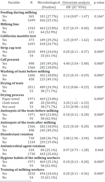
associated with bovine mastitis, according to hygiene-health management on rural properties Bahia and Pernambuco Variable N Microbiological Univariate analysis p value (Positive) ORa (ICb 95%)
Feeding during milking
Yes 365 101 (27.7%) 1.14 (0.87 – 1.47) 0.166* No 1699 426 (25.1%)
Milking line
Yes 1943 463 (23.8%) 0.27 (0.19 – 0.40) 0.000*
No 121 64 (52.9%)
California mastitis test
Yes 373 109 (29.2%) 1.25 (0.97 – 1.62) 0.042* No 1691 418 (24.7%)
Strip cup test
Yes 2010 494 (24.6%) 0.20 (0.11 – 0.37) 0.000* No 54 33 (61.1%)
Calf present
Yes 498 245 (49.2%) 4.40 (3.54 – 5.48) 0.000*
No 1566 282 (18.0%)
Washing of teats before milking
Yes 1606 302 (18.8%) 0.23 (0.19 – 0.29) 0.000* No 458 225 (49.1%)
Drying of teats
Yes 2011 489 (24.3%) 0.12 (0.06 – 0.23) 0.000* No 53 38 (71.7%)
Drying process
Paper towel 1971 469 (23.8%) - 0.000* Cloth towel 40 20 (50.0%) 3.20 (1.62 – 6.33)
Not used 53 38 (71.7%) 2.53 (0.98 – 6.56)
Antisepsis of the teats before milking
Yes 1971 469 (23.8%) 0.18 (0.12 – 0.28) 0.000* No 93 58 (62.4%)
Antisepsis of the teats after milking
Yes 1566 282 (18.0%) 0.22 (0.18 – 0.28) 0.000* No 498 245 (49.2%)
Disinfectant rotation
Yes 730 268 (36.7%) 2.40 (1.96 – 2.94) 0.000* No 1334 259 (19.4%)
Antimicrobial agent rotation
Yes 334 84 (25.1%) 0.97 (0.73 – 1.28) 0.460 No 1730 443 (25.6%)
Hygiene habits of the milking workers
Yes 1971 469 (25.1%) 0.18 (0.12 – 0.28) 0.000* No 93 58 (62.4%)
Training of milking workers
Yes 2010 494 (24.6%) 0.20 (0.11 – 0.36) 0.000* No 54 33 (61.1%)
a Odds ratio, b Confidence interval. * Association significant at 5%.
Table 5. Multivariate analysis of the risk factors associated with bovine mastitis on rural properties in Bahia and
Pernambuco
Variables p value ORa ICb 95% Coefficient SEc
Feed supplement
No/Yes 0.000* 2.92 1.59 5.60 1.096 0.320
Animal husbandry system
Extensive/Intensive 0.000* 8.13 4.17 15.83 2.095 0.340 Semi-intensive/Intensive 0.000* 4.19 3.35 5.24 1.433 0.114
Teat drying process
Cloth towel/Paper towel 0.000* 3.20 1.70 6.00 1.164 0.320 Not used/Paper towel 0.000* 8.11 4.42 14.87 2.093 0.309
Antisepsis of the teats before milking
No/Yes 0.005* 1.93 1.21 3.06 0.658 0.236
Antisepsis of the teats after milking
No/Yes 0.000* 3.90 3.09 4.93 1.362 0.119 Hygiene habits of the milking workers
No/Yes 0.000* 5.30 3.44 8.17 1.669 0.220
a Odds ratio, b Confidence interval, c Standard error of the estimate. *
showed multidrug resistance; this was observed in 90.5% and 71.7% in farms G and B, respectively. In all the herds, most (80.4%) of the bacteria were resistant to three or four antimicrobial agents simultaneously, principally from the
beta-lactam class. Multidrug resistance to five, six or seven
drugs was found in 28 (19.6%) Staphylococcus spp., 67.9% of which came from farm B. Such isolates also showed a
characteristic resistance profile for the tetracyclines and
streptomycin, which was little pronounced on the other properties and is associated with the use of these medica-tions for treatment of infectious diseases in the animals of the herd in question.
Some production characteristics were identified as risk
factors for the occurrence of mastitis on the properties. A
significantly greater frequency of positive samples was seen
on the microbiological examination for animals maintained
in an extensive production system (OR=8.13; p<0.05) or se
-mi-intensive production system (OR=4.19; p<0.05). Some authors affirmed that cows raised intensively are more sus -ceptible to the development of intramammary infections through the greater concentration of animals and exposure to organic matter and to pathogenic microorganisms (Kal-mus et al. 2006). In spite of that, we believe that the results
of this study are associated with deficiencies in nutritional
and hygiene-health management of the animals and of the facilities, with little adoption of measures for control and prevention of mastitis in the herds analyzed. In addition,
the lack of feed supplements (OR=2.92; p<0.05) was also
indicated as a risk factor for the disease, considering that
insufficient ingestion of certain vitamins and minerals may
negatively affected the immunological resistance of the cows through causing alterations in the mechanisms re-lated to the leukocyte function and to the integrity of the mammary tissue (Heinrichs et al. 2009).
Among the variables associated with milking manage-ment, the teat drying process, the lack of performing an-tisepsis of the teats and inadequate hygiene habits of the milking workers constituted risk factors for mastitis (Table 5). The use of cloth towels for drying the teats after washing is not recommended due to the possibility of transmission of microorganisms to the udder; such microorganisms may be disseminated among the animals, especially if they are used for multiple cows. On the other hand, the practices of disinfection of the teats before and after milking proved to be effective in elimination of surface agents of the mam-mary gland, contributing to reduction in the incidence of mastitis in the herds. Moreover, adjustment of the hygiene habits of the milking workers, directing them to wash their hands with soap and water before and during milking, is an essential measure for prevention of intramammary infec-tions (Fonseca & Santos 2001).
In spite of not being confirmed as risk factors, other fac
-tors that showed significant association (p<0.2) are worthy
of note. The percentage of positive microbiological exami-nations was greater for the samples in which milking was performed with the presence of the calf. According to Brito et al. (2000), sucking by the calf promotes an increase in the colonization of microorganisms from the oral cavity in the skin of the teats; nevertheless, Oliveira et al. (2011)
affirmed that this procedure may reduce the rates of masti -tis due to the removal of residual milk from the mammary gland and the antimicrobial action of the saliva. In addition, these authors observed that the feeding of animals during milking contributed to the increase in the occurrence of in-fection in cows from Minas Gerais (Souza et al. 2005) and Pernambuco (Oliveira et al. 2012) since soon after the mi-lking, the teat sphincter remains open, favoring the entran-ce of environmental pathogens.
CONCLUSIONS
On the rural properties studied, there is a predominan-ce of subclinical and clinical infections caused by Staphylo-coccus spp. and Prototheca spp. respectively.
The presence of multiresistant isolates in bovine milk shows the importance of adequate choice and use of anti-microbial agents, with a view towards success in the treat-ment of mastitis.
The risk factors identified are mainly associated with deficiencies in management during milking.
Control and prevention measures, including antisepsis of the teats and good practices for achieving hygienic mi-lking should be established for the purpose of preventing new cases of the disease in the herds.
REFERENCES
Amorim R.N.L., Souza A.O.G., Lima P.M., Bezerra F.S.B., Alves N.D. & Feijó F.M.C. 2010. Mastite clínica em bovino causada por Prototheca zopfii no estado do Ceará. Acta Vet. Bras. 4:307-311.
Barbalho T.C.F. & Mota R.A. 2001. Isolamento de agentes bacterianos en-volvidos em mastite subclínica bovina no Estado de Pernambuco. Revta Bras. Saúde Prod. Anim. 2:31-36.
Barlow J. 2011. Mastitis therapy and antimicrobial susceptibility: a multi-species review with a focus on antibiotic treatment of mastitis in dairy cattle. J. Mammary Gland. Biol. Neoplasia.16:383-407.
Bauer A.W., Kirby W.M., Sherris J.C. & Turck M. 1966. Antibiotic suscepti-bility testing by a standardized single disc method. Am. J. Clin. Pathol. 45:493-496.
Bradley A. 2002. Bovine mastitis: an envolving disease. Vet. J. 164:116-128.
Brito J.R.F., Paiva e Brito M.A.V. & Verneque R.S. 2000. Contagem bacteria-na da superfície de tetas de vacas submetidas a diferentes processos de higienização, incluindo a ordenha manual com participação do bezerro para estimular a descida do leite. Ciência Rural 30:847-850.
Bueno V.F.F., Nicolau E.S., Mesquita A.J., Ribeiro A.R., Silva J.A.B., Costa E.O., Coelho K.O. & Neves R.B. 2002. Mastite bovina clínica e subclínica na região de Pirassununga, SP: frequências e redução na produção. Ciênc. Anim. Bras. 3:47-52.
Bueno V.F.F., Mesquita A.J. & Dias Filho F.C. 2006. Prototheca zopfii: impor-tante patógeno na etiologia da mastite bovina no Brasil. Ciênc. Anim. Bras. 7:273-283.
CLSI 2008. Performance Standards for Antimicrobial Disk and Dilution Susceptibility Tests for Bacteria Isolated from Animals: approved stan-dard. 3rd ed. CLSI, Wayne.
Coentrão C.M., Souza G.N., Brito J.R.F., Paiva e Brito M.A.V. & Lilenbaum W. 2008. Fatores de risco para mastite subclínica em vacas leiteiras. Arq. Bras. Med. Vet. Zootec. 60:283-288.
Corbellini L.G., Orlemeier D., Cruz C., Dias M.M. & Ferreiro L. 2001. Bovine mastitis due to Prototheca zopfii: clinical, epidemiological and patho-logical aspects in a Brazilian dairy herd. Trop. Anim. Health Prod. 6:463-473.
Staphylococcus aureus e suas implicações em saúde pública. Ciência Ru-ral 34:1315-1320.
Ferreira J.L., Lins J.L.F.H.A., Cavalcant T.V., Macedo N.A. & Borjas A.R. 2007. Prevalência e etiologia da mastite bovina no município de Teresina, Piauí. Ciênc. Anim. Bras. 8:261-266.
Freitas M.F.L., Pinheiro Júnior J.W., Stamford T.L.M., Rabelo S.S.A., Silva D.R., Silveira Filho V.M., Santos F.G.B., Sena M.J. & Mota R.A. 2005. Perfil de sensibilidade antimicrobiana in vitro de Staphylococcus coagulase po-sitivos isolados do leite de vacas com mastite no Agreste do estado de Pernambuco. Arqs Inst. Biológico, São Paulo, 72:171-177.
Fonseca L.F. & Santos M.V. 2001. Qualidade do Leite e Controle de Mastite. Lemos Editorial, São Paulo. 175p.
Guimarães F.F. & Langoni H. 2009. Leite: alimento imprescindível, mas com riscos para a saúde pública. Vet. Zootec. 16:38-51.
Heinrichs A.J., Costello S.S. & Jones C.M. 2009. Control of heifer mastitis by nutrition. Vet. Microbiol. 134:172-176.
Hosmer D. & Lemeshow S. 1989. Applied Logistic Regression. John Wiley and Sons, New York. 322p.
IBGE 2011. Banco de dados agregados - Sistema IBGE de recuperação au-tomática. Available at <ftp://ftp.ibge.gov.br/Producao_Pecuaria/Produ -cao_da_Pecuaria_Municipal/2011/> Accessed on Dec. 20, 2012. Langoni H., Penachio D.S., Citadella J.C.C., Laurino F., Faccioli-Martins P.Y.,
Lucheis S.B., Menozzi B.D. & Silva A.V. 2011. Aspectos microbiológicos e de qualidade do leite bovino. Pesq. Vet. Bras. 31:1059-1065.
Kalmus P., Viltrop A., Aasmãe B. & Kask K. 2006. Occurrence of clinical mastitis in primiparous Estonian dairy cows in different housing condi-tions. Acta Vet. Scand. 48:21.
Kapronezai J., Melville P. & Benites N.R. 2005. Análise microbiológica, tes-te de tamis e California Mastitis Test realizados em amostras de leite de fêmeas bubalinas pertencentes a rebanhos do estado de São Paulo. Arqs Inst. Biológico, São Paulo, 72:183-187.
Martins R.P., Silva J.A.G., Nakazato L., Dutra V. & Almeida Filho E.S. 2010. Prevalência e etiologia da mastite bovina na microrregião de Cuiabá, MT. Ciênc. Anim. Bras. 11:181-187.
Medeiros E.S., Mota R.A., Santos M.V., Freitas M.F.L., Pinheiro Júnior J.W. & Teles J.A.A. 2009. Perfil de sensibilidade microbiana in vitro de linha-gens de Staphylococcus spp. isoladas de vacas com mastite subclínica. Pesq. Vet. Bras. 29:569-574.
Mota R.A., Sá M.E.P., Oliveira A.A.F., Silva L.B.G. & Souza M.I. 1999. Mastite bovina por Prototheca zopfii no Estado de Pernambuco. Anais do Encon-tro de Pesquisadores em Mastites, Botucatu, SP, p.162. (Resumo)
Mota R.A., Medeiros E.S., Santos M.V., Pinheiro Júnior J.W., Moura A.P.B.L. & Coutinho L.C.A. 2012. Participação dos Staphylococcus spp. na etiolo-gia das mastites em bovinos leiteiros no estado de Pernambuco (Brasil). Ciênc. Anim. Bras. 13:124-130.
Nader Filho A., Ferreira L.M., Amaral L.A., Rossi Junior O.D. & Oliveira R.P. 2007. Sensibilidade antimicrobiana dos Staphylococcus aureus isolados no leite de vacas com mastite. Arqs Inst. Biológico, São Paulo, 74:1-4. National Mastitis Council 1999. Laboratory Handbook and Bovine
Masti-tis. The National Mastitis Council, Arlington. 222p.
Olde Reikerink R.G., Barkema H., Kelton D. & Scholl D. 2008. Incidence rate of clinical mastitis on Canadian dairy farms. J. Dairy Sci. 91:1366-1377. Oliveira A.A., Melo C.B. & Azevedo H.C. 2009. Diagnóstico e determinação
microbiológica da mastite em rebanhos bovinos leiteiros nos tabuleiros costeiros de Sergipe. Ciênc. Anim. Bras. 10:226-230.
Oliveira C.M.C., Sousa M.G.S., Silva N.S., Mendonça C.L., Silveira J.A.S., Oai-gen R.P., Andrade S.J. & Barbosa J.D. 2011. Prevalência e etiologia da mastite bovina na bacia leiteira de Rondon do Pará, estado do Pará. Pesq. Vet. Bras. 31:104-110.
Oliveira J.M.B., Vanderlei D.R., Moraes W.S., Brandespim D.F., Mota R.A., Oli-veira A.A.F., Medeiros E.S. & Pinheiro Júnior J.W. 2012. Fatores de risco associados à mastite bovina na microrregião Garanhuns, Pernambuco. Pesq. Vet. Bras. 32:391-395.
Quinn P.J., Carter M.E., Markey B. & Carter G.R. 1994. Clinical Veterinary Microbiology. Wolfe, London. 648p.
Ribeiro M.G., Geraldo J.S., Langoni H., Lara G.H.B., Siqueira A.K., Salerno T. & Fernandes M.C. 2009. Microrganismos patogênicos, celularidade e resíduos de antimicrobianos no leite bovino produzido no sistema orgâ-nico. Pesq. Vet. Bras. 29:52-58.
Schalm O.W. & Noorlander D.O. 1957. Experiments and observations leading to development of the California mastitis test. J. Am. Vet. Med. Assoc. 130:199-204.
Souza G.N., Brito J.R.F., Moreira E.C., Brito M.A.V.P. & Bastos R.R. 2005. Fa-tores de risco associados à alta contagem de células somáticas do leite do tanque em rebanhos leiteiros da Zona da Mata de Minas Gerais. Arq. Bras. Med. Vet. Zootec. 57:251-260.
Taponen S. & Pyörälä S. 2009. Coagulase-negative staphylococci as cause of bovine mastitis - not so different from Staphylococcus aureus? Vet. Microbiol. 134:29-36.