R E S E A R C H
Open Access
Tityus serrulatus
envenoming in non-obese
diabetic mice: a risk factor for severity
Guilherme Honda de Oliveira
1, Felipe Augusto Cerni
1, Iara Aimê Cardoso
1, Eliane Candiani Arantes
1and Manuela Berto Pucca
1,2*Abstract
Background:In Brazil, accidents with venomous animals are considered a public health problem.Tityus serrulatus (Ts), popularly known as the yellow scorpion, is most frequently responsible for the severe accidents in the country. Ts envenoming can cause several signs and symptoms classified according to their clinical manifestations as mild, moderate or severe. Furthermore, the victims usually present biochemical alterations, including hyperglycemia. Nevertheless, Ts envenoming and its induced hyperglycemia were never studied or documented in a patient with diabetes mellitus (DM). Therefore, this is the first study to evaluate the glycemia during Ts envenoming using a diabetic animal model (NOD, non-obese diabetic).
Methods:Female mice (BALB/c or NOD) were challenged with a non-lethal dose of Ts venom. Blood glucose level was measured (tail blood using a glucose meter) over a 24-h period. The total glycosylated hemoglobin (HbA1c) levels were measured 30 days after Ts venom injection. Moreover, the insulin levels were analyzed at the glycemia peak.
Results:The results demonstrated that the envenomed NOD animals presented a significant increase of glycemia, glycosylated hemoglobin (HbA1c) and insulin levels compared to the envenomed BALB/c control group, corroborating that DM victims present great risk of developing severe envenoming. Moreover, the envenomed NOD animals presented highest risk of death and sequelae.
Conclusions:This study demonstrated that the diabetic victims stung by Ts scorpion should be always considered a risk group for scorpion envenoming severity.
Keywords:Tityus serrulatus, Diabetes mellitus, Scorpion venom, NOD mice, Glycemia
Background
In Brazil, accidents involving venomous animals are considered a public health problem. Tityus serrulatus (Ts), popularly known as the yellow scorpion, is most frequently responsible for these accidents. During the period from 2000 to 2015, 727,113 cases of scorpion envenoming were reported in Brazil, with 1026 deaths and a mortality rate of 0.14 % [1–4].
Envenoming by Ts can cause several signs and symptoms according to not only the content of venom, but also the victim’s body weight, the blood-brain-barrier permeability, sex, health conditions and sting location. The mild
envenoming is characterized by an intense local pain and possible paresthesia. Moderate envenoming manifests through local pain as well as nausea, sweating, vomiting, tachycardia, tachypnea and increasing of blood pressure. Severe envenoming presents the same symptoms of mod-erate followed by agitation and exhaustion, abdominal pain, stiffness and muscle spasms, convulsions, fever, dehy-dration, cardiac arrhythmias, heart failure and even coma [5–11]. Moreover, biochemical alterations are also ob-served during Ts envenoming such as hyperglycemia [6, 12–15].
Hyperglycemia is a sign constantly observed in dia-betic individuals. Diabetes mellitus (DM) currently affects about 314 million people worldwide. Solely in 2012, it was responsible for 1.5 million deaths according to the World Health Organization (WHO). Currently, based on the etiology of the disease, DM can be
* Correspondence:manu.pucca@ufrr.br
1Department of Physics and Chemistry, School of Pharmaceutical Sciences of
Ribeirão Preto, University of São Paulo (USP), Av. do Café, s/n, 14040-903 Ribeirão Preto, SP, Brazil
2Medical School of Roraima, Federal University of Roraima (UFRR), Av.
Capitão Ene Garcez, 2413, Boa Vista, RR 69310-000, Brazil
classified into two types: type 1 and type 2 [16]. Al-though the pathologies have different origins, both have similar signs and symptoms such as hyperglycemia, poly-uria, polyphagia, polydipsia, unexplained weight loss and may also include foot pain, blurred vision, frequent in-fections and even coma [17, 18].
The type 1 DM, or insulin-dependent DM, has a gen-etic etiology with the most common type being among children and juveniles. It is an autoimmune disease in which self-reactive T cells induce the B cell production of specific antibodies against beta cells of the islets of Langerhans. This autoimmune mechanism results in the destruction of these cells and consequently the decrease of insulin production [19, 20]. On the other hand, type 2 DM is the most common type of diabetes (accounts for 90 %). It is considered a chronic metabolic disorder and has been characterized by impaired insulin action and/or abnormal insulin secretion and eventual pancreatic beta-cell failure. An early abnormality in the disease is insulin resistance, which is the key linking factor for the meta-bolic syndrome disease cluster of glucose intolerance, hypertension and dyslipidemia [21–23].
The model employing non-obese diabetic (NOD) ani-mals is useful for type 1 diabetes, and presents an auto-immune genetic disease where T cells (CD4+andCD8+) become self-reactive to pancreatic islets, resulting in in-flammation during the first 3 to 4 weeks of life. How-ever, only after mice reach 4 to 6 months of age does it become possible to verify insulitis, which results in insu-lin deficiency and therefore the cinsu-linical signs of diabetes [24, 25]. Thus, based on the Ts venom-induced hypergly-cemia and the high incidence of diabetic patients, this is the first study to investigate the Ts envenoming com-plications in DM individuals, using NOD mice as the experimental model.
Methods
T. serrulatusvenom
The Ts scorpions, collected from the Ribeirão Preto re-gion, were kept in the Serpentarium of the Medical School of RibeirãoPreto (FMRP/USP). The venom extraction from 145 scorpions was performed using the telson elec-trical stimulation method─12 mV [26]. After extraction,
the pooled venom was desiccated and stored at –20 °C. The use of Ts venom was approved by the Genetic Patri-mony Management Board (CGEN/MMA), through the Access and Shipment Component of Genetic Heritage for scientific research purposes (number 010174/2014-1).
Mass assessment of the venom ofT. serrulatus
The desiccated venom was dispersed in 1 mL of ultra-pure water and centrifuged at 10,015 ×g, 4 °C for 10 min, and the supernatant was stored at 4 °C. The pel-let was resuspended using the same conditions. The total
supernatant (2 mL) resulted in soluble pooled venom without the presence of mucus.
The mass of soluble pooled venom was estimated by absorbance readings at 280 nm using the NanoDrop spectrophotometer 2000 (Thermo Scientific, USA) and the extinction coefficient of the soluble venom [27]:
ε 1 mg=mL
280 nm ¼1:65
Tricine-SDS-PAGE
The venom was analyzed using tricine sodium dodecyl sulfate polyacrylamide gel electrophoresis (Tricine-SDS-PAGE) according to the method used for ultra-low-mass proteins [28]. The 16.5 % separating gel used was over-laid by a 5 % stacking gel. Samples consist of different masses of Ts venom (10, 20 and 30μg) and the molecu-lar mass marker (M-3546, Sigma-Aldrich®, USA). The gel was stained with Coomassie Blue plus one PhastGel® R-350 (GE Healthcare, Sweden) and destained with 10 % acetic acid (V/V).
Animals
Females of BALB/c and NOD lineage (18–25 g) were ob-tained from the biotherium of the School of Pharmaceut-ical Sciences of Ribeirão Preto (FCFRP/USP) and the biotherium of the Ribeirão Preto Medical School (FMRP/ USP), respectively. The animals were kept in cages with filters in an air-conditioned environment (23 ± 1 °C, 55 ± 5 % humidity) until the blood glucose values of NOD animals became significantly higher than those of the con-trols (BALB/c), indicating the hyperglycemia characteristic of the diabetic state (15 weeks old). Food and water were provided ad libitum. Mouse experimental models are in accordance with the Ethical Principles in Animal Experi-mentation under the license number 13.1.372.53.0.
Basal blood glucose levels andT. serrulatusvenom dose The basal glucose levels of BALB/c and NOD mice were measured in tail blood using a glucose meter (One Touch Ultra®, Lifescan, USA). The dose of Ts venom capable of inducing hyperglycemia in mice (BALB/c and NOD) was also adjusted. The doses of 1 mg/kg and 0.5 mg/kg were tested in the different mouse species.
Touch Ultra®, Lifescan, USA). Blood glucose was measured over a 24-h period (at 0, 1, 2, 3, 4, 5, 6, 12 and 24 h).
Glycosylated hemoglobin (HbA1c) induced by
T. serrulatusvenom
Groups of female BALB/c or NOD mice (18–25 g,n= 4) were challenged with a non-lethal dose of Ts venom (0.5 mg/kg) using subcutaneous injection (similar to scorpion sting site), diluted in sterile physiological solu-tion (0.9 % W/V of NaCl) in a final volume of 0.2 mL. Control groups received only sterile physiological solution. After 30 days (to reflect mean glycemia for the previous 30 days), 0.5 mL of blood from the retro-orbital cavity was collected in heparinized tubes under intraperitoneal anesthesia: ketamine 60 mg/kg (Dopalen, Agripands Brasil Ltda®, Brazil) and xylazine 8 mg kg (Rompun, Bayer Animal Health®, Brazil). The blood was centrifuged at 10,000 rpm for ten minutes, at 4 °C, to obtain the plasma. The measurement of total glycosylated hemoglobin (HbA1c) levels was performed according to the manufacturer’s instructions (Doles®, Brazil).
Insulin levels induced byT. serrulatusvenom
Groups of female BALB/c or NOD mice (18–25 g,n= 4) were challenged with a non-lethal dose of Ts venom (0.5 mg/kg) using subcutaneous injection (similar to scor-pion sting site), diluted in sterile physiological solution (0.9 % W/V of NaCl) in a final volume of 0.2 mL. Control groups received only sterile physiological solution. After reaching the hyperglycemia peak (1 h after envenom-ation), 0.5 mL of blood from the retro-orbital cavity was collected in heparinized tubes under anesthesia. The blood
was centrifuged at 10,000 rpm for 10 min, at 4 °C, to obtain the plasma. The insulin assay was performed using the immunoassay method according to the manufacturer’s instructions (Ultra Sensitive Mouse Insulin ELISA kit, Crystal Chem, USA).
Results
T. serrulatusvenom mass and electrophoresis
The pooled venom obtained from 145 Ts scorpions resulted in 21 mg of soluble venom, a median of 0.14 mg per scorpion.
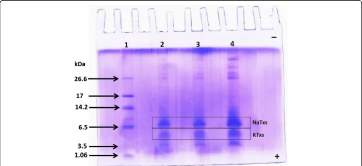
The Tricine-SDS-PAGE indicates the Ts venom pro-tein profile using different masses (10, 20 and 30 μg) (Fig. 1). Two main bands of low molecular masses were observed. The sodium-channel toxins (NaTxs; from 60 to 76 amino acid residues) are the main proteins evi-denced in the electrophoretic band of molecular mass between 6000 and 8000 Da, whereas the potassium-channel toxins (KTxs; from 22 to 47 amino acid resi-dues) are the main components of the electrophoretic band from 5000 to 4000 Da.
T. serrulatusvenom increased the glucose levels of mice The median basal glucose – time 0, before Ts venom (TsV) injection – was 82.9 and 125 mg/dL for BALB/c and NOD mice, respectively (Fig. 2a). Ideally, NOD mice are considered diabetic with glucose levels higher than 200 mg/dL. However, during the experimental design standardization using NOD mice, we observed that both the dose of Ts venom and the glucose basal levels were limiting factors to the experiment, inducing lethality of 25 to 100 % (Fig. 2b). Therefore, we decided to use
Fig. 1Electrophoretic profile of the pooled Ts venom. Molecular mass markers (lane 1); pooled Ts venom: 10, 20 and 30μg (lanes 2, 3 and 4, respectively).
NOD animals presenting glucose levels lower than 150 mg/dL, but significantly higher than glucose levels of the BALB/c control (p< 0.001) and the Ts venom con-centration of 0.5 mg/kg (100 % survival).
All mouse groups that received 0.5 mg/kg of Ts venom showed hyperglycemia 1 h after envenomation, com-pared to the respective control (Fig. 3). However, the
hyperglycemia was much more impressive in the NOD group, reaching glucose levels≥200 mg/dL. During the
analyzed time, glucose levels decrease, presenting a sig-nificant hypoglycemic period at 4 h for BALB/c and at 5 to 6 h for NOD animals. Nevertheless, the basal glucose levels for all challenged mice were reestablished after 12 h.
Fig. 2Standardization of basal glucose levels andT. serrulatusvenom dose.aBasal glucose (time 0, before TsV injection) was measured in tail
blood extracted from mice using a glucose meter (One Touch Ultra®, Lifescan, USA). Results are expressed as means ± SD (n= 4), which were analyzed by pairedttest (*p< 0.001).bNOD survival using different glucose basal levels (> 200, < 200 and < 150 mg/dL) andT. serrulatusvenom (TsV) concentrations (0.5 and 1 mg/kg)
Fig. 3Kinetic glucose level assay of BALB/c and NOD mice injected with Ts venom. Groups of mice were injected with 0.5 mg/kg ofT. serrulatus
T. serrulatusvenom affected the glycosylated hemoglobin
(HbA1c) of NOD mice
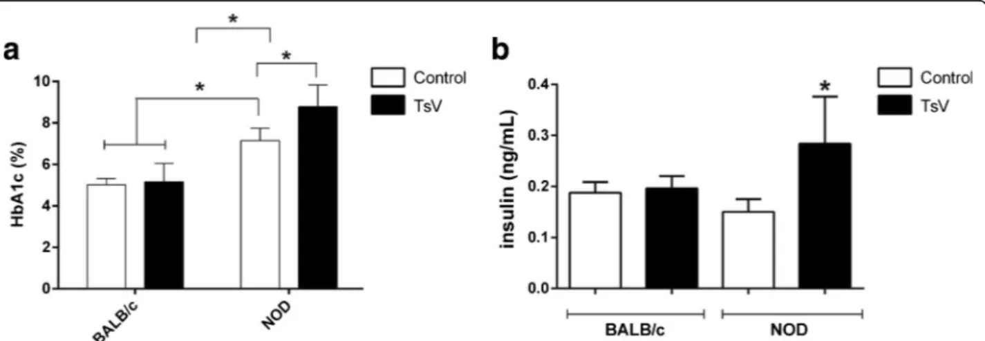
The total glycosylated hemoglobin (HbA1c) was analyzed in mouse blood 30 days after envenoming. The results demonstrate that although Ts venom had induced hyper-glycemia in BALB/c mice, it was not able to augment HbA1c (Fig. 4a). However, Ts venom caused a significant increase of HbA1c in envenomed NOD animals. Further-more, the NOD group control also presented a significant increase of HbA1, indicating the diabetic condition of the mice.
T. serrulatusvenom increased insulin levels of NOD mice The insulin levels were analyzed in mouse plasma during the peak of hyperglycemia (1 h after envenoming). The re-sults demonstrate that Ts venom did not change insulin levels in BALB/c mice (Fig. 4b). As to the NOD group, a significant increase of insulin was observed in the enve-nomed mice. On the other hand, no significant changes on insulin levels were found in the NOD control group.
Discussion
The venom ofT. serrulatus(Ts) is widely studied espe-cially because of its biologically active compounds including the neurotoxins, a class of peptides mostly specific to voltage-gated sodium (Nav) or potassium (Kv) channels [29]. So far, the actions of Ts neurotoxins on Nav and Kv channels have demonstrated a gamut of physiological responses, including the increasing of plasma glucose levels [14, 15, 30–40]. Although the mechanism of hyperglycemia induced by scorpions’ venom is not clearly understood, there are studies demonstrating that it can occur through the excessive release of catecholamine, increases in glucagon and cortisol, and alterations in thyroid hormone levels or
insulin secretion [41–43]. Furthermore, an isolated α -toxin from Ts, denominated Ts5, demonstrated a direct effect on isolated islets of Langerhans (not provided by catecholamine’s action), enhancing β-cell membrane depolarization and significantly potentiating glucose-induced insulin secretion [43]. Therefore, this pioneer-ing study aimed to elucidate the effect of hyperglycemia induced by Ts venom in diabetic individuals, using a NOD mouse model.
The Ts pooled venom used in the study showed a protein profile similar to others previous described, presenting protein masses corresponding to neuro-toxins specific to sodium (NaTxs) and potassium (KTxs) channels [27, 44].
The in vivo assays demonstrated that blood glucose levels significantly increase 1 h after Ts injection (0.5 mg/kg), independently of the mouse model. Indeed, the highest glucose level at 1 h after envenoming has already been described in Wistar rats using the same dose (0.5 mg/kg) and intraperitoneal (i.p.) injection, and in BALB/c mice using 1 mg/kg of Ts venom and sub-cutaneous (s.c.) injection [14, 15]. A peculiar observation during our experimental design standardization was that either the highest dose of Ts venom (1 mg/kg) or the highest glucose basal levels (≥200 mg/dL) cause NOD
mice lethality. Furthermore, the surviving mouse that presented basal glucose≥200 mg/dL and received
0.5 mg/kg of Ts venom (n= 1) demonstrated a clinical sequela represented by an ocular disease with partial loss of vision, probably retinopathy (Additional file 1).
The retina is an insulin-sensitive tissue and excess glucose or lipids may exert their noxious effects, accel-erating retinal cell death [45]. NOD mice frequently present diabetic retinopathy, with histological analysis showing loss of retinal microvessels and reduced
Fig. 4Blood glycosylated hemoglobin (HbA1c) percentage (%) and insulin plasma levels from BALB/c and NOD mice injected with Ts venom. Groups
perfusion of the retina, which result in hypoxia with evidence of disordered focal proliferation of new ves-sels [46]. Definitely, diabetic retinopathy is a frequent cause of blindness developed by diabetic individuals after macular edema [47]. Nevertheless, the retinop-athy induced by Ts venom in diabetic mice requires further investigation.
During the Ts venom-induced glucose kinetic assay, we also observed a period in which the glucose reached levels lower than basal glycemia, which was previously described as a hypoglycemic period [14, 15]. BALB/c mice reduced glucose levels at 4 h following Ts venom injection, while NOD demonstrated a prolonged de-crease of glucose levels at 5 to 6 h following Ts venom injection. It is known that beta cells of the islets of Langerhans sense changes in the plasma glucose levels and adjust the rate of insulin production aiming to maintain the homeostatic glucose plasma concentration [48]. Thus, we assume that the production of insulin to control hyperglycemia induced by such envenoming re-mains high and, along with a decrease in glycogenolysis (due to hepatic glycogen depletion), causes a reduction of glucose levels.
Regarding NOD animals, the delayed hypoglycemia can be explained by the higher glucose levels required to be controlled, and the prolonged hypoglycemia (2 h) by the higher rates of insulin production. In-deed, a significant increasing of insulin was observed in NOD animals challenged with Ts venom. Although the increasing of this hormone in NOD mice seems peculiar, we assume that their diabetic disease was not sufficiently advanced to impair the insulin pro-duction significantly (the decrease of basal insulin of NOD control animals was not statistically significant compared to BALB/c control). In this sense, the ani-mals still present a number of suitable insulin-producing beta cells in the islets of Langerhans.
According to the literature, the NOD model acquires insulin deficit after the age of 14 weeks (with high variety among individuals), that is, before this period, despite pre-senting high glucose levels, the animals still produce insu-lin. On the other hand, when these mice become overtly diabetic, they quickly lose weight and require insulin treat-ment [49]. The effect of high insulin levels observed in NOD-envenomed mice is somewhat controversial. This effect may be beneficial, since insulin treatment in scor-pion sting victims is known as a metabolic support, which controls the adverse metabolic response produced by cate-cholamines and other counter-regulatory hormones [50]; or it could be unfavorable, since the injection of insulin after Ts envenoming can enhance the venom’s lethality [15]. Based on the latter, the lethality induced by Ts sting among diabetic individuals should be higher than that in healthy humans. In any case, insulin therapy
after Ts envenoming should be further investigated in diabetic individuals, especially insulin-dependent ones.
On the other hand, BALB/c envenomed group did not present differences in insulin production compared to BALB/c control, although hyperinsulinemia has been re-ported previously during envenoming by the scorpion Mesobuthus tamulus concanesisand even byT. serrulatus [15, 42]. However, in the Ts study, the authors used a rat model and i.p. injection, which may justify such differences. We also evaluated the glycosylated hemoglobin (HbA1c) percentage, which is the primary method recommended before initiating therapy in diabetes pa-tients [51]. Our results demonstrated that NOD con-trol animals surely presented a diabetic clinical condition showing HbA1c higher than 7 % while the NOD envenomed group displayed a significant eleva-tion, with levels higher than 8 %. The augmentation of HbA1c is considered a risk factor for diabetic neuropathy and retinopathy [52]. This also explains why we observed only an ocular alteration in the envenomed group of NOD mice.
Currently, the treatment used for Ts accidents varies according to the clinical severity, which depends on the signs and symptoms manifested by the patient. Mild and moderate cases of Ts envenoming consist mostly of pain relief through analgesics at the sting site, orally or parenterally. On the other hand, severe scorpion envenoming cases require the mandatory use of the specific antivenom. In Brazil, the available venoms used for Ts envenoming are the scorpion anti-venom (SAE or soro antiescorpiônico in Portuguese) and the arachnid antivenom (SAAr orsoro antiaracní-deo in Portuguese). Their use is also compulsory in children under 7 years and in adults with previous health problems (e.g. hypertension and cardiovascular problems) even if they present mild or moderate clin-ical manifestations [1, 53]. In this regard, our results support the hypothesis that diabetic victims present a higher risk of developing severe envenoming. Therefore, we also advise consideration of the use of antivenom in cases of Ts envenoming in diabetic persons ─ a risk
group for Ts envenoming severity.
Conclusion
Additional file
Additional file 1:Representative comparison of eyeball of Ts envenomed
and non-envenomed mice. Mice were injected with Ts venom (1 mg/kg). (A) Envenomed NOD mice with glucose basal level≥200 mg/dL, retinopathy indication. (B) Envenomed BALB/c mice, typical ptosis. (C) BALB/c mice control (non-envenomed), normal eyeball. (PPTX 81 kb)
Abbreviations
DM:Diabetes mellitus; HbA1c: Glycosylated hemoglobin; i.p.: Intraperitoneal injection; KTx: Neurotoxin specific potassium channels; Kv: Voltage-gated potassium channel; NaTx: Neurotoxin specific sodium channels; Nav: Voltage-gated sodium channel; NOD: Non-obese diabetic; SDS: Sodium dodecyl sulfate; Ts:Tityus serrulatus
Acknowledgements
Thanks to the Center for the Study of Venoms and Venomous Animals (CEVAP) of UNESP for enabling the publication of this paper (CAPES–
grant n. 23038.006285/2011-21, AUXPE–Toxinologia–1219/2011).
Funding
This study received financial support from the State of São Paulo Research Foundation (FAPESP–scholarships to MBP 2012/12954-6 and FAC 2012/ 13590-8), the National Council for Scientific and Technological Development (CNPq–scholarship to GHO) and NAP-TOXAN-USP (grant n. 12e125432.1.3).
Authors’contributions
GHO was the baccalaureate student (Iniciação Científica) responsible for the project, and was involved in the organization and execution of the experimentation section, data acquisition, statistical analysis and data interpretation, as well as drafting and revising the manuscript. MBP and ECA were the advisor researchers of this project and critically revised the manuscript for its content. FAC and IAC substantially contributed to the experimental execution and revision of the manuscript. All authors read and approved the final manuscript.
Competing interests
The authors declare that they have no competing interests.
Ethics approval and consent to participate
The present study was approved by the Ethics Commission for the Use of Animals (CEUA) at the USP Campus in Ribeirão Preto (protocol number 13.1.372.53.0). The use of Ts venom was approved by the Genetic Patrimony Management Board (CGEN/MMA), through the Access and Shipment Component of Genetic Heritage for scientific research purposes (number 010174/2014-1).
Received: 29 March 2016 Accepted: 2 September 2016
References
1. Pucca MB, Oliveira FN, Schwartz EF, Arantes EC, da Silva RM L. Scorpionism and dangerous species of Brazil. In: Gopalakrishnakone P, editor. Scorpion Venoms. Netherlands: Springer; 2015. p. 299–324.
2. SUS, Portal da Saúde: Situação Epidemiológica - Dados. 2016. http:// portalsaude.saude.gov.br. Accessed 25 Jan 2016.
3. Reckziegel GC, Pinto VL. Scorpionism in Brazil in the years 2000 to 2012. J Venom Anim Toxins incl Trop Dis. 2014;20:20–46.
4. Chippaux JP. Epidemiology of envenomations by terrestrial venomous animals in Brazil based on case reporting: from obvious facts to contingencies. J Venom Anim Toxins incl Trop Dis. 2015;21:13. 5. Bucaretchi F, Fernandes LC, Fernandes CB, Branco MM, Prado CC, Vieira RJ,
et al. Clinical consequences ofTityus bahiensisandTityus serrulatusscorpion stings in the region of Campinas, southeastern Brazil. Toxicon. 2014;89:17–25. 6. Amitai Y. Clinical manifestations and management of scorpion envenomation.
Public Health Rev. 1998;26(3):257–63.
7. Guidine PA, Cash D, Drumond LE, de Souza ERGH, Massensini AR, Willilams SC, et al. Brainstem structures are primarily affected in an experimental model of severe scorpion envenomation. Toxicol Sci. 2014;137(1):147–57. 8. Amaral CF, de Rezende NA, Freire-Maia L. Acute pulmonary edema after
Tityus serrulatusscorpion sting in children. Am J Cardiol. 1993;71(2):242–5.
9. Bahloul M, Chaari A, Dammak H, Algia NB, Bouaziz M. Nosocomial scorpion envenomation: an unusual mode of scorpion sting. Clin Toxicol (Phila). 2010;48(9):962.
10. Nishioka SA, Silveria PV, Pereira CA. Scorpion sting on the penis. J Urol. 1993;150(5 Pt 1):1501.
11. Nunan EA, Moraes MF, Cardoso VN, Moraes-Santos T. Effect of age on body distribution of Tityustoxin fromTityus serrulatusscorpion venom in rats. Life Sci. 2003;73(3):319–25.
12. Fukuhara YD, Reis ML, Dellalibera-Joviliano R, Cunha FQ, Donadi EA. Increased plasma levels of IL-1beta, IL-6, IL-8, IL-10 and TNF-alpha in patients moderately or severely envenomed byTityus serrulatusscorpion sting. Toxicon. 2003;41(1):49–55.
13. Cusinato DA, Souza AM, Vasconcelos F, Guimarães LF, Leite FP, Gregório ZM, et al. Assessment of biochemical and hematological parameters in rats injected withTityus serrulatusscorpion venom. Toxicon. 2010;56(8):1477–86. 14. Pucca MB, Zoccal KF, Roncolato EC, Bertolini TB, Campos LB, Cologna CT, et
al. Serrumab: a human monoclonal antibody that counters the biochemical and immunological effects ofTityus serrulatusvenom. J Immunotoxicol. 2012;9(2):173–83.
15. Vasconcelos F, Sampaio SV, Garófalo MA, Guimarães LF, Giglio JR, Arantes EC. Insulin-like effects ofBauhinia forficataaqueous extract uponTityus serrulatusscorpion envenoming. J Ethnopharmacol. 2004;95(2–3):385–92. 16. American Diabetes Association. Diagnosis and classification of Diabetes
Mellitus. In Diabetes Care. 2004. p. S5,S6,S7,S8,S9,S10.
17. Greene JA. A classification of the different types of diabetes mellitus with a discussion of the diagnosis and treatment of each. Ariz Med. 1946;3:158–61. 18. World Health Organization (WHO). Global status report on
noncommunicable diseases 2014. 2014 cited 2015; Available from: http:// www.who.int/nmh/publications/ncd-status-report-2014/en/. Accessed 25 Jan 2016.
19. Fukuda H. Report on a case of infantile diabetes. Showa Igakkai Zasshi. 1962;22:223-6. Article in Japanese.
20. Pugliese A. Advances in the etiology and mechanisms of type 1 diabetes. Discov Med. 2014;18(98):141–50.
21. Kasuga M. Molecular basis for the development of type 2 diabetes mellitus. Nihon Rinsho. 2002;60 Suppl 7:468–76. Article in Japanese.
22. Olokoba AB, Obateru OA, Olokoba LB. Type 2 diabetes mellitus: a review of current trends. Oman Med J. 2012;27(4):269–73.
23. Kahn CR. Banting Lecture. Insulin action, diabetogenes, and the cause of type II diabetes. Diabetes. 1994;43(8):1066–84.
24. Makino S, Kunimoto K, Muraoka Y, Mizushima Y, Katagiri K, Tochino Y. Breeding of a non-obese, diabetic strain of mice. Jikken Dobutsu. 1980;29(1):1–13. 25. Chhabra G, Dixit A. Structure modeling and antidiabetic activity of a seed
protein of Momordica charantia in non-obese diabetic (NOD) mice. Bioinformation. 2013;9(15):766–70.
26. Lowe RM, Farrell PM. A portable device for the electrical extraction of scorpion venom. Toxicon. 2011;57(2):244–7.
27. Pucca MB, Amorim FG, Cerni FA, Bordon KC, Cardoso IA, Anjolette FA, et al. Influence of post-starvation extraction time and prey-specific diet inTityus serrulatusscorpion venom composition and hyaluronidase activity. Toxicon. 2014;90:326–36.
28. Schagger H, von Jagow G. Tricine-sodium dodecyl sulfate-polyacrylamide gel electrophoresis for the separation of proteins in the range from 1 to 100 kDa. Anal Biochem. 1987;166(2):368–79.
29. Ortiz E, Gurrola GB, Schwartz EF, Possani LD. Scorpion venom components as potential candidates for drug development. Toxicon. 2015;93:125–35. 30. Pucca MB, Peigneur S, Cologna CT, Cerni FA, Zoccal KF, Bordon Kde C, et al.
Electrophysiological characterization of the firstTityus serrulatusalpha-like toxin, Ts5: Evidence of a pro-inflammatory toxin on macrophages. Biochimie. 2015;115:8–16.
31. Peigneur S, Cologna CT, Cremonez CM, Mille BG, Pucca MB, Cuypers E, et al. A gamut of undiscovered electrophysiological effects produced byTityus serrulatustoxin 1 on NaV-type isoforms. Neuropharmacology. 2015;95:269–77. 32. Cologna CT, Peigneur S, Rustiquel JK, Nonato MC, Tytgat J, Arantes EC. Investigation of the relationship between the structure and function of Ts2, a neurotoxin fromTityus serrulatusvenom. FEBS J. 2012;279(8): 1495–504.
33. Cerni FA, Pucca MB, Peigneur S, Cremonez CM, Bordon KC, Tytgat J, et al. Electrophysiological characterization of Ts6 and Ts7, K+channel toxins
34. Pucca MB, Cerni FA, Peigneur S, Bordon KC, Tytgat J, Arantes EC. Revealing the function and the structural model of Ts4: insights into the“non-toxic”
toxin fromTityus serrulatusvenom. Toxins (Basel). 2015;7(7):2534–50. 35. Cologna CT, Peigneur S, Rosa JC, Selistre de Araujo HS, Varanda WA, Tytgat J,
et al. Purification and characterization of Ts15, the first member of a new alpha-KTX subfamily from the venom of the Brazilian scorpionTityus serrulatus. Toxicon. 2011;58(1):54–61.
36. Zoccal KF, Bitencourt CS, Secatto A, Sorgi CA, Bordon KC, Sampaio SV, et al.
Tityus serrulatusvenom and toxins Ts1, Ts2 and Ts6 induce macrophage activation and production of immune mediators. Toxicon. 2011;57(7–8):1101–8. 37. Pessini AC, de Souza AM, Faccioli LH, Gregório ZM, Arantes EC. Time course
of acute-phase response induced byTityus serrulatusvenom and TsTX-I in mice. Int Immunopharmacol. 2003;3(5):765–74.
38. Cupo P, Jurca M, Azevedo Marques MM, Oliveira JS, Hering SE. Severe scorpion envenomation in Brazil. Clinical, laboratory and anatomopathological aspects. Rev Inst Med Trop Sao Paulo. 1994;36(1):67–76.
39. D’Suze G, Moncada S, González C, Sevcik C, Aguilar V, Alagón A. Relationship between plasmatic levels of various cytokines, tumour necrosis factor, enzymes, glucose and venom concentration following Tityus scorpion sting. Toxicon. 2003;41(3):367–75.
40. Ribeiro EL, Pinto MC, Labarrere CR, Paes Leme FO, Chávez Olórtegui C, Melo MM. Biochemical profile of dogs experimentally envenomed with
Tityus serrulatusscorpion venom. Toxicon. 2010;55(6):1125–31. 41. Murthy KR, Zare MA. Effect of Indian red scorpion (Mesobuthus tamulus
concanesis, Pocock) venom on thyroxine and triiodothyronine in experimental acute myocarditis and its reversal by species specific antivenom. Indian J Exp Biol. 1998;36(1):16–21.
42. Murthy KRK, Haghnazari L. The blood levels of glucagon, cortisol and insulin following the injection of venom by the scorpion (Mesobuthus tamulus concanesis, Pocock) in dogs. J Venom Anim Toxins. 1999;5(1):47–55. 43. Goncalves AA, Toyama MH, Carneiro EM, Marangoni S, Arantes EC, Giglio JR,
et al. Participation of Na(+) channels in the potentiation byTityus serrulatus
alpha-toxin TsTx-V of glucose-induced electrical activity and insulin secretion in rodent islet beta-cells. Toxicon. 2003;41(8):1039–45.
44. Pucca MB, Roncolato EC, Campos LB, Fernandes FS, Mendes GR, Bertolini TB, et al. ExperimentalTityus serrulatusscorpion envenomation: age- and sex-related differences in symptoms and mortality in mice. J Venom Anim Toxins incl Trop Dis. 2011;17(3):325–32.
45. Antonetti DA, Barber AJ, Bronson SK, Freeman WM, Gardner TW, Jefferson LS, et al. Diabetic retinopathy: seeing beyond glucose-induced microvascular disease. Diabetes. 2006;55(9):2401–11.
46. Shaw SG, Boden JP, Biecker E, Reichen J, Rothen B. Endothelin antagonism prevents diabetic retinopathy in NOD mice: a potential role of the angiogenic factor adrenomedullin. Exp Biol Med (Maywood). 2006;231(6):1101–5. 47. Munoz de Escalona-Rojas JE, Quereda-Castaneda A, Garcia-Garcia O. Update
of diabetic retinopathy for Primary Care physicians: Towards an improvement of telematic medicine. Semergen. 2016;42(3):172–6. Article in Spanish. 48. Docherty K, Clark AR. Nutrient regulation of insulin gene expression. FASEB J.
1994;8(1):20–7.
49. King AJ. The use of animal models in diabetes research. Br J Pharmacol. 2012;166(3):877–94.
50. Murthy KR, Hase NK. Scorpion envenoming and the role of insulin. Toxicon. 1994;32(9):1041–4.
51. American Diabetes Association (ADA): standards of medical care for patients with diabetes mellitus. Diabetes Care. 1999;22(Supple. 1):32–41.
52. McCarter RJ, Hempe JM, Gomez R, Chalew SA. Biological variation in HbA1c predicts risk of retinopathy and nephropathy in type 1 diabetes. Diabetes Care. 2004;27(6):1259–64.
53. Pucca MB, Cerni FA, Pinheiro Junior EL, Bordon KC, Amorim FG, Cordeiro FA, et al.Tityus serrulatusvenom - A lethal cocktail. Toxicon. 2015;108:272–84.
• We accept pre-submission inquiries
• Our selector tool helps you to find the most relevant journal
• We provide round the clock customer support
• Convenient online submission
• Thorough peer review
• Inclusion in PubMed and all major indexing services
• Maximum visibility for your research
Submit your manuscript at www.biomedcentral.com/submit