Full paper published online: May 30, 2010 ISSN 1678-9199.
Cardiorespiratory evaluation of juvenile rats experimentally envenomed with Tityus serrulatus venom
Pinto MCL (1), Melo MB (1), Cruz ML (1), Verçosa Junior D (1), Melo MM (1)
(1) School of Veterinary Medicine, Federal University of Minas Gerais, UFMG, Belo
Horizonte, Minas Gerais State, Brazil.
ABSTRACT: Accidental envenomation caused by Tityus serrulatus scorpions is very common in Brazil and may result in serious cardiorespiratory alterations that are frequently fatal to children. In the present study, the effects of T. serrulatus venom on the cardiorespiratory system of recently weaned male Wistar rats were evaluated. Fifteen animals were distributed into three groups (n = 5). The control group A received 400 μL ultrapure water by subcutaneous injection, while the experimental groups B and C were injected with scorpion venom (100 and 450 μg, respectively, in 400 μL water). Electrocardiogram (ECG) traces were obtained prior to the experiment, at five-minute intervals up to 30 minutes after treatment. At 40 minutes after envenomation, the animals had severe acute symptoms and were subsequently anesthetized for blood collection by means of intracardiac puncture. Biochemical profiles for the cardiac muscle were established by colorimetric analysis of creatine kinase (CK) and CK-MB isoenzyme. Semiquantitative analysis of troponin was performed using the immunochromatographic assay. Following euthanasia, the lungs and hearts were removed and subjected to histopathological examination. All experimental animals had ECG alterations compatible with electrolytic imbalance, myocarditis and alterations of the cardiac conduction system. Envenomed animals had accentuated bradycardia at 25 and 30 minutes after venom inoculation. All experimental animals had myocardial lesions, which were confirmed by increased serum levels of CK and CK-MB, although there were no alterations in the serum concentration of troponin. Pulmonary hemorrhage was detected in whole lungs and microscopically confirmed by the presence of congested capillaries and erythrocytes in the alveolar parenchyma. In conclusion, T. serrulatus venom caused great cardiorespiratory damage to weaned rats.
KEY WORDS: Tityus serrulatus, rats, electrocardiography, CK, CK-MB, troponin, pulmonary hemorrhages.
CONFLICTS OF INTEREST: There is no conflict.
CORRESPONDENCE TO:
MARILIA MARTINS MELO, Escola de Veterinária, Universidade Federal de Minas
Gerais, Belo Horizonte, MG, 30.123-970, Brasil. Phone: +55 31 3409 2229. Fax: +55
INTRODUCTION
In tropical and subtropical countries, scorpion envenomation is very common and
often evolves into a severe condition in children. According to the Brazilian Ministry of
Health (1), 10,000 cases of human accidental envenomation by scorpions were
recorded in Brazil in 1998; this number increased to 18,000 in 2001 and doubled to
36,000 cases in 2009. In general, these cases were characterized by a total mortality
rate of 1.1%, although the rate among children exceeded 10%.
The clinical manifestations of envenomation are attributed to the venom action on ion
channels of neuronal membranes and subsequent release of neurotransmitters,
especially catecholamines from autonomic nerve terminals. These adrenergic and
cholinergic releases produce a systemic response that is characterized by increased
levels of different inflammatory mediators (2).
Alterations in the cardiorespiratory system resulting from the toxic effects of
catecholamines are of particular significance due to the fatal consequences they may
engender. Such manifestations include hypertension followed by hypotensive shock,
arrhythmias, tachycardia and/or bradycardia, which may culminate in pulmonary
edema (3-7). The action of catecholamines on cardiac tissue is similar to that
induced by pheochromocytoma, a tumor that affects the chromaffin cells present in
the medulla of the adrenal glands and leads to great concentrations of adrenaline (6,
8).
However, some myocardial lesions have shown to be compatible not only with
catecholamine release, suggesting a direct cardiotoxic effect of the scorpion venom
(6). Additionally, the heart contractile function was sustained when sympathetic nerve
terminals were blocked (9). Respiratory manifestations caused by scorpionism
include coughing, sneezing, rhinorrhea, pulmonary rales, dyspnea and pulmonary
edema, which may have cardiogenic or non-cardiogenic origin (10, 11). Furthermore,
pulmonary lesions characterized by bilateral congestion of the capillaries, lung alveoli
filled with amorphous and proteinaceous material, and red blood cells in focal areas
have been detected following scorpion envenomation (12).
In Brazil, the predominant scorpion species is Tityus serrulatus, especially in urban
areas where optimal environmental habits abound (13-15). In view of the high
incidence of infant fatality following scorpion envenomation, it is crucial that the
cardiorespiratory alterations induced by T. serrulatus venom in young individuals can
experimental envenomation with T. serrulatus venom on the cardiorespiratory system
of recently weaned rats were evaluated.
MATERIALS AND METHODS
This research project, which received the protocol number 171/2008, was approved
by the Ethical Committee on Animal Experimentation of the Federal University of
Minas Gerais (UFMG) on March 14, 2008.
Fifteen recently weaned male Wistar rats with mean weight of 130 g (range 110-150
g) were supplied by the animal facility of the Institute of Biological Sciences at
UFMG. The animals were kept in cages (40 x 45 x 45 cm) under appropriate
conditions in the Laboratory of Toxicology of the School of Veterinary Medicine of
UFMG and received commercial animal food (Labina®, Purina, Brazil) and water ad
libitum. Venom was manually extracted from T. serrulatus scorpions kindly provided
by the Zoonosis Control Center (Ituiutaba, Minas Gerais State, Brazil); then, it was
lyophilized and incorporated with ultrapure water at the time of use.
The study animals were distributed into three equal groups (n = 5). Rats in the control
group (group A) received 400 µL ultrapure water as placebo, while those in the
experimental groups received 400 µL of a solution containing 100 µg (group B) or
450 µg (group C) scorpion venom; such doses were previously determined in a pilot
experiment and were based on the LD50 of T. serrulatus venom for rats (16, 17). In
all cases, administration occurred via subcutaneous injection into the interscapular
region using a hypodermic syringe.
Electrocardiography (ECG) was carried out using a model ECG40® instrument
(Funbec, Brazil) and traces were recorded on thermal paper (48 mm x 30 m;
Controles Gráficos Daru, Brazil) at speeds of 25 and 50 mm/s and sensitivities of N
and 2N. ECG electrodes were connected to the animals through disposable
acupuncture needles inserted into appropriate sites. Electrocardiograms were
obtained prior to the experiment and at 5, 10, 15, 20, 25 and 30 minutes after
placebo or venom administration. Throughout the experiment, the animals were
maintained under general anesthesia induced by isoflurane inhalation through a
Metalvet Plus® anesthetic inhaler (Metalvet, Brazil). Preanesthetic medication to help
general anesthesia induction included morphine (2.5 mg/kg) and diazepam (2.5
At 40 min after placebo or venom administration, the animals were anesthetized by
means of intramuscular injection of a mixture containing xylazine hydrochloride (10
mg/kg) and ketamine (75 mg/kg), as recommended by the Ethics Committee on
Animal Experimentation, and blood was collected by intracardiac puncture. Blood
samples were stored in flasks without anticoagulant. Biochemical profiles of blood
from the cardiac muscle were established based on colorimetric analysis of creatine
kinase (CK) and CK-MB isoenzyme using a Cobas Mira Classic® chemical analyzer
(Roche, USA) and commercial kits from Bioclin (Belo Horizonte, Brazil).
Semiquantitative analysis of troponin was performed using the
immunochromatographic Hexagon Troponin Plus® test (Human International,
Germany).
The animals were euthanized by hypovolemia under anesthesia and subjected to
necropsy. Hearts and lungs were removed, fixed in 10% buffered formalin and
embedded in paraffin (19). Histological sections (4-μm thick) were stained with
hematoxylin-eosin (HE) and analyzed under an optical microscope.
The experiment followed a random design. Data were subjected to Lillifors,
Kolmogorov-Smirnov and Shapiro-Wilk normality tests. Analysis of variance
(ANOVA) was applied to the variables, the mean values of which were compared
using the SNK test. Statistical analyses were carried out with the aid of SAS (Cary,
USA) and “Sistema para Análises Estatísticas e Genéticas” (20) software.
RESULTS AND DISCUSSION
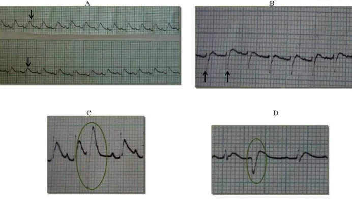
Alterations in the ECG traces were observed from five minutes after the
envenomation of experimental animals with T. serrulatus venom, and such variations
were more severe in the later stages of the experiment. The animals of groups B and
C had changes that were compatible with electrolytic imbalance, including increased
T wave (Figure 1A) and presence of an rS wave (Figure 1B). Such changes are
typically caused by electrolytic, respiratory and gastrointestinal losses.
Scorpion venom is known to produce alterations in the serum concentration of ions
resulting from its direct action on ion channels of the plasma membrane. In addition,
respiratory and metabolic acidosis, together with increased H+ excretion by the
stomach, has also been described as a direct effect of envenomation (6, 21, 22). An
rS wave may indicate a modified repolarization axis caused by an increase in the
suggests that the right ventricle of envenomed animals overworked due to the
presence of extensive hemorrhagic pulmonary areas, detected by macroscopic and
histopathological examination. Under such circumstances, impaired blood flow could
have accounted for the cardiac complications observed.
Additionally, ECG traces revealed the presence of premature ventricular complexes
(PVC) (Figure 1) and T waves of varying morphology (data not shown). PVC are
impulses generated in the heart ventricles due to the abnormal automaticity of
myocytes and may cause direct effects on the cardiovascular system or secondary
effects resulting from inadequate tissue perfusion. According to various authors, PVC
are associated with myocardial abnormalities caused by catecholamine and cytokine
release following scorpion envenomation (6, 9, 22-27).
Figure 1. Electrocardiographic traces for animals subjected to experimental envenomation using Tityus serrulatus venom; (A) increased T wave at ten minutes for animals in both envenomed groups, (B) presence of an rS wave at ten minutes for animals in groups B and C, and (C-D) premature ventricular complexes at 15 and 20 minutes for all envenomed animals (PVC; circled). Speed of 50 mm/s and sensitivity
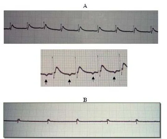
Initially, all experimental animals had a discreet increase in the cardiac rate [heart
rates rising from 400 beats per min (bpm) to 460-480 bpm], which was caused by
pain stimulus and adrenaline release, a positive chronotropic effect. At 25 minutes
after the venom administration, envenomed animals had accentuated bradycardia
and inverted T wave (Figure 2A), and their heart rate fell typically to 200-240 bpm.
Bradycardia was caused by increased vagal tone resulting from cholinergic release,
together with the acute renal failure (ARF) developed by the animals (demonstrated
by laboratory analysis). Hypercalcemia, which may be intensified by the low renal
perfusion typical of ARF, also contributes to bradycardia since it depresses the
cardiac conduction. At 30 minutes after envenomation, bradycardia intensified, falling
to 80-140 bpm (Figure 2B), as demonstrated by third-degree atrioventricular (AV)
blocks (data not shown), which reflected a serious hemodynamic disorder that was
characterized by the complete dissociation between atrial and ventricular beats
caused by cell inhibition or inactivity in the AV junction or in the right and left bundle
branches. Escape rhythm was also detected, indicating that the frequency of the
sinoatrial (SA) node was reduced or even stopped, and the activity of AV junction or
ventricle pacemakers assumed the cardiac rhythm. Escape rhythm is associated with
bradycardia and electrolytic imbalance, as observed in scorpion envenomation
Figure 2. Electrocardiographic traces of animals in group C, which were subjected to experimental envenomation using Tityus serrulatus venom; (A) bradycardia at 25 minutes and inverted P wave (arrowed), and (B) accentuated bradycardia at 30 minutes. Speed of 25 mm/s and sensitivity of N.
Sinus arrhythmia was observed together with inversion and increased amplitude of T,
P and Q waves in dogs subjected to experimental envenomation using T. serrulatus
venom (28). Similarly, sinus tachycardia, ectopic pacemaker, ventricular
extrasystoles and alterations in the ST segment were observed in a dog accidentally
envenomed by T. bahiensis (29). The ECG traces obtained in the present study were
comparable with those for scorpionism in general. However, the changes in ECG
parameters were more severe among the animals in group C, which received a lower
venom dose.
Cardiac muscle function was assessed by quantifying the activities of serum CK,
CK-MB isoenzyme and troponin I following envenomation (Table 1). CK levels in the
experimental animals of groups B and C were significantly higher (P < 0.05) than that
of group A (control), indicating that the venom induced muscle lesions. CK-MB
control group; however, such increases were only statistically valid for group C due to
an unusually low activity presented by one animal in group B.
Previous studies have shown that lesions in the heart are a consequence of
catecholamine overload. Indeed, inhibition of ion channels by scorpion venom
triggers the release of neuronal and adrenal catecholamines, leading to increased
oxygen consumption by the myocytes, myocardium hypoxia and tissue degeneration
(30). Moreover, a study using electron microscopy identified myocardial anomalies
that were different from those attributed to excessive catecholamine levels (6).
Although CK and CK-MB activities peaked later than the blood collection time in the
present study, the increased levels determined for both enzymes indicate an acute
Table 1. Activities of creatine kinase (CK) and cardiac isoenzyme (CK-MB) in the serum of individual animals in the control group A (injected with 400 µL ultrapure
water) and in the experimental groups B (injected with 400 µL of a solution containing
100 µg scorpion venom) and C (injected with 400 µL of a solution containing 450 µg
scorpion venom)
Group Animals CK (U/L) CK-MB (U/L) Troponin
1 1613 591 Negative
2 1748 461 Negative
3 1293 232 Negative
4 907 223 Negative
5 904 560 Negative
A
Mean 1293 ± 390a 413 ± 176a
1 1480 400 Negative
2 5060 1160 Negative
3 2920 920 Negative
4 5620 1460 Negative
5 3770 3540 Negative
B
Mean 3770 ± 1662b 1496 ± 1206ab
1 4340 2020 Negative
2 4120 1140 Negative
3 1260 740 Negative
4 5860 1500 Negative
5 8240 2060 Negative
C
Mean 4764 ± 2557b 1492 ± 568b
Mean values (± standard error) are shown for the five animals in each group. Within each column,
mean values followed by different lowercase letters are significantly different at P < 0.05 according to
analysis of variance and SNK test
Although increased levels of troponin I (> 50-fold) have been reported for humans
observed for the experimental animals in the present study (11, 12, 32). However,
this result is not entirely reliable since the amounts of released troponin I could have
been below the detection limits (0.5 ng/mL) of the semiquantitative test employed.
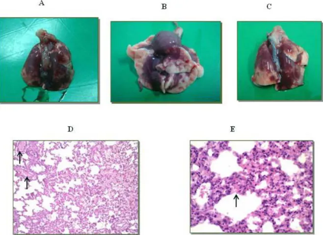
Macroscopically, extensive diffuse hemorrhagic areas were found in all lung lobes of
the animals in experimental groups B and C (Figure 3 – A to C), although no
noteworthy alterations were observed in their hearts. Histological analysis of the lung
tissue of envenomed animals showed moderate congestion of the alveolar capillaries
and large quantities of erythrocytes distributed diffusely in the interstitial and
intra-alveolar spaces (Figure 3 – D and E). Such hemorrhagic indications demonstrate the
direct action of the venom. Histological analysis of the heart tissue from experimental
animals revealed no histopathological alterations.
Kinetic studies concerning the distribution of T. serrulatus venom, determined by
ELISA, have demonstrated the great affinity of toxins for heart, lungs, spleen and
serum (33, 34). Venom levels were maximal at 30 minutes after administration; after
two hours of experiment these concentrations plummeted, and no venom could be
detected in any tissue at eight hours after envenomation. Radioactivity tracer
experiments using technetium-99 showed that Mesobuthus tamulus venom could be
detected in the lungs of rats at five minutes after envenomation (35). Such a rapid
distribution of the venom in the lungs, together with intense pulmonary hemorrhage,
support the hypothesis that the venom has a direct action on organs and ultimately
leads to development of pulmonary edema and impairment of the cardiac function, as
observed in classical scorpion envenomation syndrome.
Although cardiac lesions could not be detected by optical microscopy, the molecular
structure of the tissue was affected, which was demonstrated by the significant
increases in CK and CK-MB activities. Several researchers have explained the
severity of scorpion envenomation in children by demonstrating the occurrence of
myocarditis, high blood pressure, tachyarrhythmia followed by bradycardia, serious
pulmonary edema, congestive heart failure and respiratory insufficiency in victims
(10, 32, 36-38). A study evaluated 41 Egyptian children who were accidentally
envenomed by scorpions and reported the occurrence of severe clinical
manifestations, in addition to myocarditis (17 patients), increased CK and CK-MB
activities, increased troponin-I levels, and a mortality rate of 12.5% (23). The
influence of age on the pharmacokinetics and biodistribution of isolated toxins from T.
serrulatus venom in rats has been investigated and the results indicate that toxin
distribution and maximum concentration in the brain, heart and liver occur very
rapidly in young animals compared to adult ones, whereas clearance and elimination
are slower (39, 40). The results of the present study corroborate previous findings
regarding the susceptibility of young animals to scorpion venom.
CONCLUSIONS
T. serrulatus venom, at doses of 100 and 450 µg/animal, induced acute alterations in
the electrocardiographic, biochemical and histopathological parameters of recently
weaned rats, indicating the great vulnerability of young animals to scorpion
REFERENCES
1. Brasil. Ministério da Saúde. Portal da saúde. Brasília, DF: Secretaria do Ministério
da Saúde. [update 2010 Feb 5; cited 2007 Feb 5]. Available from:
http://portal.saude.gov.br/portal/arquivos/pdf/casos_ac_escorpioes_bra_00a09_tabel
a.pdf.
2. D'Suze G, Moncada S, González C, Sevcik C, Aguilar V, Alagón A. Relationship
between plasmatic levels of various cytokines, tumor necrosis factor, enzymes,
glucose and venom concentration following Tityus scorpion sting. Toxicon.
2003;41(3):367-75.
3. Freire-Maia L, Campos JA. On the treatment of the cardiovascular manifestations
of scorpion envenomation. Toxicon. 1987;25(1):125-30.
4. Gueron M, Ovsyshcher I. What is the treatment for the cardiovascular
manifestations for scorpion envenomation? Toxicon. 1987;25(1):121-4.
5. Gueron M, Ilia R, Sofer S. The cardiovascular system after scorpion
envenomation. A review. J Toxicol Clin Toxicol. 1992;30(1):245-58.
6. Ismail M. The scorpion envenoming syndrome. Toxicon. 1995;33(7):825-58.
7. Ismail M, Abd-Elsalam MA. Are the toxicological effects of scorpion envenomation
related to tissue venom concentration? Toxicon. 1988;26(3):233-56.
8. Faiçal S, Shiota D. Feocromocitoma: atualização diagnóstica e terapêutica. Rev
Assoc Med Brasil. 1997;43(3):237-44.
9. Teixeira Jr AL, Fontoura BF, Freire-Maia L, Machado CRS, Camargos ERS,
Teixeira MM. Evidence for a direct action of Tityus serrulatus scorpion venom on the
cardiac muscle. Toxicon. 2001;39(5):703-9.
10. Campos JA, Silva AO, Lopez M, Freire-Maia L. Signs, symptoms and treatment
of severe scorpion sting in children. Toxicon. 1979;17(1):1-21.
11. Cupo P, Azevedo-Marques MM, Hering SE. Escorpionismo. In: Cardoso JLC,
editor. Animais peçonhentos no Brasil: biologia clínica e terapêutica dos acidentes.
São Paulo: Salvier; 2003. 198-208 p.
12. Cupo P, Jurca M, Azevedo-Marques MM, Oliveira JSM, Hering SE. Severe
scorpion envenomation in Brazil: clinical, laboratorial and anatomopathological
aspects. Rev Inst Med Trop São Paulo. 1994;36(1):67-76.
13. Fukuhara YDM, Dellalibera-Joviliano R, Cunha FQC, Reis ML, Donadi EA. The
kinin system in the envenomation caused by the Tityus serrulatus scorpion sting.
14. Lourenço WR, Cloudsley-Thompson JL, Cuellar O, Von Eickstedt VRD ,
Barraviera B , Knox MB. The evolution of scorpionism in Brazil in recent years. J
Venom Anim Toxins. 1996;2(2):121-34.
15. Freire-Maia L, Campos JA, Amaral CF. Approaches to the treatment of scorpion
envenoming. Toxicon. 1994;32(9):1009-14.
16. Nunan EA, Cardoso VN, Moraes-Santos T. Lethal effect of the scorpion Tityus
serrulatus venom: comparative study on adult and weanling rats. Rev Bras Ciênc
Farm. 2001;37(1):39-44.
17. Vasconcelos F, Sampaio SV, Garófalo MA, Guimarães LF, Giglio JR, Arantes
EC. Insulin-like effects of Bauhinia forficata aqueous extract upon Tityus serrulatus
scorpion envenoming. J Ethnopharmacol. 2004;95(2-3):385-92.
18. Flecknell P. Laboratory animal anaesthesia. 2nd ed. San Diego: Academic Press;
1996. 274 p.
19. Prophet EB, Mills R. Afip laboratory methods in histotechnology. Washington,
DC: American Registry of Pathology; 1992. 278 p.
20. Fundação Arthur Bernardes. Sistema para análises estatísticas e genéticas
(SAEG). Version 9.1. Viçosa: Universidade Federal de Viçosa; 2007.
21. El-Asmar MF. Metabolic effect of scorpion venom. In: Tu AT, editor. Handbook of
natural toxins, insects, poisons, allergens and other invertebrate venoms. New York,
NY: Marcel Dekker; 1984. 551-75 p. 2 vol.
22. Meki ARMA, Mohey El-Dean ZM. Serum interleukin-1β, interleukin-6, nitric oxide
and α1-antitrypsin in scorpion envenomed children. Toxicon. 1998;36(12):1727-48.
23. Magalhães MM, Pereira ME, Amaral CF, Rezende NA, Campolina D, Bucaretchi
F, et al. Serum levels of cytokines in patients envenomed by Tityus serrulatus
scorpion sting. Toxicon. 1999;37(8):1155-64.
24. Meki AR, Mohamed ZM, El-Deen HM. Significance of assessment of serum
cardiac troponin I and interleukin-8 in scorpion envenomed children. Toxicon. 2003;
41(2):129-37.
25. Murthy KR, Hase NK. Scorpion envenoming and the role of insulin. Toxicon.
1994; 32(9):1041-4.
26. Raab W. Key position of catecholamines in functional and degenerative
cardiovascular pathology. Am J Cardiol. 1960;5(1):571-8.
27. Sofer S, Gueron M, White RM, Lifshitz M, Apte RN. Interleukin-6 release
28. Cordeiro FF, Sakate M, Fernandes V, Cuyumjian PR. Clinical and cardiovascular
alterations produced by scorpion envenomation in dogs. J Venom Anim Toxins incl
Trop Dis. 2006;12(1):19-43.
29. Cardoso MJL, Sakate M, Ciampolini P, Moutinho FQ, Cherubini AL.
Envenomation by scorpion in dog – case report. J Venom Anim Toxins incl Trop Dis.
2004;10(1):98-105.
30. Possani LD, Becerril B, Delepierre M, Tytgat J. Scorpion toxins specific for Na+
-channels. Eur J Biochem. 1999;264(2):287-300.
31. Thrall MA. Veterinary hematology and clinical chemistry. Philadelphia: Lippincott
Willians & Wilkins; 2004. 518 p.
32. Bucaretchi F, Baracat ECE, Nogueira RJN, Chaves A, Zambrone FAD, Fonseca
MRCC, Tourinho FS. A comparative study of severe scorpion envenomation in
children caused by Tityus bahiensis and Tityus serrulatus. Rev Inst Med Trop São
Paulo. 1995;37(4):331-6.
33. Revelo MP, Bambirra EA, Ferreira AP, Diniz CR, Chávez-Olórtegui C. Body
distribution of Tityus serrulatus scorpion venom in mice and effects of scorpion
antivenom. Toxicon. 1996;34(10):1119-25.
34. Santana GC, Freire AC, Ferreira AP, Cháves-Olórtegui C, Diniz CR, Freire-Maia
L. Pharmacokinetics of Tityus serrulatus scorpion venom determined by
enzyme-linked immunosorbent assay in the rat. Toxicon. 1996;34(9):1063-6.
35. Murugesan S, Murthy KRK, Noronha OPD, Samuel AM. Tc 99m-Scorpion
venom: labelling, biodistribution and scintiimaging. J Venom Anim Toxins.
1999;5(1):35-46.
36. Amaral CF, Lopes JA, Magalhães RA, de Rezende NA. Electrocardiographic,
enzymatic and echocardiographic evidence of myocardial damage after Tityus
serrulatus scorpion poisoning. Am J Cardiol. 1991;67(7):655-7.
37. El-Amin EO. Issues in management of scorpion sting in children. Toxicon. 1992;
30(1):111-5.
38. Sofer S, Gueron M. Respiratory failure in children following envenomation by the
scorpion Leiurus quinquestriatus: hemodynamic and neurological aspects. Toxicon.
1988;26(1):931-9.
39. Nunan EA, Moraes MF, Cardoso VN, Moraes-Santos, T. Effect of age on body
distribution of tityustoxin from Tityus serrulatus scorpion venom in rats. Life Sci.
40. Nunan EA, Arya V, Hochhaus G, Cardoso VN, Moraes-Santos T. Age effects on
the pharmacokinetics of tityustoxin from Tityus serrulatus scorpion venom in rats.