Universidade de Lisboa
Faculdade de Farmácia
INVESTIGATION OF LANA: A HUMAN VIRUS TUMOR GENE
Anita Marisa Pinto Sousa
Dissertation supervised by Professor João Pedro Monteiro e Louro Machado de
Simas and co-supervised by Professor Maria João Carlos da Silva Gama.
Master’s degree in Biopharmaceutical Sciences
3
Resumo
Os herpesvírus pertencem à grande família de vírus, Herpesviridae e distinguem-se pela sua capacidade de infetar uma enorme variedade de hospedeiros. Até à data, foram identificados oito herpesvírus capazes de infetar humanos e estima-se que quase toda a população adulta esteja infetada por pelo menos um dos vírus pertencentes a esta família. Os os herpesvírus humanos são capazes de se adaptar ao meio intracelular do hospedeiro e escapar ao seu sistema imunitário estabelecendo uma infeção crónica que pode permanecer em estado de latência durante toda a vida do hospedeiro. No entanto, estes vírus normalmente não são causadores de doenças graves, a não ser que o sistema imunitário do hospedeiro esteja extremamente debilitado. A família Herpesviridae pode ser dividida em 3 subfamílias: alfa, beta e gama. E, tendo em conta os vírus capazes de infetar humanos, a família alfa inclui os herpesvirus simplex (HHV-1 e HHV-2) e o vírus varicela-zóster (HHV-3). Os herpesvírus pertencentes à subfamília beta são o citomegalovírus (HHV-5), HHV-6 (variantes A e B) e ainda o HHV-7. Finalmente, os membros da família gama são o vírus Epstein-Barr (HHV-4) e o herpesvírus associado ao sarcoma de Kaposi (HHV-8) e são associados a alguns tipos de doenças cancerígenas. Estes vírus são capazes de induzir a proliferação de células B infetadas através de reações que ocorrem no centro germinativo e expandir a latência viral às células B de memória, que são o maior reservatório de latência dos gamaherpesvírus. Em particular, o herpesvírus associado ao sarcoma de Kaposi (KSHV), um dos sete vírus causadores de cancro até hoje conhecidos, evoluiu no sentido de perturbar os mecanismos celulares normais do hospedeiro como o crescimento celular, a apoptose e até mesmo o sistema imunitário bem como as respostas antivirais. Esta contínua desregulação celular ao longo dos anos pode eventualmente levar ao desenvolvimento de doenças neoplásticas, principalmente em indivíduos imunocomprometidos. Sabe-se que o KSHV é o agente etiológico responsável pelo linfoma primário de efusão primário, pela doença de Castleman multicêntrica e pelo sarcoma de Kaposi, que é o tipo de cancro mais comum em pacientes infetados com o vírus da imunodeficiência humana (HIV).
O ciclo de vida do KSHV, tal como os outros herpesvírus, pode ser distinguido em duas fases distintas: uma fase lítica e uma fase de latência. Após a infeção inicial, os genes de ambas as fases são expressos mas após alguns ciclos de replicação viral, a fase lítica decresce e a fase de latência é estabelecida. Para que o genoma viral se mantenha nas células em divisão, o epissoma viral associa-se às histonas e estabelece-se como um cromossoma extra no núcleo
4
das células infetadas com expressão genética muito limitada. Esta reduzida expressão genética ajuda o vírus a escapar ao sistema imunitário do hospedeiro e ao mesmo tempo garante a sua sobrevivência e persistência viral. Uma das principais características deste tipo de vírus é sua capacidade de persistir no hospedeiro através da manutenção da latência viral, o que requer proteínas virais e celulares. Cerca de 90% das células infetadas possui o programa genético necessário à manutenção da latência viral. O programa de latência traduz-se num locus, onde são expressos vários genes que codificam para proteínas essenciais à latência viral, tal como a LANA (Latency-associated Nuclear Antigen).
A LANA é considerada a proteína responsável pela persistência viral e, consequentemente pela latência viral. É uma proteína multifuncional, sendo maioritariamente responsável pela segregação, replicação e manutenção do epissoma. No entanto, a LANA pode também interferir com os mecanismos de transcrição e interagir com diversas proteínas celulares e interferir com os mecanismos anti-tumorais do hospedeiro. A LANA, atuando como uma ubiquitina ligase E3, é capaz de regular a transcrição e sinalizar para degradação proteossomal supressores tumorais tais como o von Hippel-Lindau (VHL), o p53 ou ainda o nuclear factor-kappa B (NF-B). A ubiquitinação está presente em todas as células eucarióticas e é um mecanismo regulatório essencial no ciclo celular, na apoptose, na endocitose ou ainda na resposta imunitária. Sendo assim, o KSHV, desenvolveu mecanismos com o objetivo de modular o seu funcionamento a seu favor. A ubiquitinação é um processo que ocorre através de uma cascata enzimática constituída por 3 enzimas: E1 (enzima ativadora da ubiquitina), E2 (enzima conjugadora) e E3 (ubiquitina ligase). Numa primeira etapa há a ligação da ubiquitina à E1 que, depois de ativada, é transferida para uma E2 que, juntamente com uma E3, transferem a ubiquitina para o substrato proteico para que seja degradado via proteossoma. A enzima E3 parece ser a responsável pelo reconhecimento específico do substrato e existem vários tipos de E3, tal como as Ellongin BC-Cullin5-SOCS (EC5S). As EC5S são complexos proteicos constituídos por várias subunidades, incluindo uma Cullin5 que se liga a uma Rbx1, formando assim o módulo Cullin 5-Rbx1, um heterodímero Elongin BC e uma proteína de reconhecimento de substrato SOCS (supressor of cytokine signaling). Todas as proteínas SOCS possuem uma homologia de sequência de 40 aminoácidos conservada – motivo box. Este motivo SOCS-box é o responsável pela interação entre a Elongin BC e a Cullin5, estabelecendo a ligação entre o substrato de ubiquitinação e a E2. O complexo Elongin BC é um regulador positivo da RNA polimerase II e possui duas subunidades reguladoras (B e C). Este complexo desempenha um papel importante na regulação da transcrição e ainda ajuda a estabelecer a ligação entre as proteínas SOCS e as Cullin 5. Curiosamente, a proteína viral LANA possui uma SOCS-box
5
bipartida: BC-box + Cullin-box, localizadas na região C-terminal e N-terminal da proteína, respetivamente. Esta sequência proteica BC-box interage com uma Elongin C e estabelece um complexo proteico LANA-Elongin BC, que por sua vez ao ligar-se a um complexo Cullin5/Rbx reconstitui o complexo proteico: Ellongin BC-Cullin5-SOCS. Este complexo assemelha-se ao EC5S, funcionando como uma ubiquitina ligase, modulando os mecanismos de ubiquitinação do hospedeiro a favor do vírus.
A infeção causada pelo KSHV é naturalmente limitada a humanos, por isso é muito importante o estabelecimento de um modelo animal de infeção para que o seu mecanismo de patogénese viral possa ser estudado in vivo. Visto que o herpesvirus murídeo 68 (MHV68), um vírus endémico que infeta o rato-do-campo Europeu e é capaz de infetar ratinhos de laboratório (Mus musculus), partilha alguma homologia de sequência genómica com o KSHV e codifica uma proteína homóloga à LANA do KSHV (mLANA), pode ser utilizado para a infeção de ratinhos de laboratório, providenciando um excelente modelo de estudo para o KSHV. A mLANA também possui uma região C-terminal, é expressa nas células B do centro germinativo e está envolvida na persistência do epissoma viral. Por último, a mLANA também é capaz de regular a transcrição genómica através de um complexo proteico EC5S, que é mediado por uma “SOCS-box” viral que partilha alguma homologia com a SOCS-box presente na kLANA.
Neste projeto, tirando partido do modelo de infeção já estabelecido no laboratório, foram gerados vírus quimera (v-kLANA Elo-BC 46 e v-KM Elo-BC 34) com mutações na BC-box da kLANA (T212A, L213A, N214A, P215A, I216A, C217A) com o objetivo de avaliar a importância deste motivo na patogénese viral in vivo. O v-kLANA é um vírus quimera em que a mLANA foi substituída pela kLANA completa, incluindo a sua região 5´UTR e o v-KM é um vírus quimera que contém uma proteína de fusão entre a kLANA e a mLANA, ou seja, possui a kLANA mas com a região C-terminal da mLANA. Após mutagénese e reconstituição dos vírus quimera mutantes, 80 ratinhos foram divididos em 4 grupos distintos de infeção e inoculados com o vírus correspondente: 2 grupos com os vírus controlo (v-kLANA e v-KM) e 2 grupos com os vírus mutantes (v-kLANA Elo-BC 46 e v-KM Elo-BC 34). Aos dias 5, 7 e 10 pós infeção os ratinhos foram sacrificados e os pulmões foram devidamente extraídos e aos dias 10, 14 e 21 pós infeção os baços foram cirurgicamente removidos, post mortem. Após o processamento dos pulmões e baços, foi possível calcular os títulos virais na fase lítica e latente, respetivamente, e ainda averiguar o número de células positivas nos baços para DNA viral.
Os resultados demonstraram que as mutações inseridas na BC-box da kLANA não afetaram a expressão proteica e, no geral, também não afetaram a fase lítica do vírus nos pulmões. Estes resultados permitem concluir que os vírus mutantes v-kLANA Elo-BC 46 e v-KM
6
Elo-BC 34 são ambos viáveis. No entanto, os resultados in vivo não permitiram tirar conclusões acerca da importância e possível função da BC-box na latência, o que significa que estudos futuros terão de ser feitos no sentido de confirmar ou refutar os fenótipos virais descritos neste projeto.
7
Abstract
Kaposi’s sarcoma-associated herpesvirus (KSHV) is one of the seven recognized human tumor viruses. It is the etiologic agent underlying Kaposi sarcoma, primary effusion lymphoma and multicentric Castleman’s disease. This gammaherpesvirus establishes a lifelong persistence infection in the host and displays a lifecycle with two distinct phases: a short productive lytic phase and a prolonged latent phase. Latency-associated nuclear antigen (LANA) is the major latent gene and is strongly expressed in all forms of KSHV-associated malignancies. LANA mediates episomal replication, segregation and persistence, which is required for long-term maintenance of viral DNA. LANA proteins are also modulators of transcription through E3-ubiquitin ligase activity. Ubiquitination is an essential regulatory mechanism in eukaryotes, controlling a wide range of cellular pathways, including protein degradation. KSHV, like other viruses, has evolved mechanisms to hijack protein degradation pathways in order to establish an environment that favors its propagation. LANA protein has a bipartite SOCS-box motif divided into a Cullin-box and a BC-box located within its C-terminal and N-terminal regions, respectively. The BC-box interacts with an Elongin C and establishes a LANA-Elongin BC complex, which associates with a Cullin/Rbx1 module thus reconstituting a complex protein with E3 ubiquitin-ligase activity. This complex protein assembles an EC5S ubiquitin ligase, which enables the virus to hijack the E3 ligase components of the cell, thus modulating the ubiquitination pathway of its host. In this project, by taking advantage of the MHV68 in vivo model to study KSHV pathogenesis, chimeric viruses with engineered mutations within the BC-box were generated in order to assess the importance of the BC-box motif within the kLANA N-terminal region. Results showed that mutations within kLANA BC-box neither affect protein expression or viral growth in vivo and viral lytic phase in the lungs was not significantly affected. Latency results were not conclusive and therefore the importance and role of kLANA BC-box is still unclear and viral phenotypes need to be reassessed.
8
Aknowledgements
Em primeiro lugar, agradeço ao Doutor Pedro Simas, por me ter dado a oportunidade de desenvolver a minha tese de mestrado no seu laboratório, por ter partilhado o seu vasto conhecimento científico comigo e pela disponibilidade, atenção e simpatia que sempre demonstrou ao longo deste ano. Gostaria também de agradecer à Doutora Marta Miranda, por todo o acompanhamento científico e conselhos dados durante o meu percurso no laboratório, foram sem dúvida indispensáveis para atingir os meus objetivos.
Queria agradecer à Doutora Cecília Rodrigues, a coordenadora do Mestrado em Ciências Biofarmacêuticas e à Doutora Maria João Gama, a minha orientadora interna, pela disponibilidade e simpatia que sempre demonstraram para esclarecer todas as minhas dúvidas.
Agradeço à Doutora Andreia Mósca, por toda a orientação científica, paciência e calma (muita calma), mesmo nos momentos mais difíceis e, acima de tudo, pela amizade.
Gostaria de agradecer à minha colega de laboratório, Catarina Costa, que conheci no início deste ano letivo, mas que com certeza levarei comigo por muitos mais anos. Obrigada pelo apoio incondicional, pela ansiedade e pânico partilhados, pelos serões no iMM, pelas gargalhadas até doer a barriga e por teres sido a minha pessoa no laboratório durante este ano.
Queria agradecer à minha amiga, Catarina Ferreira, que esteve comigo desde o início desta longa e difícil jornada que é a vida académica. Obrigada pelo companheirismo, pela cumplicidade, amizade, carinho e compreensão. Sei que, mesmo quando estamos juntas no mesmo barco (estamos sempre, infelizmente) de uma coisa posso ter a certeza, no meio do pânico e do caos, é sempre melhor estar contigo. Obrigada Cacá por estares sempre presente nos bons e maus momentos, és uma amiga incansável e com quem sei que poderei sempre contar.
Obrigada ao Filipe Rodrigues, o meu melhor amigo e o meu “partner in crime”. Agradeço-te por me Agradeço-teres ouvido sempre que precisei, por me Agradeço-ter queixado (quase) sem parar ao longo deste ano e por, mesmo assim, não te teres fartado e teres sempre dito que ia tudo correr bem e que acreditas em mim. Obrigada por estares presente na minha vida, por sentires quando é que preciso de uma ida ao café, de um jantar, de dançar ou simplesmente de uma chamada telefónica para me animar, obrigada por seres tu.
9
Um obrigada muito especial à minha irmã, Priscila Sousa Gonçalves, que é também a minha melhor amiga e a melhor conselheira do mundo. Agradeço-te por todo o teu apoio incondicional, por me protegeres desde que nasci, por todos os miminhos, abraços, carinhos, almoços, jantares e festas. Obrigada pelas nossas conversas infinitas, por conheceres e perceberes cada particularidade minha e por partilhares esta personalidade peculiar comigo (assim não me sinto sozinha no mundo). Queria agradecer também ao teu marido, Élvio Sousa Gonçalves, que muito tem aturado ao longo destes 15 anos e que é como um irmão para mim.
À minha mãe, Ana Sousa, que é a melhor pessoa que eu conheço e o melhor exemplo feminino que poderia ter. Obrigada por seres o pilar da família Sousa: és a nossa calma na tempestade, o quentinho no Inverno e o sol no Verão. Obrigada por me ouvires, mesmo não percebendo tudo, e por me apoiares incondicionalmente. És a minha “personal cheerleader” e melhoras a minha vida todos os dias, obrigada mami. Ao meu pai, Francisco Sousa, que é o maior sonhador que eu alguma vez conheci. Ensinou-me que podemos e devemos ter muitos sonhos e que não faz mal que nem todos se concretizem, basta apenas alguns. E, nem sempre os sonhos se realizam exatamente da forma que idealizámos, mas não faz mal, às vezes são ainda melhores. Ensinou-me também, através da sua garra, luta, perseverança e inteligência que podemos alcançar aquilo que planeamos para nós e que “nunca se sabe onde é que estaremos daqui a 10 anos” (é um otimista no fundo). Obrigada por teres sido, muitas vezes, o impulsionador de alguns sonhos e aventuras minhas e por todo o apoio subentendido nas tuas palavras e ações. Com a vossa ajuda consegui encerrar mais um capítulo da minha vida. Obrigada.
Finalmente, queria agradecer ao meu namorado, Joaquim Torres, que é o melhor companheiro de aventuras que alguma vez tive. Obrigada por teres sido incansável ao longo deste ano e por me fazeres rir a toda a hora. Dizem que “rir é o melhor remédio” e não podia estar mais de acordo. Não és da minha área, mas durante este ano ouviste-me a toda a hora, todos os dias e tentaste aconselhar-me da melhor forma, mesmo quando tu próprio tiveste dias menos bons. Obrigada por acreditares em mim mais do que eu própria (às vezes) e por me fazeres sentir que, contigo na minha vida, vai sempre correr tudo bem.
10
Table of contents
Chapter 1: Introduction ...16
1. Cancer virology ...16
2. Herpesviridae family ...17
2.1 Gammaherpesvirus subfamily: Kaposi’s sarcoma-associated herpesvirus ...18
2.1.1 Clinical diseases associated with KSHV infection ...19
2.1.2 KSHV infection and the host immune response ...20
2.1.3 KSHV viral lifecycle ...22
2.1.4 The KSHV latency-associated nuclear antigen ...23
2.1.5 LANA and its role during ubiquitination ...25
2.2 MHV68: a KSHV study model ...26
2.2.1 Chimeric virus – v-kLANA ...28
2.2.2 Chimeric virus – v-KM ...28
Significance ...29
Chapter 2: Materials and Methods ...30
1. Generation of MHV-68 recombinant viruses ...30
1.1 Plasmids...30
1.2 DNA: isolation and analysis ...31
1.2.1 Plasmid DNA purification ...31
1.2.2 Quantification and sequencing of DNA ...32
1.2.3 Restriction endonuclease reactions ...32
1.2.4 DNA ligation ...32
1.2.5 Analysis and isolation of DNA by gel electrophoresis ...33
1.3 Bacterial strains ...33
1.3.1 Super Competent E.coli cells preparation ...33
1.3.2 Transformation of competent cells ...34
1.4 Cloning procedures to engineer v-kLANA Elo-BC and v-swap Elo-BC viruses ...34
1.4.1 Mutagenesis: Polymerase Chain Reaction (PCR) to introduce mutations into pSP72_PCR1_PCR3 ...34
1.4.2 Subcloning the pSP72_PCR1_3 Elo-BC into pSP72_PCR1_5 and pSP72_994swap ...36
1.4.3 Subcloning pSP72_swap Elo-BC and pSP72_PCR1_5 Elo-BC into BamHI-shuttle vector ...36
11
1.5.1 BAC DNA preps ...38
1.6 Cell lines...39
1.7 Viruses ...39
1.8 Reconstitution of MHV-68 viruses...40
1.8.1 Virus reconstitution on BHK-21 cells ...40
1.8.2 Passage through NIH3T3-Cre cells to remove BAC sequences ...41
1.9 Production of viral stocks: working stock media (WSM) and cell working stock (CWS) .41 1.10 Virus titration using the suspension method: plaque assay ...41
2. Protein expression analysis ...42
2.1 Sodium dodecyl sulfate-polyacrylamide gel electrophoresis (SDS-PAGE) ...42
2.2 Transference of proteins into nitrocellulose membranes ...43
2.3 Western blot ...43
3. Ethics statement ...44
4. Mice ...44
5. In vivo assays ...44
5.1 Infection of mice ...44
5.2 Lung viral titration ...45
5.3 Single cell suspensions ...45
5.4 Infectious Center Assay (ex vivo explant co-culture assay) ...46
5.5 Limiting dilution assay and real-time PCR of viral DNA positive cells ...46
6. Statistical analysis ...47
Chapter 4: Results and Discussion ...47
1. Generation and characterization of MHV68 recombinant virus ...47
2. In vitro assays ...50
2.3 Viral titration ...50
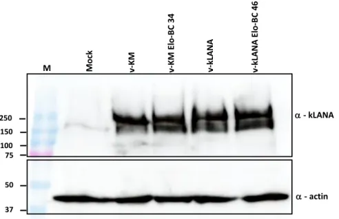
2.4 Expression of kLANA mutant proteins ...51
3. In vivo assays ...52
3.1 Lung viral titres ...52
3.2 Infectious center assay and quantification of viral DNA-positive total splenocytes ...53
Chapter 5: Conclusion and future perspectives ...56
References ...58
12
Table index
Table 1 - Human herpesviruses classification ...18
Table 2 - Primers used for sequencing ...32
Table 3 - Primers used in the mutagenesis strategy ...36
Table 4 - Primers used in the colony PCR ...38
Table 5 - List of MHV68 recombinant virus constructed during this project ...40
Table 6 - Antibodies used in the western blot assay ...43
Table 7 - in vivo experiments ...45
Table 8 - Viral stock titers ...51
13
Figure index
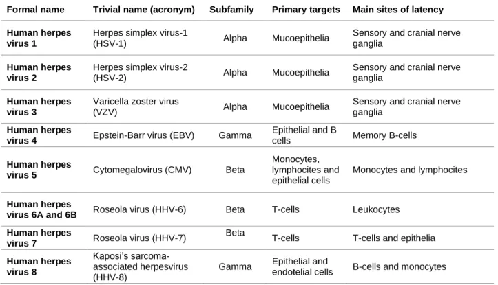
Figure 1 - Episome segregation model ...24
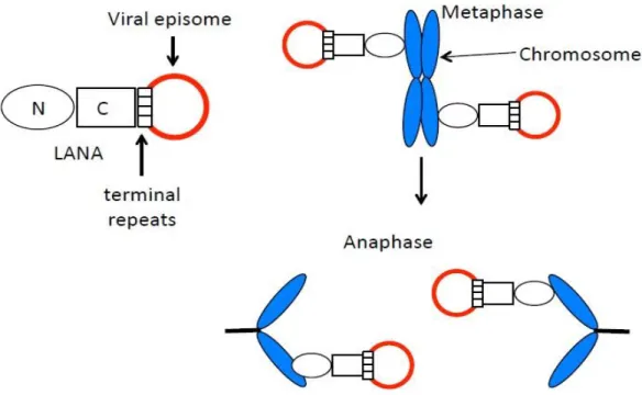
Figure 2 - A model for KSHV LANA assembles EC5S ubiquitin complex to target downstream substrates for degradation ...26
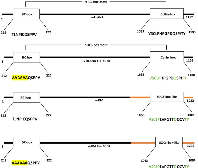
Figure 3 - Schematic representation of LANA and ORF73. Proteins with the putative SOCS-box motifs indicated ...27
Figure 4 - Representation of kLANA proteins expressed by each virus, highlighting the kLANA SOCS-box motif ...48
Figure 5 - Restriction profiles of non-yfp BAC plasmids and schematic representation of MHV68 genome with each restriction enzyme sites ...50
Figure 6 - BAC cassette removal through NHI-3T3 cells expressing Cre recombinase ...50
Figure 7 - Expression detection of viral protein kLANA ...51
Figure 8 - Lung viral titration. BALB/cByJ mice were intranasally inoculated with 104 PFU ...52
Figure 9 - Assessment of latent infection in the spleens of infected mice ...54
Figure 10 - Subcloning the pSP72_PCR1_3 Elo-BC into pSP72_PCR1_5 (A) and pSP72_994swap (B) ...63
Figure 11 - Subcloning pSP72_PCR1_5 (A) and Elo-BC pSP72_swap Elo-BC (B) into BamHI-shuttle vector ...64
Figure 12 - BAC mutagenesis strategy ...64
14
Abbreviations
AmpR: Ampicilin resistance
BAC: Bacterial artificial chromosome
BHK: Baby hamster kidney
CMV: Cytomegalovirus
CRL: Cullin RING ligase
CPE: Cytopathic effect
CWS: Cell working stock
DBD: DNA binding domain
DMEM: Dulbecco’s modified Eagle medium DPI: Days post-infection
dsDNA: Double-stranded DNA
EBV:Epstein-Barr virus
EC5S: ElonginBC-Cullin5-SOCS
EQE: Glutamate- and glutamine-rich region
FBS: Fetal bovine serum
GC: Germinal center
GFP: Green fluorescent protein
HAART: highly active antiretroviral therapy
HBV: Hepatitis B virus
HCV: Hepatitis C virus
HHV-1 to 8: Human herpesvirus 1 to 8
HIF: Hypoxia-inducible factor 1-alpha HIV: Human immunodeficiency virus
HPV: Human papillomaviruses
HSV: Herpes simplex virus
HTLV-1: Human T-lymphotropic virus-I
HUVECs: Human endothelial cells
IARC: Internation Agency for Research on Cacer
ICA: Infectious center assay
IE: Immediate early
IFN: Interferon
IRFs: Interferon regulatory factors
KanR: Kanamycin resistance
KS: Kaposi’s sarcoma
KSHV: Kaposi’s sarcoma-associated virus LANA: Latency-associated nuclear antigen
LB: Luria-Bertani
LBS: LANA binding sites
15
MHV68: Murine gammaherpesvirus 68
miRNA: micro RNA
MOI: Multiplicity of infection
NF-B: Nuclear factor kappa B NLRs: NOD-like receptors
O/N: Overnight
ORF: Open reading frame
PAMPs: Pathogen-associated molecular patterns
PCR: Polymerase chain reaction
PEL: Primary effusion lymphoma
PFU: Plaque forming unit
PRRs: Pattern recognition receptors
RBL: Red blood lysis buffer
RLRs: Retinoic acid-like receptors
RT: Room temperature
RTA: Replication and transcription activator
SOCS: Suppressor of cytokine signaling protein
ssDNA: Single-stranded DNA
TERT: telomerase reverse transcriptase
TGF: Transforming growth factor beta TLRs: Toll-like receptors
TPA: 12-O-tetradecanoyphorbol-13-acetate
TR: Terminal repeats
UTR: Untranslated region
VHL: von Hippel-Lindau
vIRFs: Viral interferon regulatory factors
VZV: Varicella-zoster virus
WB: Western blot
WSM: Working stock media
WT: Wild type
16
Chapter 1: Introduction
1. Cancer virologyIt is widely accepted that viruses play a significant role in the development of particular cancers in many different animals, including humans (Cooper, 1995). Indeed, according to the International Agency for Research on Cancer (IARC) approximately 12% to 20% of human cancers are caused by viruses. Viruses have been key elements to modern cancer research and provide profound insights into possible different cancer causes, whether they are infectious or non-infectious (Moore and Chang, 2010). The first described human tumor virus was the Epstein-Barr virus (EBV) in 1964 and, until now, six more human cancers have been described: Hepatitis B virus (HBV) (1965), Human T-lymphotropic virus-I (HTLV-I) (1980), High-risk human papillomaviruses (HPV) (1983-1984), Hepatitis C virus (HCV) (1989), Kaposi’s sarcoma-associated herpesvirus (KSHV) (1994) and finally Merkel cell polymarirus (MCV), in 2008 (Moore and Chang, 2010). However, there is no obvious molecular rule able to firmly establish or eliminate an agent as a potential human tumor virus a priori, and almost all of the tumor viruses have very close relatives that do not cause cancer in humans (Moore and Chang, 2010). Thus, why do some viruses actually cause cancer? Since tumors do not increase the virus transmissibility or its fitness, one possible explanation is that cancers caused by viruses are biological accidents. Moreover, tumors are “dead-ends” for viruses and only a few people infected with any of the existing tumor viruses eventually develop cancer.
Viruses can cause cancer by interacting with host proteins, proliferating when the host immune system is weakened, and hijacking proliferating host cells (Wen and Damania, 2010). Compared to other viruses, human tumor viruses are unusual because they infect, but do not kill, their host cells. This allows human tumor viruses to establish persistent chronic inflammation leading to tissue damage, which is one of the known mechanisms of carcinogenesis (Goossens and Hoshida, 2015). Moreover, tumor viruses are strongly selected to encode oncogenes able to initiate tumorigenesis, such as oncoproteins that target p53, telomerase reverse transcriptase (TERT), cytoplasmic PI3K/AKT/mTOR, nuclear factor-B (NF-B), -catenin or interferon signaling pathways (Mesri, Cesarman and Boshoff, 2010; Moore and Chang, 2010; Dissinger and Damania, 2016; Dittmer et al., 2016). It is thought that these viral oncogenes target cellular tumor suppressor pathways to re-initiate the cell cycle entry of differentiated cells to promote viral replication. The result is cellular genomic instability and aneuploidy, which contributes to carcinogenesis (Moore and Chang, 2010). The other theory is based on large DNA tumor viruses, such as EBV or KSHV. This theory suggests a more complex interaction between viral oncogenes
17
and host, where the virus main goal is to evade immune responses during the latency program rather than assuring viral genome replication (Moore and Chang, 2003, 2010).
Despite the already recognized importance of tumor causing viruses by the scientific community, there is still a considerable amount of work to do in this research area. For instance, EBV has been discovered 55 years ago and there is still no vaccine. Moreover, KSHV has emerged as a leading cause of adult cancer in sub-Saharan Africa and the Kaposi sarcoma (KS) is the most common cancer in individuals living with HIV nowadays (Schulz and Cesarman, 2015). Although no movement has yet been made for the pursuit of clinical interventions. Thus, the development of anti-latent viral strategies and immunological therapies against these cancer viruses is critical.
2. Herpesviridae family
Human herpesviruses have large (100-200 nm) and double stranded DNA (ds-DNA) genomes enclosed inside the viral protein cage (capsid), surrounded by a tegument layer which is enclosed in a lipid envelope constituted with glycoproteins (Chakraborty, Veettil and Chandran, 2012; Grinde, 2013). The family name Herpesviridae is derived from the Greek word herpein meaning "to creep", which refers to the latent, recurring infections, typical of this family of viruses. All human herpesviruses are able to adapt very well to the cellular milieu of the host and evade his immune system, establishing life-long latent infection (Wen and Damania, 2010). Considering gene diversity, herpesviruses are among the most complex human viruses due to the 70-200 open reading frames (ORFs) of their genomes (Grinde, 2013). This large repertoire of herpes genes allows a persistent infection in the host through cell manipulation and immune evasion.
Up to date, there are eight known herpesviruses that commonly infect humans and approximately all of the adult population is infected with at least one of these (Grinde, 2013). Although, these viruses are highly unlikely to cause severe disease, unless the host immune system is already compromised. The Herpesviridae family can be classified into 3 different subfamilies, according to their host range, replication cycle and cell tropism: alpha, beta and gamma (Roizman B, Carmichael LE, Deinhardt F, de-The G, Nahmias AJ, Plowright W, Rapp F, Sheldrick P, Takahashi M, 1981). The human alpha subfamily includes herpes simplex viruses (HHV-1 and HHV-2) and varicella-zoster virus (HHV-3). Human herpesviruses belonging to the betaherpesvirinae subfamily are cytomegalovirus (HHV-5), HHV-6 variants A and B and HHV-7. Finally, the members of the gamma subfamily are strongly associated with neoplastic diseases:
18
Epstein-Barr virus (HHV-4) and Kaposi’s sarcoma-associated herpesvirus (HHV-8) (Boshoff and Chang, 2001; Wen and Damania, 2010).
Table 1 - Human herpesviruses classification
Formal name Trivial name (acronym) Subfamily Primary targets Main sites of latency Human herpes
virus 1
Herpes simplex virus-1
(HSV-1) Alpha Mucoepithelia
Sensory and cranial nerve ganglia
Human herpes virus 2
Herpes simplex virus-2
(HSV-2) Alpha Mucoepithelia
Sensory and cranial nerve ganglia
Human herpes virus 3
Varicella zoster virus
(VZV) Alpha Mucoepithelia
Sensory and cranial nerve ganglia
Human herpes
virus 4 Epstein-Barr virus (EBV) Gamma
Epithelial and B
cells Memory B-cells
Human herpes
virus 5 Cytomegalovirus (CMV) Beta
Monocytes, lymphocites and epithelial cells
Monocytes and lymphocites
Human herpes
virus 6A and 6B Roseola virus (HHV-6) Beta T-cells Leukocytes Human herpes
virus 7 Roseola virus (HHV-7)
Beta
T-cells T-cells and epithelia
Human herpes virus 8
Kaposi’s sarcoma-associated herpesvirus (HHV-8)
Gamma Epithelial and
endotelial cells B-cells and monocytes
2.1 Gammaherpesvirus subfamily: Kaposi’s sarcoma-associated herpesvirus
The gammaherpesviruses persistent infection is highly associated with several human malignancies, including some types of tumors (Dirk P. Dittmer, 2013). These viruses are able to induce proliferation of latently infected B cells through reactions that occur in the germinal center (GC), expanding latency and gaining access to memory B cells, the major reservoir of gammaherpesviruses latency (Cerqueira et al., 2016).
Kaposi’s sarcoma-associated herpesvirus, also known as herpesvirus 8 (HHV-8), is one of the seven recognized human cancer-causing viruses that has evolved to subvert the normal cellular pathways of its host through a plethora of viral gene products (Moore and Chang, 2010). The KSHV genome consists of ~165 kb, encompassing the several KSHV ORFs, flanked by 30-kb terminal repeat (TR) sequences (Juillard et al., 2016; Purushothaman et al., 2016).
Upon infection, the linear KSHV genome is transported to the nucleus, circularizes and is maintained as an episome (Purushothaman et al., 2016). This gammaherpesvirus is able to
19
dysregulate the cell cycle progression, apoptosis, immune surveillance and even antiviral responses of the host (Dittmer et al., 2016). This disturbance contributes to host cell transformation and eventually to the development of neoplastic disorders, most frequently in immunodeficient individuals (Wen and Damania, 2010). Although the International Agency for Research on Cancer classified KSHV as a class I carcinogen, the majority of KSHV infections have no immediate clinical symptoms and, as with other human neoplastic viruses, cancer only emerges decades after infection (Thakker and Verma, 2016).
2.1.1 Clinical diseases associated with KSHV infection
KSHV is the etiologic agent underlying primary effusion lymphoma (PEL), multicentric Castleman’s disease (MCD) and Kaposi’s sarcoma (KS), the leading AIDS malignancy (Schulz and Cesarman, 2015).
Kaposi’s sarcoma
In 1872, the Austrian-Hungarian dermatologist Moritz Kaposi described five cases of a lethal and previously unknown “idiopathic multiple pigmented sarcomas of the skin”. This form of disease was designated Kaposi’s sarcoma only in 1891 (Boshoff and Chang, 2001). In 1994, hespesvirus-like DNA sequences were identified in KS tissue through analysis of DNA fragments obtained from KS biopsies (Chang et al., 1994). This newly identified virus was then named Kaposi’s sarcoma-associated herpesvirus (KSHV) or human herpesvirus-8 (HHV-8), the eighth member of the Herpesviridae family. KS can be classified into 4 distinct clinical subtypes, based on the epidemiological and clinical outcomes: classic/sporadic, endemic/African, epidemic/AIDS-associated, and iatrogenic/post-transplant (Wen and Damania, 2010). Classic KS, the tumor first described by Kaposi, affects mainly elderly males of Mediterranean or eastern European origin (Gramolelli and Schulz, 2015). Endemic KS is clinically more aggressive, with more lymph node involvement than classic KS, and is commonly seen in children of sub-Saharan Africa and is associated with high mortality rates (Wen and Damania, 2010; Dirk P. Dittmer, 2013). AIDS-associated KS, is the most clinically aggressive variant found in patients, however through highly active antiretroviral therapy (HARRT) and the resulting immune reconstitution, the incidence and mortality of AIDS-associated KS have dramatically dropped (Wen and Damania, 2010). Finally, the iatrogenic KS, develops in solid organ transplant recipients, because it is associated with immunosuppressive therapy after organ transplant (Dirk P. Dittmer, 2013). Renal transplant patients are the most likely group to develop this form of KS and usually, iatrogenic KS regresses
20
upon reduction or withdrawal of immunosuppressive therapy (Wen and Damania, 2010; Gramolelli and Schulz, 2015).
Primary effusion lymphoma
The primary effusion lymphoma (PEL) is a diffuse large B cell lymphoma of post germinal center origin (Dittmer et al., 2016). Different from KS, PEL presents as a lymphomatous effusion tumor contained in various body cavities such as the pericardium, pleuron, and the peritoneum. PEL is a very aggressive tumor with rapid progress and a very poor prognosis associated with high mortality rates and a median survival time of two to six months (Wen and Damania, 2010). Cells with PEL contain many KSHV episomes (50 to 150 viral copies per cell) and they can either be KSHV single-positive or KSHV/EBV double positive (Wen and Damania, 2010; Dittmer et al., 2016).
Multicentric Castleman’s disease
Multicentric Castleman’s disease (MCD) is a lymphoproliferative disorder, affecting multiple sets of lymph nodes and other tissues throughout the body (Dittmer et al., 2016). MCD has two known variants: plasmablastic, which is highly associated with KSHV, and hyaline, which is not (Wen and Damania, 2010). In MCD caused by KSHV, there is a cytokine over activity caused by viral products, leading to atypical lymphoproliferations and potential progression to lymphoma. Plasmablastic MCD can be very aggressive and rapidly progress, thus causing high fatality (Wen and Damania, 2010). KSHV genomes, similar to KS and PEL patients, are detectable in almost all HIV-positive MCD cases and in about 50% of HIV-negative MCD cases (Moore and Chang, 2010).
2.1.2 KSHV infection and the host immune response
The human immune system works like a shield, designed and equipped to suppress a variety of diseases and yet, can never eliminate the Kaposi’s sarcoma-associated herpesvirus. This is due to the fact that KSHV, like all the other members of his family, is able to establish lifelong infection in the host and evade viral clearance (Wen and Damania, 2010).
KSHV is thought to enter cells predominantly through the endocytic pathway, which involves envelope glycoproteins that mediate virus attachment, entry, assembly, and exit of virus (Chakraborty, Veettil and Chandran, 2012). Upon infection, KSHV leads to changes in cell morphology, glucose metabolism, growth rate, lifespan and gene expression (Mesri, Cesarman
21
and Boshoff, 2010). This gammaherpesvirus encodes approximately 86 ORFs, including several potential immunomodulators (K3, k5, K9, K11.1), and anti-apoptotic protein (K7) which regulates cytokine secretion, antagonizes host interferon (IFN)-mediated antiviral responses, and regulates immune evasion (Purushothaman et al., 2016). KSHV is capable of infecting different cell types, hence possesses multiple viral receptors to enter the host cell (Dittmer et al., 2016). During KSHV entry into the host cells, it encounters several innate immune patrol cells that trigger an antiviral response. Remarkably, it is likely that this innate immune response activation, during primary infection, induces the viral latent phase (Akula et al., 2002; Wang et al., 2003; Rappocciolo et al., 2006).
The innate immune response is triggered through pattern recognition receptors (PRRs) that are able to recognize pathogen-associated molecular patterns (PAMPs). These PRRs are unique, meaning that each cell type expresses its own, and lead to the production of interferon and pro-inflammatory cytokines (Dittmer et al., 2016). Thus, there is a multitude of PRRs in human cells including toll-like receptors (TLRs), retinoic acid-like receptors (RLRs) and NOD-like receptors (NLRs) (Dittmer et al., 2016; Ma and Damania, 2016). During primary KSHV infection, activation of the different TLRs in monocytes results in interferon production and upregulation of both cytokines and chemokines (West and Damania, 2008; West et al., 2011). Following de novo infection with KSHV and during viral reactivation, the cytosolic RIG-I-like receptor induces IFN-β production and suppresses viral gene expression (Inn et al., 2011). As the members of the NLR family can sense PAMPs, they form inflammasomes which leads to cleavage and production of IL-1 and IL-18, two well-known pro-inflammatory cytokines (Jacobs and Damania, 2012).
Studies suggest a very thin and delicate equilibrium between viral infection and host response. Although the innate immune system activation induces KSHV into a latent state, a high degree of innate response also facilitates killing of the infected cells and tries to prevent latency. Thus, KSHV encodes several genes to escape the innate immune signaling pathways, which include viral interferon regulatory factors (vIRFs), complement regulatory proteins, tegument proteins and DNA-binding proteins (Dittmer et al., 2016). KSHV encodes four different vIRFs, some ablate cellular IRF signaling and others inhibit IFN production (Jacobs and Damania, 2011). All herpesviruses encode for tegument proteins, which are early deposited into the cytoplasm following virion fusion and capsid release. ORF45 and ORF64 are tegument proteins that are able to block IRF7 phosphorylation and activation of type I IFN responses and encode potent deubiquitinating activity, respectively (Inn et al., 2011; Dittmer et al., 2016). ORF50 is a DNA-binding protein and a transcription factor, although it can also induce the degradation of innate immune responders such as IRF7 or TLR3. Lastly, latency-associated nuclear antigen (LANA) is
22
another DNA-binding protein that also inhibits IFN- induction, through binding to its promotor, and the transcription of IFN-gamma-inducible genes (Cloutier and Flamand, 2010; Dittmer et al., 2016). The adaptive immune system also plays an important role during KSHV infection. This herpesvirus expresses proteins that can affect either the antigen presentation, the B cell targeting, the MHC class I display, and even the neutrophil and basophil activation (Dittmer et al., 2016).
2.1.3 KSHV viral lifecycle
Like other herpesviruses, KSHV comprehends two different lifecycle phases: latent and lytic. Following primary infection, the genes from both phases are expressed, but after some rounds of viral replication, the lytic gene expression decreases, and latency is established. Although, both lytic and latent cycle replications are important for the long-term maintenance of the virus in the host, and the gene products from both expression programs have critical roles in the pathogenesis of KSHV-associated diseases (Purushothaman et al., 2016)(Fig.1).
KSHV viral episome associates with cellular histones and exists as an extra-chromosome in the nucleus of infected cells with the expression of limited viral genes. This allow the virus to maintain the genome in dividing cells and to escape the host immune responses while, at the same time, enhancing cell survival and virus persistence (Purushothaman et al., 2016). A key feature of all herpesviruses infection is their lifelong persistence in the host by maintaining viral latency, which requires not only viral but also cellular proteins (Correia et al., 2013; Dissinger and Damania, 2016). The latent transcriptional program is carried out in more than 90% of infected cells and has led to the characterization of a latency locus that is abundantly and consistently transcribed in all the KSHV infected cells. This latency locus contains several latency associated proteins such as vCyclin, vFLIP, miRNAs and LANA (Purushothaman et al., 2016). Furthermore, to maintain latency, it is crucial that only latent genes are expressed while lytic gene transcription is repressed (Prasad et al., 2012). In fact, studies have demonstrated that in actively transcribed latency regions, the chromatin presents an open configuration, lacks nucleosomes, and exhibits active histone marks, while lytic genes are present in the condensed chromatin regions (Toth et al., 2010; Dissinger and Damania, 2016). This indicates that epigenetics plays a crucial role in the maintenance of latency through the interaction of histone modifiers and chromatin, and several other proteins such as LANA (Hu et al., 2014). Latent KSHV expresses a very small subset of proteins in the infected cells, and no functional or infectious viral particles are produced. The lytic cycle replication is characterized by the expression of linear viral genomes, and can be divided into three different phases: immediate early (IE), early, and late gene expression (Sun et al., 1999). This process follows a highly orchestrated temporal order. The transcription of
23
IE genes does not require prior protein synthesis and are crucial for regulating the subsequent transcriptional cascade (Wen and Damania, 2010). Early gene expression requires proteins encoded by IE genes and late genes expression follows DNA replication. Late genes encode for structural proteins necessary for the envelope glycoproteins and also for the assembly of new virions (Dissinger and Damania, 2016). The lytic viral lifecycle is crucial for viral propagation and tumorigenesis through replication and constant infection of new cells (Aneja and Yuan, 2017).
The latent phase can be induced into the lytic phase with the addition of chemicals that activate the expression of IE genes and transcription activator (RTA), encoded by ORF50, which is the master regulator of KSHV lytic replication (Krishnan et al., 2004). Those chemicals include 12-O-tetradecanoyphorbol-13-acetate (TPA) and sodium butyrate (Aneja and Yuan, 2017). These agents acting as extracellular stimuli, can also affect chromatin regulatory factors and as a result, the viral episome gradually relaxes from its compact chromatin structure, leading to the expression of all viral genes and the production of infectious virion particles from the lytic replication phase (Purushothaman et al., 2016). However, the majority of infected cells bear the virus in the latent phase, and only 3-5% of the cells exhibit the productive lytic infection phase (Chudasama et al., 2015). It is the balance between KSHV latent and lytic viral life cycles that enables the virus to escape immune surveillance and maintain persistent infection and propagate in the individual and maybe even spread to other individuals (Aneja and Yuan, 2017).
2.1.4 The KSHV latency-associated nuclear antigen
KSHV latency-associated nuclear antigen is a 1162 amino acid protein encoded by ORF73 of the viral genome (Juillard et al., 2016). It is considered the viral protein responsible for viral persistence and the major gene of the latent viral lifecycle (Correia et al., 2013; Purushothaman et al., 2016). LANA mediates episomal replication, segregation and persistence, which is required for long-term maintenance of viral DNA into the daughter cells during mitosis (Correia et al., 2013). The C-terminal region of kLANA harbors a DNA-binding domain (DBD) that self-associates and cooperatively binds to two adjacent sites in each terminal repeat (TR) DNA sequences of the viral episome, LANA-binding sites (LBSs) 1 and 2 (LBS1, LBS2) (Juillard et al., 2016). LBS1 is a high affinity site which helps the binding of LANA to LBS2 and both sites contribute to LANA’s ability to suppress transcription and facilitate DNA replication (Garber, Hu and Renne, 2002; Correia et al., 2013). N-terminal kLANA region binds histones H2A and H2B on the nucleosome surface to attach to mitotic chromosomes (Barbera et al., 2006; Juillard et al., 2016). These binding properties together, allow LANA to form a molecular tethering apparatus
24
which ensures that viral genomes are segregated to daughter cell nuclei following mitosis (Correia et al., 2013; Habison et al., 2017) (Fig.1).
Although the main function of LANA is to maintain the viral episome, this is a multifunctional nuclear protein that can also interfere with important anti-tumorigenic pathways (Si and Robertson, 2006; Mesri, Cesarman and Boshoff, 2010). Moreover, LANA has been shown to dysregulate Wnt signaling, through stabilization of -catenin, and to inhibit anti-proliferative transforming growth factor- (TGF 1), by epigenetic suppression of TGF receptors (Fujimuro et al., 2003; Di Bartolo et al., 2008). Indeed, LANA might also contribute to angiogenesis, a crucial step during the oncogenesis process, by stabilizing hypoxia-inducible factor 1 (HIF1) (Cai et al., 2006). LANA was also shown to upregulate the human telomerase reverse transcriptase (hTERT) gene expression and increase the lifespan of primary human umbilical vascular endothelial cells (HUVECs) (Watanabe et al., 2003). This nuclear antigen, is also able to interfere with transcription and interact with several cellular proteins to modulate host functions involved in latency regulation and oncogenesis (Rodrigues et al., 2009). Moreover, LANA is able to stabilize Myc, establishing a link between virus induced lymphoproliferation and disease (Rodrigues et al., 2013). Finally, LANA is able to regulate transcription through E3-ubiquitin ligase activity (Correia et al., 2013). LANA functions as a component of an ubiquitin complex that targets tumor
Figure 1 - Episome segregation model. C-terminal LANA (C) binds terminal repeat DNA sequences in the episome. N-terminal (N) associates with the nucleosome. These binding characteristics allow tethering of the episome to chromosomes during mitosis and segregation of episomes to daughter cells in anaphase.
25
suppressors such as the von Hippel-Lindau (VHL), p53 and nuclear factor-kappa B (NF-B) for degradation (Cai et al., 2006; Rodrigues et al., 2009).
2.1.5 LANA and its role during ubiquitination
Ubiquitination is an essential regulatory mechanism and is involved in nearly all aspects of eukaryotic biology such as signal transduction, development, apoptosis, cell cycle progression, endocytosis and even immune response (Cai et al., 2006). Thus, KSHV, like other viruses, has developed mechanisms to modulate ubiquitination in its favor (Correia et al., 2013). The ubiquitination process can be divided into two steps: (1) the covalent attachment of multiple ubiquitin molecules to the protein substrate and (2) the degradation of the ubiquitylated protein by the 26S proteasome complex (Cai et al., 2006). This process occurs through a three-enzyme cascade involving E1 ubiquitin-activating enzyme, an E2 ubiquitin-conjugating enzyme, and an E3 ubiquitin-ligase enzyme. The process of ubiquitin activation uses ATP as a substrate to the peptide bond formation between ubiquitin and E1. Then, the activated ubiquitin is transferred to an E2, which together with an E3 transfers ubiquitin to the substrate protein to be degraded by the proteasome (Callis, 2014). The E3 ubiquitin-ligase is believed to be the main responsible for substrate specific recognition and, to date, there are several E3 ligases already described. One such protein is the EC5S (ElonginBC-Cullin5-SOCS) (Rodrigues et al., 2009), which is a multi-subunit complex containing a scaffold protein (Cullin 5) attached to a RING finger protein (Rbx1)(Cullin 5-Rbx module), an adaptor heterodimer (Elongin BC), and a substrate recognition protein (suppressor of cytokine signaling [SOCS] box protein) (Cerqueira et al., 2016). SOCS proteins share a 40-amino-acid sequence that is known as the SOCS-box motif, which is responsible for the interaction with Elongin BC and Cullin 5 modules establishing the bridge between the substrate of ubiquitination and the E2 ubiquitin-conjugating enzyme (Yoshimura, Naka and Kubo, 2007). The Elongin BC complex is a positive regulator of RNA polymerase II and has a transcriptionally active subunit A, and two regulatory subunits B and C. This complex has an important role in transcriptional regulation and acts as an adaptor connecting Cullin 5 and SOCS-box proteins (Okumura et al., 2012) (Fig.3). Moreover, an Elongin BC complex containing a member of the Cullin RING ligase (CRL) superfamily plays a crucial role in some cellular processes such as tumorigenesis, signal transduction, cell mobility and differentiation (Okumura et al., 2012).
26
Curiously, the LANA protein encoded by KSHV has a bipartite SOCS-box motif divided into a Cullin-box and a BC-box located within its C-terminal and N-terminal regions, respectively (Fig.2). This BC-box interacts with the host Elongin C creating a LANA-Elongin BC complex which is capable of assembling with a Cullin5/Rbx1 module and reconstitute a complex protein with E3
ubiquitin-ligase activity (Cai et al., 2006). This complex protein assembles an EC5S ubiquitin ligase, which enables the virus to hijack the E3 ligase components of the cell, thus modulating the ubiquitination pathway of its host (Cerqueira et al., 2016).
2.2 MHV68: a KSHV study model
The KSHV infection is naturally limited to humans, so it is of much importance to establish a small animal model of infection in order to study viral pathogenesis in vivo (Habison et al., 2017). Dittmer et al., showed that KSHV infection into SCID mice implanted with human fetal thymus and liver grafts led to lytic and latent infection, mostly in B cells (Dittmer et al., 1999). In 2006, Parsons and his team proved that injection into NOD/SCID mice resulted in latent and lytic infection in multiple cell types, including B cells and macrophages, for several months (Parsons et al., 2006). Furthermore, oral, intraperitoneal or vaginal inoculation into a humanized NOD/SCID/IL2R gamma mouse implanted with human fetal liver and thymus also induced KSHV infection of both
Figure 2 - A model for KSHV LANA assembles EC5S ubiquitin complex to target downstream substrates for degradation.
LANA is predicted to form a complex with Cul5/Rbx1 that interacts with Elongin BC. LANA acts as adapter to link substrates
which bind at its amino (1) or carboxyl (2) -terminal domain (like VHL and p53) to EC5S ubiquitin complex and induces the
pathway of ubiquitin E1 activation, E2 conjugation, and substrate polyubiquitylation as well as 26S proteasome–mediated degradation (Cai et al., 2006).
27
B cells and macrophages (Wang et al., 2014). However, despite the obvious advantage of allowing direct investigation of KSHV, these models are limited because they all require immune suppressed mice (Habison et al., 2017). Thus, the deepest insights about KSHV biology mostly come from studying the murine gammaherpesvirus 68 (MHV68), a gamma-2 herpesvirus.
The MHV-68 is a naturally occurring virus that was first isolated from Slovakian bank voles (Clethironomys glareolus) and is endemic in European wood mice (Dong, Forrest and Liang, 2017). MHV68 genome consists of 118 kb, flanked by terminal repeat sequences of 1.2 kb and encodes approximately 80 genes (Pedro Simas and Efstathiou, 1998). This gammaherpesvirus is genetically related to KSHV and EBV and, a genomic organization showed that their genomes are collinear and contain large blocks of conserved genes (Pedro Simas and Efstathiou, 1998). Moreover, MHV68 is able to infect laboratory mice (Mus musculus) and establish a long persistent infection, contrarily to KSHV (Miranda et al., 2018). Thus, MHV68 infection of mice provides a complementary, well-characterized model for KSHV investigation. It defines the distinction between establishment of latency and persistence of latency, which is the long-term survival of infected cells that are capable of reactivation upon the right stimuli (Dittmer et al., 2016).
Following intranasal infection of mice, MHV68 undergoes acute productive (lytic) infection in the lungs and nasal epithelium (Dong, Forrest and Liang, 2017). During this phase, the virus replicates and disseminates progeny virions resolving by approximately 2 weeks post-infection (Purushothaman et al., 2016; Dong, Forrest and Liang, 2017). After lytic infection in the lungs, MHV68 disseminates to lymphoid organs where it establishes latency and drives proliferation of the B cells germinal center in the spleen (Habison et al., 2017). Latency is characterized by minimal viral gene expression and viral genome maintenance as an episome in the nucleus of the host cell (Purushothaman et al., 2016).
This viral infection model shares sequence homology and has a genome that is generally colinear with KSHV and, most importantly, it encodes a LANA homolog (mLANA). mLANA, despite being smaller than kLANA, has also a C-terminal DNA binding domain and is expressed in germinal center B cells (Virgin et al., 1997; Habison et al., 2017) (Fig.3). MHV68 LANA also mediates episome persistence by interacting with the TR sequences of the virus, which is
BC-box Cullin-box SOCS-box SOCS-box motif 1162 aa 1162 aa 314 aa 314 aa LANA (KSHV) KSHV LANA (MHV68) Figure 3 - Subcloning the pSP72_PCR1 _3 Elo-BC into pSP72_PCR1 _5 (A) and pSP72_994sw ap (B)MHV68
Figure 3- Schematic representation of LANA and ORF73. Proteins with the putative SOCS-box motifs indicated. The two proteins are aligned according to the shared primary sequence homology, corresponding to amino acids 95–274 and 963– 1162 of ORF73 and LANA, respectively.
28
essential for efficient establishment of MHV68 latency in vivo (Correia et al., 2013). Lastly, mLANA is also able to regulate transcription through E3-ligase activity by the assembly of an EC5S complex which is mediated by a viral SOCS-box-like motif, homologous to the one present in the C-terminus kLANA (Correia et al., 2013).
2.2.1 Chimeric virus – v-kLANA
In 2017, Aline C. Habison et. al, investigated the possibility of inter-species functionality between MHV68 and KSHV LANA for the investigation of KSHV persistent infection in vivo. They engineered a chimeric virus (v-kLANA) in which the mLANA was replaced with full-length kLANA ORF, including its 5´ untranslated region (UTR) (Fig.4). What they discovered was that kLANA was able to support episome persistence of plasmids containing TR elements from mLANA (mTR) and similarly, that mLANA can support episome persistence by acting with TR elements from kLANA (kTR) (Habison et al., 2017). Moreover, they found that this chimeric virus, was capable of rescuing mLANA deficienct MHV68 and establish latent infection in mice. Finally, both wild type and chimeric MHV68 infected cells had similar viral genome copy numbers. These findings combined prove that mLANA and kLANA act reciprocally on TR DNA, and that kLANA functionally substitutes for mLANA, providing a novel infection model system for LANA investigation in vivo (Habison et al., 2017).
2.2.2 Chimeric virus – v-KM
Both mLANA and kLANA have a C-terminal region that harbors a conserved DNA binding domain (DBD) that associates with TR DNA sequences and, along with other protein interactions, ensure the segregation and persistence of viral episomes during mitosis (Miranda et al., 2018). However, the kLANA and mLANA DBDs differ in sequence and mode of oligomerization, which means that both proteins bind DNA in considerably different ways. mLANA oligomers bind DNA in a rigid and linear form, while kLANA oligomers are flexible and bent (Miranda et al., 2018). Thus, Miranda et. al, generated a new chimeric MHV68 virus expressing kLANA with the C-terminal region of mLANA instead of that of kLANA. Indeed, this replacement resulted in a chimeric virus (v-KM) able to establish in vivo infection at higher levels compared with the previously described kLANA chimeric virus (v-kLANA) (Miranda et al., 2018).
29
Significance
Despite more than 60 million years of evolutionary divergence between KSHV and MHV68, kLANA can functionally substitute for mLANA in MHV68 chimeric virus. kLANA is able to rescue mLANA deficient virus to enable a chimeric virus to establish latent infection in mice. This system allows in vivo investigation of kLANA in a well-established small animal model of infection.
In this project, both chimeric viruses (v-kLANA and v-KM) were used as a study model to assess KSHV infection in vivo. Viral mutations within the BC-box motif (T212A, L213A, N214A, P215A, I216A, C217A)) were engineered in order to achieve complete loss of E3-ubiquitin ligase without affecting episomal function, which lies in the C-terminus region of the protein.
Importantly, this work successfully tests the concept of functional substitution of a human pathogen gene into a model pathogen to allow in vivo investigation. Previously, such chimeric viruses were felt to be non-viable due to evolutionary divergence. These findings also have the potential to be applied to other human pathogens, which lack small animal models.
30
Chapter 2: Materials and Methods
1. Generation of MHV-68 recombinant viruses
1.1 Plasmids
pSP72_PCR1_3 (ampicillin resistance – ampR) contains DNA that encodes N-terminal kLANA (residues 123808-126473) and it’s 5´UTR. This plasmid was constructed by Dr. Marta Miranda (Habison et al., 2017).
pSP72_PCR1_3 Elo-BC (ampR) was generated in this project. It contains DNA encoding the same kLANA region as pSP72_PCR1_3, except for amino acid residues 212 to 217. Each of these residues was substituted for an alanine (g.212T>A, g.213L>A, g.214N>A, g.215P>A, g.216I>A and g.217C>A).
pSP72_PCR1_5 (ampR) contains DNA encoding the full length kLANA as well as its 5´UTR, flanked by MHV-68 sequences. This plasmid was constructed by Dr. Marta Miranda (Habison et al., 2017).
pSP72_PCR1_5 EloBC (ampR) was generated in this project by subcloning a DNA fragment encompassing the mutations made in the pSP72_PCR1_3 Elo-BC (g.212T>A, g.213L>A, g.214N>A, g.215P>A, g.216I>A and g.217C>A) into the pSP72_PCR1_5 plasmid using BamHI/SacI restriction sites.
pSP72_994swap (ampR) contains DNA encoding the kLANA protein with the C-terminal region of mLANA in place of that same region of kLANA. This is a fusion protein in which the kLANA residues 1 to 994 were preserved and the C-terminal region (residues 995 to 1162) was replaced by the entire C-terminal mLANA (residues 118 to 314). This plasmid was also constructed by Dr. Marta Miranda (Miranda et al., 2018).
pSP72_994swap EloBC (ampR) was generated in the course of this project by subcloning the DNA region encompassing the previous mutations made in the pSP72_PCR1_3 Elo-BC into the pSP72_994swap plasmid using BamHI/SacI restriction sites.
BamHI-G shuttle plasmid (kanamycin resistance – kanR) contains the MHV-68 genomic BamHI-G fragment (genomic coordinates 101653-106902) cloned into the pST76KSR plasmid, which is a shuttle vector. This plasmid was constructed by Dr. Sofia Marques. BamHI-G is the MHV-68 genome fragment that contains the mLANA coding sequence (genome coordinates 103925-104869).
BamHI-G shuttle_PCR1_5 Elo-BC (kanR) was generated during this project by subcloning the DNA region of pSP72_PCR1_5 EloBC between the two BglII sites. This subcloned plasmid
31
region contains DNA encoding the full length kLANA ORF (with the previously engineered mutations), its 5´UTR and an upper and lower flank of MHV68 sequences.
The BamHI-G Shuttle_994swap Elo-BC (kanR) was also generated during this project by subcloning the DNA region of pSP72_994swap EloBC between the two BglII sites. This subcloned plasmid region contains DNA encoding: the kLANA protein with the C-terminal region of mLANA in place of that same region of kLANA (with the previously engineered mutations); the 5´untranslated region as well as an upper and lower flanks of the MHV68 virus.
1.2 DNA: isolation and analysis
1.2.1 Plasmid DNA purification
All plasmid DNA was isolated from plasmid-containing E.coli strains (section1.3). Bacteria as grown in Luria-Bertani (LB) broth (NZYtech) containing the appropriate antibiotic and plasmid DNA was then isolated using an alkaline lysis method according to the scale of the preparation and known copy number of the plasmid.
Small scale plasmid preparations
For small scale plasmid preparations (minipreps), a single colony was inoculated in 10 mL of LB broth containing the appropriate antibiotic and incubated overnight with shaking (220 rpm) at 37C. The next day cultures were centrifuged at 3059xg for 10min at room temperature (RT), and the pellet was processed using Promega’s Wizard Plus SV minipreps DNA 13 Purification System, according to the manufacturer’s instructions. Plasmid DNA was eluted in 70 L of MiliQ nuclease free water and stored at -20C.
Medium scale plasmid preparations
For medium scale plasmid preparations (midipreps), single colonies were inoculated in 50 mL of LB broth containing the appropriate antibiotic and incubated overnight with shaking (220 rpm) at 37C or 30C (for shuttle plasmid). The next day cultures were centrifuged at 6000 x g for 15 min at 4°C, and the pellet was processed using the QIAGEN Plasmid Purification kit, according to the manufacturer’s instructions. Plasmid DNA was eluted in 100 L of MiliQ nuclease free water and stored at -20C.
32 Large scale plasmid preparations
For large scale plasmid preparations (maxipreps), single colonies were inoculated in 200 mL of LB broth containing the appropriate antibiotic and incubated overnight with shaking (220 rpm) at 37C or 30C (for shuttle plasmid). The next day cultures were centrifuged at 6000 x g for 10 min at 4°C, and the pellet was processed using the NZYTech NZYMaxiprep kit, using the manufacturer’s instructions. Plasmid DNA was eluted in 150 L of MiliQ nuclease free water and stored at -20C.
1.2.2 Quantification and sequencing of DNA
DNA was quantified by ultraviolet spectrophotometry at 260 nm using a ND-2000 spectrophotometer (Thermo Scientific). When needed, in order to confirm if the desired DNA sequence was correct, DNA plasmid minipreps were sequenced at Eurofins using the Sanger method. The sequences were then analyzed with SnapGene 4.0.7 software.
Table 2 - Primers used for sequencing
OLIGONUCLEOTIDES SEQUENCE (5´ – 3´)
F4 KLANA CCGGGAATGCGCCTGAGGTCG
IMM TR8 ATCACCCCAGGATCCCTCAGAC
KLANA SEQ1 GTAAAGTAGGACTAGACAC
1.2.3 Restriction endonuclease reactions
Restriction endonuclease reactions were performed to obtain either inserts and/or linear vectors and for the screening of the correct clones after BAC mutagenesis. All reactions were performed using approximately 500-4000 ng of DNA and 1-20 U of restriction enzyme (New England Biolabs (NEB) or Roche) in a total volume of 30-50 μL and incubated for 1 hour (h) to overnight (O/N), at the temperature recommended by the manufacturer.
1.2.4 DNA ligation
For DNA ligations both vector and insert were digested, purified and ligated using T4 DNA ligase (NEB). Approximately 100 ng of vector DNA was ligated with 3-fold molar excess of insert vector in a 20 μL reaction containing 2 μL 10× T4 DNA ligase buffer, 1 U T4 DNA ligase and sterile MiliQ water. The reactions were incubated O/N at 16ºC and, in the next day, heat inactivated at 65°C for 10 minutes.
33
1.2.5 Analysis and isolation of DNA by gel electrophoresis
DNA fragments were separated by size on 0.8%-1.2% agarose (NZYTech) gels, in 1 × Tris Acetate-EDTA (TAE) buffer (40 mM Tris-acetate, 1 mM EDTA, pH 8.0) and DNA was stained with red safe (iNtRON Biotechnology) or gel red (Biotium). Loading buffer (10 mM EDTA, 5% glycerol, 0.025% bromophenol blue, 0.025% xylene cyanol) was mixed with the samples before loading them into the gel (1/10 of the final reaction volume). Electrophoresis was performed during 1 h to O/N, at 50 V to 100 V (Bio-Rad Laboratories power supplies), depending on the size of the DNA bands and on the assay. DNA fragments were visualized by UV transillumination, and images acquired using the ChemiDoc XRS+ system. DNA fragments were further analyzed by comparison with the molecular weight ladder (1 kb Plus DNA Ladder, Invitrogen). In case of DNA band excision from gel, the DNA was purified using QIAquick gel extraction kit (QIAGEN), following the manufacturer’s instructions. A small amount of purified DNA was analyzed on agarose gel to check DNA purification.
1.3 Bacterial strains
E. coli XL 10-Gold Ultracompetent Cells (Agilent Technologies) were used to transform the ligation reactions to obtain pSP72_PCR1_3 Elo-BC, pSP72_PCR1_5 EloBC and pSP72_994swap EloBC.
Competent E. coli DH5 prepared in the laboratory were used to transform ligation reaction to obtain the last two plasmids: BamHI-G shuttle_PCR1_5 Elo-BC and BamHI-G Shuttle_994swap Elo-BC, and to transform other plasmids, when needed.
E. coli DH10B, harboring the MHV68 cloned in a bacterial artificial chromosome (BAC), were provided by Dr. Heiko Adler and Dr. Ulrich Koszinowski (Adler et al., 2000) and were used to generate the non-yfp recombinant viruses of this project.
E. coli DH10B yfp MHV68 BAC was kindly provided by Dr. Samuel Speck and were used to generate the yfp recombinant viruses. Collins et. al, cloned an expression cassette that expresses a fusion protein consisting of histone H2B and EYFP and cloned it into the region between ORFs 27 and 29b (Collins and Speck, 2012). This allows tracking of the MHV-68 infection of germinal center B cells in vivo.
1.3.1 Super Competent E.coli cells preparation
The desired E. coli strain was grown in LB broth medium with the required antibiotic selective pressure (ampicillin) at 18 C, with shaking at 150-200 rpm until a final OD550nm of 0.4-0.5 (24-36 hours). The next day cells were incubated on ice for 15 minutes and harvested by