EFFECTS OF SHORTWAVE DIATHERMY ON THE QUADRICEPS
FEMORIS MUSCLE TORQUE DURING NEUROMUSCULAR
ELECTRICAL STIMULATION AND VOLUNTARY
CONTRACTION IN HEALTHY INDIVIDUALS
ORIGINAL ARTICLE
LOCOMOTOR APPARATUS IN EXERCISE AND SPORTS
Fábio Chittero Boldrini1 Alexandre Dias Lopes1 Richard Eloin Liebano1
1. University of São Paulo (UNICID) – São Paulo, SP, Brazil.
Mailing address:
Richard Eloin Liebano
Departamento de Fisioterapia da Universidade Cidade de São Paulo (UNICID) - Rua Cesário Galeno, 448/475 - 03071-000 – São Paulo, SP, Brasil. [email protected]
ABSTRACT
Introduction: The influence of the heat effects on muscle torque in males and females is still unk-nown, especially when associated with electrical stimulation. Objectives: To assess the effects of shor-twave diathermy (SWD) on voluntary and electrically induced torque in healthy males and females, and to assess the discomfort produced by electrical stimulation. Methods: Twenty-six subjects participated in the study. Voluntary and electrically induced torque was assessed using an isokinetic dynamometer. The subjects were asked to attend 4 different sessions: measurement of maximal voluntary contrac-tion (MVC); MVC after SWD; maximal electrically induced torque (MEIT); MEIT after SWD. Discomfort during MEIT was measured using a visual analogue scale. Results: MEIT was higher in males after SWD (p = 0.030). Females presented more discomfort during MEIT after SWD application when compared with male subjects (p = 0.044). Conclusion: SWD did not affect the MVC; however, there was increase in MEIT after SWD in males and discomfort increase in females.
Keywords:electrical stimulation, physical therapy modalities, muscle contraction.
INTRODUCTION
Temperature increase in the muscle has presented effects in pro-perties which contribute to the determination of muscle strength as blood flow, creatine phosphate, oxygen contribution, increase in velocity of nervous conduction, release of calcium and acetylcholine and metabolic activity1-4, besides being able to provide boosting
of the muscle function, such as muscle strength 3,5. On the other
hand, in the muscular system, temperature increase promotes de-crease in the firing rate of the type II afferent and gamma efferent fibers6 of the muscle spindle and increase in the firing rate of the Ib
fibers of the Golgi tendon organ7,8. These alterations could lead to
reduction in the capacity to generate muscler tension, contradicting the hypothesis suggested by the researchers previously mentioned.
The use of warm water jets in lower limbs of athletes from many sports such as swimming, basketball and gymnastics, promoted reduction of muscle strength, with recovery of contractile capa-city after two hours from the application3,9. Cyclists submitted to
immersion in hot water, presented immediate increase of 11% in peak torque10. The same way of heat application, on the triceps
surae muscle this time, derived reports of dynamic performance im-provement in cyclists and jumpers11. Results of increase in strength
were also observed when shortwave diathermy was used on the quadriceps femoris muscle, and after two hours from the end of the application, muscular strength increased and remained above the level prior to the application4.
Neuromuscular electrical stimulation (NMES) is commonly used in rehabilitation by physiotherapists with analgesic aim, for imvement of the muscular properties related to training such as pro-duction of torque of the quadriceps muscle, intramuscular blood flow and also as prevention measure12-14. The excito-motor current,
more popularly known for performance of NMES, is called Russian current, which is medium-frequency alternated current (2.500 Hz) modulated in bursts of 50 Hz with duty cycles of 50%. This current was initially described by Yakov Kots in the 70’s decade, who repor-ted strength gain in healthy muscles of elite athletes15. Currently, this
current has been widely used in studies which evaluate maximal electrically-induced torque16.
Physical agents such as heat an electrical stimulation have been massively used in the physiotherapeutic treatment for analgesic purposes, and benefits in the associated use of these devices have been observed when compared with the individual application of the modalities6. However, studies which present heat isolate
effects concerning muscular strength are scarce and their results have been contradictory3,4,9-11. Moreover, no studies which associate
neuromuscular electrical stimulation with thermal agents for torque production have been found. Studies about the influence of heat in the capacity of generating strength, as well as the association between heat and neuromuscular electrical stimulation, are extre-mely important, since they could contribute to the optimization of the strength gain programs, which are commonly used both in the clinical practice and in patients with hypotrophy due to extended immobilization time after surgical intervention.
The production of local heat in the muscle could present positive effects to NMES due to hemodynamic and neuromuscular alterations such as increase of blood flow, better oxygen contribution, ATP provision, optimization of nervous conduction and contractile components velocity, producing hence better conditions for use of the electrical stimulation, and consequently, improvement in muscular performance8. Furthermore, the analgesic effect produced
stimulation and make it better accepted by the patients and allowing the use of higher current intensities. Thus, the aims of this investigation were to analyze the short-wave diathermy effect in the torque of the quadriceps femoris muscle generated by voluntary contraction and in the electrically-induced torque in healthy individuals, and to evaluate the short-wave diathermy effects on the sensory discomfort caused by neuromuscular electrical stimulation during the production of electrically-induced torque.
METHODS
Participants
28 volunteers, out of which, 13 male and 15 female, aged be-tween 18 and 25 years, BMI bebe-tween 19 and 25 kg/m², who did not present history of surgical intervention, injuries or musculoske-tal and neuromuscular diseases of the dominant lower limb were selected for participation in this study. Two female volunteers who reported severe discomfort during the electrical current application were excluded from the study. Before the study performance, all volunteers received information about the procedures to be carried out, and signed a consent form accepting the participation. The present study was approved by the Ethics Committee in Research of the University of São Paulo under protocol n° 13.323.922.
Instruments
Torque was determined with the use of an isokinetic dyna-mometer (Cybex® Norm 6000), previously calibrated. Application
of deep heat was possible with the use of short wave device Diatermax 350P (KLD® – Biosistemas Equipamentos Eletrônicos
Ltda.). Motor point of the rectus femoris and vastus medialis mus-cles was determined with the use of the device Nemesys941P (Quark® – Produtos Médicos), while for neuromuscular electrical
stimulation, the Endophasys device was used (KLD® – Biosistemas
Equipamentos Eletrônicos Ltda.). The discomfort generated by the electrical current was measured through the application of the visual analog scale (VAS).
Procedures
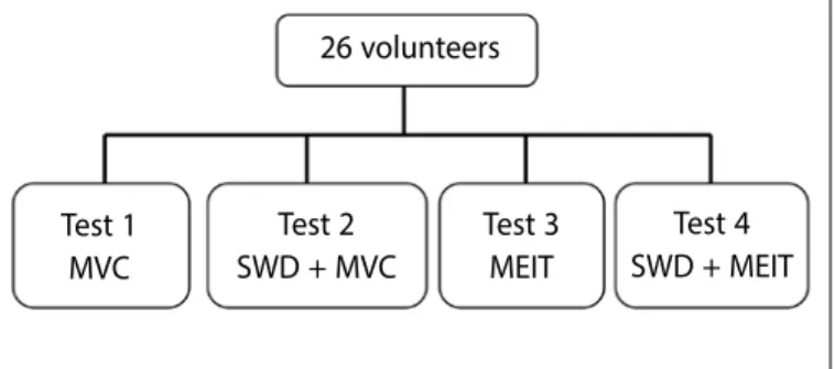
All individuals were analyzed in four moments, randomly placed through a draw with an opaque and sealed envelope, on diffe-rent days of the week, respecting an interval of two to seven days between tests, always performed between 1 and 6 o’clock in the afternoon (figure 1). Moreover, in order to determine the voluntary and electrically-induced torque (MEIT), three measurements were performed, with the highest of the three found values being used for the analyses18,19.
Torque was determined with the volunteers positioned on the dynamometer’s chair, hips immobilized with a belt, at 85° of flexion, and non-dominant limb immobilized with a double cushion on the distal third tibia, 3 cm close to the ankle joint20. The trunk was also
stabilized with belts in order to avoid possible muscular compen-sations. The dynamometer was programed at angle velocity of 0° per second (isometric mode), and the knee joint of the dominant leg was positioned at 60º of flexion, and the dynamometer’s arm rest was placed on the distal region of the volunteer’s leg, which allowed a complete dorsiflexion arch12,21-23.
During the tests 1 and 2, the volunteers were verbally encouraged
Figure 1. Chart of the groups and their respective procedures.
by the examiner to perform the highest strength as possible. Prior to tests 3 and 4, five values proof moments were performed, in which amplitude of electrical current was gradually increased until the maximum the individual could bear. Thus, the highest value the individual could bear was used in all the measurements which applied the neuromuscular electrical stimulation, when the volunteer remained still and with the evaluated limb relaxed for the measurements.
The percentage of the maximal voluntary contraction (MVC) obtained in test 1 was used for normalization of the data obtained in tests 3 and 4. In all tests, before the measurement and in the intervals between measurements, a three-minute time was ne-cessary for the individuals’ positioning. This time was nene-cessary so as to avoid fatigue between repetitions. In the intervals between electrically-induced contractions, the visual analog scale (VAS) was shown to the volunteers so that they could identify the discomfort generated by the electrical current. The VAS was a 10-centimeter line where the term “no discomfort” was written on the left extremity and the term “maximal discomfort possible” on the right extremity. Three measurements were analyzed in each test which consisted of three repetitions each and used for analysis the discomfort index presented during the production of the highest MEIT observed between the three repetitions.
Test 1 – Maximal voluntary contraction (MVC)
Prior to the beginning of the test, the volunteers were told that whenever asked, they should perform the highest strength as pos-sible to extend knee, enabling hence that torque of the quadriceps femoris muscle could be determined through MVC. This procedure was performed three times, with nine seconds of contraction each, with three-minute interval between measurements, in which the individuals remained relaxed. The volunteers were verbally encou-raged by the examiner during the tests’ performance 24.
Test 2 – Short wave diathermy (SWD) + maximal voluntary con-traction (MVC)
The volunteers were submitted to short wave diathermy application in continuous mode before the MVC evaluation, through coplanar technique during a period of 20 minutes. Capacitive electrodes were used and one of them was placed 3 cm below the antero superior iliac spine and the other 3 cm above the patella base. The volunteers were told to inform the
26 volunteers
Test 1 MVC
Test 2 SWD + MVC
Test 4 SWD + MEIT Test 3
MEIT
researcher when the thermal sensation generated by the short wave device was moderate heat (dose III), being this dose kept during the 20 minutes of application.
After this procedure, the individuals were positioned on the isokinetic dynamometer and told that when command was given, they should perform the highest strength as possible so that torque of the quadriceps femoris muscle could be determined (MVC). Three contractions with nine seconds each and interval of three minutes between them were performed. During the tests, the volunteers were verbally encouraged by the examiner.
Test 3 – Maximal electrically-induced torque (MEIT)
Before the test began, the individuals were positioned on the isokinetic dynamometer and they were asked to maintain the evalu-ated limb relaxed in order to avoid voluntary contractions during the electrical stimulation until the three minutes of pre-test inter-val were completed. The torque inter-values obtained were normalized through the percentage of the maximal voluntary contraction ob-tained in test 1. MEIT was generated with the Endophasys device (KLD® Biosistemas Equipamentos Eletrônicos Ltda.) which emitted
rectangular alternating current with carrier frequency of 2,500 Hz, modulated in bursts of 50 Hz and duty cycle of 20%. The self-ad-hesive electrodes (7.5 x 13 cm) (Axelgaard®, Valutrode) were placed
on the motor point of the oblique vastus medialis muscle and on the motor point of the femoral nerve. These points were localized with the use of a universal current generator (Nemesys941P). Each electrically-induced contraction had duration of nine seconds, with three minutes of interval, being the amplitude (intensity) increased until the previously determined value.
Test 4 – Short wave diathermy (SWD) + maximal electrically-induced torque (MEIT)
The individuals were submitted to the short wave diathermy application before the test, during 20-minute period, with electro-des being placed according to information from test 2. Following the SWD application, the individuals were placed on the isokinetic dynamometer and told to maintain the evaluated limb relaxed so as to avoid voluntary contractions during the electric stimulation. The electrodes of the electric stimulator were positioned on the motor point of the vastus medialis muscle and on the motor point of the femoral nerve, as previously described in test 3. Each electrically--induced contraction had duration of nine seconds, with three mi-nutes of interval, being the current amplitude increased until the previously determined value.
Statistical analysis
All tests were performed considering bilateral hypotheses and adopting a α = 0.05. Descriptive analysis was initially used to evaluate frequency, mean and standard deviation when data presented normal distribution verified through the Shapiro-Wilk test, median and inter-quartile interval (IQ) for data with no normal distribution. Student’s t
test was used for the comparison of the torque values means during the MVC and NMES when the groups presented normal distribution, and the Wilcoxon test for the groups which did not present data nor-mal distribution. Discomfort analysis was assessed with the Wilcoxon test. Data was analyzed with the statistical package SPSS, version 12.
RESULTS
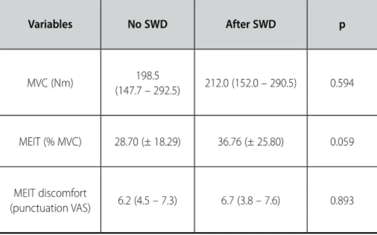
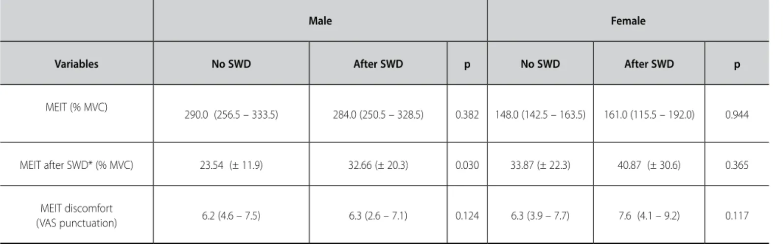
No statistically differences were found in any of the assessed variables (MVC, MEIT, (% of MVC) and sensory discomfort) compared with previous heat application or absence of it (table 1). However, when the subjects were stratified by gender, higher discomfort was observed in the female subjects during MEIT after application of SWD when compared with the male subjects (p = 0.044) (table 2). Increase of MEIT was only observed in male individuals after heat application (p = 0.030) (table 3).
Table 1. Comparison of the MVC, MEIT and sensory discomfort among the individuals who did not undergo application of SWD before the evaluations and after SWD application.
Variables No SWD After SWD p
MVC (Nm) 198.5
(147.7 – 292.5) 212.0 (152.0 – 290.5) 0.594
MEIT (% MVC) 28.70 (± 18.29) 36.76 (± 25.80) 0.059
MEIT discomfort
(punctuation VAS) 6.2 (4.5 – 7.3) 6.7 (3.8 – 7.6) 0.893
Data which did not present normal distribution are expressed in median and interquartile interval. Data which presented normal distribution are expressed in mean and standard deviation values. Subtitle: MVC – maximal isometric voluntary contraction; MEIT – maximal electrically-induced torque.
Table 2. MEIT comparison and sensory discomfort among the male and female in-dividuals who did not perform SWD application before the evaluations and after SWD application.
Variables Male Female p
MEIT (% of MVC) 23.54 (± 11.93) 33.87 (± 22.29) 0.154
MEIT after SWD
(% of MVC) 32.66 (± 20.34) 40.87 (± 30.59) 0.428
MEIT discomfort
(VAS punctuation) 6.2 (4.6 – 7.4) 6.3 (3.9 – 7.7) 0.687
MEIT discomfort after SWD
(VAS punctuation)* 6.3 (2.6 – 7.0) 7.6 (4.1 – 9.2) 0.044
Data which did not present normal distribution are expressed in median and interquartile interval. Data which presented normal distribution are expressed in median and standard deviation values. Subtitles: MVC – maximal voluntary contraction; MEIT– maximal electrically-induced torque. * p < 0.05.x
DISCUSSION
male individuals, a fact which was not observed in the women. Moreover, the women presented increase in sensory discomfort during MEIT production after the heat application.
Torque did not change during maximal voluntary contraction aassociated with heat produced by SWD. Such fact can be explained due to the fact there is no need of greater blood, nutrients, ATP or oxygen contribution, since it is a voluntary phasic muscular con-traction of high intensity and short duration, in which the proposed interval is sufficient to totally recover the energetic supplies24. It is
known that heat decreases the firing rate of the type II afferent fibers of the muscle spindle and increases the firing rate of the lb fibers of the Golgi tendon organs, leading to reduction of firing rate of the alpha and gamma motoneurons, decreasing hence the con-tractile activity7,8,25. These alterations could theoretically reduce the
voluntary torque; however, the remaining heat physiological effects, such as metabolism and velocity of nervous conduction increase, should be considered as a whole. The results of the present study corroborate previous research which, despite using different heating methods11,18, have not found statistically significant differences in
the strength voluntarily produced with previous heat application, being possible to observe that the muscular attitude did not suffer great influence of the tissue heating. Only one study using the same heat modality applied in the present study (SWD) was found in the literature4. In this investigation, decrease of voluntary torque was
observed immediately after the heat application. After 50 minutes, the strength levels reached baseline values and after two hours, they reached values higher than the ones initially measured; that is to say, before the heat application. A possible methodological flaw was the fact that the authors had performed all the strength measurements on the same day. In that case, the onset of fatigue or neuromuscular facilitation could have interfered in the results.
Increase of the neuromuscular system activation threshold due to the increase of blood flow in the adipose tissue may explain the higher discomfort reported by women during the MEIT after heat application when compared with men26-28. The discomfort
index among men and women during MEIT with no previous heat application was similar, which is in agreement with previous studies which verified higher discomfort in the female sex when the electrical stimulation was performed at the motor threshold;
however, discomfort between genders was similar when the stimulation was performed at the supramotor threshold29.
When the data are analyzed as a group, no alteration in the MEIT with the application was observed. Nevertheless, when the indivi-duals were stratified by sex, MEIT increase after SWD application was observed in the male individuals, a fact which was not observed in the female group. MEIT increase in the male individuals after heat application may be related to the increase of local metabolism and increase in velocity of nervous conduction. A hypothesis for the MEIT absence of alteration after SWD application in women may be attributed to the fact that men are considered more electrically excitable, exactly for presenting higher quantity of muscular mass and lower quantity of adipose tissue in the gluteofemoral region in comparison with women30. We believe that SWD use may have
generated higher thermal load in the adipose tissue compared with the muscle tissue, especially in women 8. This higher fat heating
may have led to increase in blood flow and electrical conduciveness in that tissue, causing the electrical current to concentrate even more in that region and increasing hence the activation threshold of the neuromuscular system28.
The main limitation presented in this study was the lack of measurement of the body composition of the participants. Further studies which investigate the association between body compo-sition and capacity of generating electrically-induced torque after heat application should be carried out. Moreover, studies which investigate whether or not MEIT increase after heat application will generate clinically significant strength increase in these individuals become necessary.
CONCLUSION
Short-wave diathermy did not influence on the torque gene-rated by voluntary contraction. However, previous application of deep heat produced increase in the electrically-induced torque in male individuals. Additionally, the discomfort produced during electrical stimulation was higher in female individuals after heat application.
All authors have declared there is not any potential conflict of interests concerning this article.
Table 3. Comparison of the MVC, MEIT and sensory discomfort among individuals from the same sex who did not had SWD application before the evaluations and after SWD application.
Male Female
Variables No SWD After SWD p No SWD After SWD p
MEIT (% MVC) 290.0 (256.5 – 333.5) 284.0 (250.5 – 328.5) 0.382 148.0 (142.5 – 163.5) 161.0 (115.5 – 192.0) 0.944
MEIT after SWD* (% MVC) 23.54 (± 11.9) 32.66 (± 20.3) 0.030 33.87 (± 22.3) 40.87 (± 30.6) 0.365
MEIT discomfort
(VAS punctuation) 6.2 (4.6 – 7.5) 6.3 (2.6 – 7.1) 0.124 6.3 (3.9 – 7.7) 7.6 (4.1 – 9.2) 0.117
REFERENCES
1. Abramson DI, Chu LS, Tuck S, Lee SW, Richardson G, Levin M. Effect of tissue temperatures and blood flow on motor nerve conduction velocity. JAMA 1966;198:1082-8.
2. Currier DP, Nelson RM. Changes in motor conduction velocity induced by exercise and diathermy. Phys Ther 1969;49:146-52.
3. Edwards RH, Harris RC, Hultman E, Kaijser L, Koh D, Nordesjo L. Effect of temperature on muscle energy metabolism and endurance during successive isometric contractions, sustained to fatigue, of the quadriceps muscle in man. Physiol 1972;220:335-52.
4. Chastain PB. The effect of deep heat on isometric strength. Phys Ther 1978;58:543-6.
5. Barnes S, Larson MR. Effects of localized hyper and hypothermia on maximal isometric grip strength. Am J Phys Med 1985;64:305-14.
6. Solomon J, Shebshacvich V, Adler R, Vulfsons S, Rosenbach A, Eisenberg E. The effects of TENS, heat, and cold on the pain thresholds induced by mechanical pressure in healthy volunteers. Neuromo-dulation 2003;6:102-7.
7. Mense S. Effects of temperature on the discharges of muscle spindles and tendon organs. Pflugers Arch 1978;374:159-66.
8. Cameron MH. Diathermy. In: Cameron HM. Physical agents in rehabilitation – from research to practice. 3rd ed. St. Louis: Saunders Elsevier, 2009;385-404.
9. Wickstrom R, Polk C. Effect of whirlpool on the strength and endurance of the quadriceps muscle in trained mal adolescents. Am J Phys Med 1961;40:91-5.
10. Sargeant AJ. Effect of muscle temperature on leg extension force and short-term power output in humans. Eur J Appl Physiol Occup Physiol1987;56:693-8.
11. Davies CT, Young K. Effect of temperature on the contractile properties and muscle power of triceps surae in humans. J Appl Physiol 1983;55:191-5.
12. Lyons LC, Robb BJ, Irrgang JJ, Fitzgerald KG. Differences in quadriceps femoris muscle torque when using a clinical electrical stimulator versus a portable electrical stimulator. Phys Ther 2005;85:44-51. 13. Bax L, Staes F, Verhagen A. Does neuromuscular electrical stimulation strengthen quadriceps femoris.
Sports Med 2005;35:191-212.
14. Ward RA, Robertson JV, Ioannou H. The effect of duty cycle and frequency on muscle torque production using kilohertz frequency range alternating current. Med Eng Phys 2004;26:569-79.
15. Ward RA, Shkuratova N. Russian electrical stimulation: The early experiments. Phys Ther 2002;82:1019-30. 16. Bellew JW, Beiswanger Z, Freeman E, Gaerte C, Trafton J. Interferential and burst-modulated biphasic
pulsed currents yield greater muscular force than Russian. Physiother Theory Pract 2011 Dec 2. [Epub ahead of print].
17. Cetin N, Aytar A, Atalay A, Akman MN.Comparing hot pack, short-wave diathermy, ultrasound, and TENS on isokinetic strength, pain, and functional status of women with osteoarthritic knees: a single--blind, randomized, controlled trial. Am J Phys Med Rehabil 2008;87:443-51.
18. Thornley LJ, Maxwell NS, Cheung SS. Local tissue temperature effects on peak torque and muscular endurance during isometric knee extension. Eur J Appl Physiol 2003;90:588-94.
19. Stewart D, Macaluso A, Vito GD. The effect of an active warm-up on surface EMG and muscle perfor-mance in healthy humans. Eur J Appl Physiol 2003;89:509-13.
20. Mendler HM. Effect of stabilization on maximum isometric knee extensor force. Phys Ther 1976;47:375-9. 21. Snyder-Mackler L, Garrett M, Roberts M. A comparison of torque generating capabilities of three
different electrical stimulating currents. J Orthop Sports Phys Ther 1989;10:297-302.
22. Brasileiro JS, Castro CES, Parizotto NA, Sandoval MC. Estúdio Comparativo entre la capacidad de ge-neración de torque y la incomodidad sensorial producidos por dos formas de estimulación electrica neuromuscular em sujetos sanos. Rev Iberoam Fisioter Kinesiol 2000;3:56-65.
23. McLoda TA, Carmack JA. Optimal burst duration during a facilitated quadriceps femoris contraction. J Athl Train 2000;35:145-50.
24. Billeter R, Hoppeler H. Muscular basis of strength. In: Komi P. Strength and power in sport. Oxford: Blackwell Scientific Publications, 1996;39-63.
25. Morrison SA, Sleivert GG, Cheung SS. Passive hyperthermia reduces voluntary activation and isometric force production. Eur J Appl Physiol 2004;91:729-36.
26. Morrissey MC. Electromyostimulation from a clinical perspective. A review. Sports Med 1988;6:29-41.
27. Lee SJ, Janssen I, Heymsfield SB, Ross R. Relation between whole-body and regional measures of human skeletal muscle. Am J Clin Nutr 2004;80:1215-21.
28. Petrofsky JS, Suh HJ, Gunda S, Prowse M, Batt J. Interrelationships between body fat and skin blood flow and the current required for electrical stimulation of human muscle. Med Eng Phys 2008;30:931-6. 29. Maffiuletti NA, Herrero AJ, Jubeau M, Impellizzeri FM, Bizzini M. Differences in electrical stimulation
thresholds between men and women. Ann Neurol 2008;63:507-12.