DOI: http://dx.doi.org/10.1590/2446-4740.03616
*e-mail: [email protected]
Received: 10 May 2016 / Accepted: 01 July 2016
Macrophages adhesion rate on Ti-6Al-4V substrates: polishing and
DLC coating effects
Everton Diniz dos Santos*, Gerson Luqueta, Ramu Rajasekaran, Thaisa Baesso dos Santos, Anelise Cristina Osorio Cesar Doria, Polyana Alves Radi, Rodrigo Savio Pessoa, Lucia Vieira, Homero Santiago Maciel
Abstract Introduction: Various works have shown that diamond-like carbon (DLC) coatings are able to improve the cells adhesion on prosthesis material and also cause protection against the physical wear. On the other hand there are reports about the effect of substrate polishing, in evidence of that roughness can enhance cell adhesion. In order to compare and quantify the joint effects of both factors, i.e, polishing and DLC coating, a commonly prosthesis material, the Ti-6Al-4V alloy, was used as raw material for substrates in our studies of macrophage cell adhesion rate on rough and polished samples, coated and uncoated with DLC. Methods: The ilms were produced by PECVD technique on Ti-6Al-4V substrates and characterized by optical proilometry, scanning electron microscopy and Raman spectroscopy. The amount of cells was measured by particle analysis in IMAGE J software. Cytotoxicity tests were also carried out to infer the biocompatibility of the samples. Results: The results showed that higher the surface roughness of the alloy, higher are the cells ixing on the samples surface, moreover group of samples with DLC favored the cell adhesion more than their respective uncoated groups. The cytotoxity tests conirmed that all samples were biocompatible independently of being polished or coated with DLC. Conclusion: From the observed results, it was found that the rougher substrate coated with DLC showed a higher cell adhesion than the polished samples, either coated or uncoated with the ilm. It is concluded that the roughness of the Ti-6Al-4V alloy and the DLC coating act complementary to enhance cell adhesion.
Keywords: DLC, RMS roughness, Cell adhesion, Ti-6Al-4V, Macrophages J774, Biomedical prosthesis.
Introduction
According to the survey of World Health Organization (WHO) in 2014, the average life time expectancy has increased substantially in the last years. Consequently, the number of elders in the world has increased (World…, 2014). According to the report of the Geography and Statistics Brazilian Institute (IBGE), the proportion of elder people in the population was estimated to be 8.17% in 2015 and 6.22% in 2006 (Instituto…, 2016), an increase of about 30% of elders in the population of Brazil in the last 10 years.
Many health problems are likely to appear during the elder life stage, often requiring a considerable amount of money for heath treatments. Osteoarthritis is one of the most common diseases that cause joint pain and weakness during physical activity, especially with elder people (Kawtar et al., 2013). This disease is characterized by the degeneration of articular cartilage, overgrowth of bone cells and remodeling of the bone (Benz et al., 2015). Medicines are widely used for symptom relief in the osteoarthritis at the early stage, but they do not work very well in complicate
cases. Major surgical interventions, such as total joint replacement, are the only effective options for people with severe osteoarthritis (Dieppe, 2005).
The medical expenses associated with surgery for
placement of orthopedic implants grew signiicantly.
of the prosthesis are harmful to the body and induce
inlammation process (Grill, 2003; Hinüber et al., 2010). Many studies have shown that Co–, Cr–, and Mo– ions are considered toxic, carcinogenic and
also able to promote inlammation (Andrews et al., 2011; Hinüber et al., 2010; Jakobsen et al., 2007). Aluminum is also considered to be a risk factor for the pathogenesis of Alzheimer’s disease (McLachlan et al., 1990; Rusina et al., 2011). However, Ti-6Al-4V has an advantage over Co-28Cr-26Mo because of the presence of Vanadium. Vanadium may reduce memory
deicits in individuals exposed continuously for a long
time (Imtiaz et al., 2015). Due to these advantages, Ti-6Al-4V is considered better than Co-28Cr-6Mo in prosthesis production.
Different materials have been used as coatings on implants in order to improve cell adhesion and to
prevent their wear, such as: ultra-ine hydroxyapatite
powders (Kaya, 2008), carbon nanotubes (Kaya, 2008), Ag–ZrCN ilms (Ferreri et al., 2015) and
DLC ilms (Casiraghi et al., 2005). All these coatings have shown good results, however only the DLC
thin ilm has been considered able to promote a
reduction on metal friction at the same time that increases the wear resistance, the hardness, and the cell adhesion rate (Grill et al., 1988; Grill, 2003; Ianno et al., 1995; Jelinek et al., 2016; Khatir et al., 2015; Marciano et al., 2009). In addition, DLC ilms may have antibactericidal effect (Marciano et al., 2009). All these properties make DLC suitable for a number of applications ranging from the coating of stents, heart valves, and prostheses in biomedical industry (Hauert and Müller, 2003). By other hand some researchers are also trying to improve the cell adhesion by only promoting changes in the material roughness, for example by surface etching or polishing, without the use of coatings on the prosthesis alloy. The results are so promising as are the works based on nanostructured coatings (Degasne et al., 1999; Musilkova et al., 2015; Safiullin et al., 2015; Varoni et al., 2015; Vrekhem et al., 2015).
Based on these, our study aimed to evaluate the effectiveness of macrophage cell adhesion on prosthesis samples based on Ti-6Al-4V alloy, correlating the results to the surface roughness of the sample and discriminating the effect of polishing and DLC coating.
Methods
Ti-6Al-V alloy was taken as the experimental material from which 16 samples were prepared as discs of 1.7 cm diameter and 3 mm thick, all them with a standard surface roughness of 300 nm. These sixteen discs were grouped under four categories of samples, namely: rougher alloy (RA), polished alloy (PA), rougher alloy with DLC coating (RA-DLC) and polished alloy with DLC coating (PA-DLC), with four discs composing each group category, such that the statistical parameters of the measurements could be evaluated.
For the samples of groups PA and PA-DLC the titanium alloy was polished using commercially available sand paper with granulation size ranging from 180 to 2000, followed by felt polishing using diamond paste of two microns. Prior to DLC deposition and cell culture, the samples were immersed in a beaker with acetone and subjected to ultrasound for 10 min. Finally, they were placed inside the oven at 40 °C for the evaporation of acetone.
DLC coating process
This section explains the DLC coating process. This process comprises three steps which includes (I) cleaning of the substrate by argon plasma, (II) deposition of silicon interlayer using hexa-methyl-disiloxane (HDMSO4) diluted in argon plasma to improve DLC adhesion on Ti-6Al-4V (Grill et al., 1988; Ianno et al., 1995) and, (III) deposition of DLC coatings by methane plasma. Table 1 shows the experimental parameters
maintained at each step of the DLC ilm deposition to produce a ilm of ~0.4 μm thickness. The coating
were produced by plasma enhanced chemical vapor deposition (PECVD) in a commercial reactor
Table 1. Experimental parameters for the DLC coating.
Parameters Substrate cleaning Interlayer deposition DLC deposition
Time (min) 10 30 120
Power (W) 200 200 200
Pressure (Torr) 3×10⁻² 3×10⁻² 3×10⁻²
Pulse (kHz) 100 130 130
Reverse pulse (μs) 0.8 3.2 3.2
Voltage (V) 650 600 600
Current (A) 0.6 0.5 0.5
Temperature range (°C) 18 to 22 300 18 to 70
manufactured by Nanomaster Inc. model NPE-4000 using a pulsed DC power supply.
Film characterization
Different techniques were used for surface characterization of the samples. High-resolution images of the samples surface, coated and uncoated with DLC, were obtained by scanning electron microscopy (SEM, FEI - Inspect F50), operated in secondary electron
mode. The ilm thickness as well as the surface
roughness of the Ti-6Al-4V samples was obtained
by optical proiler Veeco model Wiko - NT9000 with 20× magniication objective. The structure and degree of disorder of the DLC ilms were obtained by Raman
spectroscopy (Renishaw 2000) with an Ar ion laser
(λ = 514 nm), operated with backscattering geometry
at room temperature. The diameter of the laser spot
was maintained as 2.5 μm and the power at 0.6 mW on the surface of the ilm (Marciano et al., 2009).
Cells culture and cytotoxicity test
J774 macrophages from murine cell line (ATCC) were grown in culture bottles, maintained in RPMI (Roswell Park Memorial Institute) medium plus 10% fetal bovine serum and antibiotics penicillin/streptomycin. The culture bottles were kept half closed at 37 °C in
humidiied atmosphere with 5% CO2. The samples were placed in a Corning plate (24 wells) (NY- USA) and 5 ml of the medium with the cells was dripped.
The system remained untouched until the seventh day when it was found the formation of a monolayer of macrophages. At the end of the period, the medium was removed and images were taken by SEM.
To check the cytotoxicity of the coated and un-coated samples, J774 macrophages were used to make the neutral red dye uptake assay. The macrophages at concentration of 3.0 × 10⁵ cells/ml were seeded in Petri dishes (15 × 60 mm) at a total volume of 5 ml of agar-overlay. They were incubated for 48 h at 37 °C
in a humidiied atmosphere with 5% CO2. A sample of each experimental group was placed on the agar
before their complete solidiication. Two glass slides
were taken as controls and under one of them 100 µl of phenol was dropped, to act as a positive control, the other acting as a negative control. The plates were then examined macroscopically and microscopically to check the cytotoxicity and cellular integrity around the sample (Allen et al., 1994; 2001).
Image processing and statistical analysis
The amount of adhered cells was determined using ImageJ software and the mean and standard deviation (S.D.) values were calculated. Differences between the number of cells adhered on each group
were individually analyzed by unpaired t-student tests
in the Minitab 17.3 software. Signiicant interactions
between the roughness and cells adhesion were also
evaluated within a 95% conidence interval.
Results
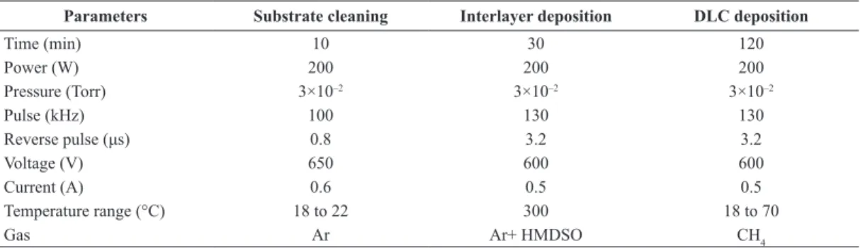
Figure 1 shows the Raman spectrum of the DLC
ilms (blue line). The red and black spectra represent
the deconvolution of the original spectrum. From
this igure, it is observed that the D band and the G
band were centered around 1350 cm–1 and 1580 cm–1
respectively. These bands are typical of DLC ilms.
The ratio of the intensities of the D and G bands (ID/IG)
was calculated to be 0.2, indicating that the ilm is a
hydrogenated DLC.
Figure 2 depicts the surface images of the four
groups of samples, obtained from proilometry
analysis, from which the root-mean-square (RMS) surface roughness was inferred. It is quite remarkable that the four sample groups ended up with a large variation of roughness, ranging from 15 nm to 997 nm, depending on the raw material polishing and DLC coating. DLC alone provides a substantial increase, by a factor about 3.5, of the roughness.
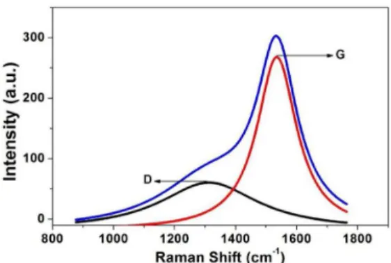
Figure 3 displays results of the cytotoxic studies to verify the cells biocompatibility with respect to RMS roughness of the Ti-6Al-4V samples covered by DLC. It is observed that the red-neutral dye was phagocytosed by the macrophages and remained inside the cells indicating that the DLC did not cause disruption of the J774 macrophages cell membrane. From the cytotoxicity studies using a biocompatible glass slides, it is observed that the positive control makes a roseate halo which indicates the cytotoxicity and the negative control did not make any cytotoxicity
Figure 1. Raman spectrum of the DLC ilms (Blue line) from coated
effect (Xuan et al., 1990) (Figure 3B). Comparing Figure 3A, B, even after 24 hours of contact with
ilms, it is clear that the dye was not released, thereby
indicating no cytotoxicity with regard to all samples. In addition to the cytotoxic study, the shape of cell grown under the sample discs was observed by optical microscopy (Figure 4A), and by SEM (Figure 4B). By this exam it was observed that the cells did not
undergo any alteration or structural changes that could affect their activity.
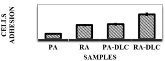
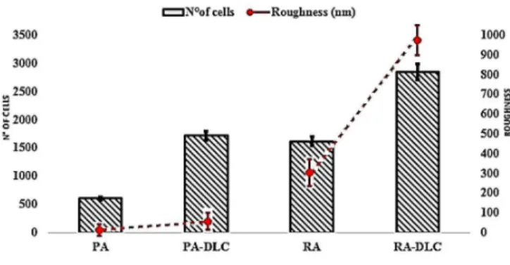
Figure 5 displays the amount of adherent cells in the four groups of samples, as measured by Image J
software. It can be inferred from the igure that cell
adhesion is higher in the rougher surface (RA) than on polished one (PA) and DLC coating further enhances the cell adhesion by more than 75%.
Figure 2. Sample groups images obtained on optical proiler with 20× magniication.
Discussion
The improvements on mechanical, chemical and biocompatibility characteristics of prosthesis are currently a focus of great interest in the biomedical sector, attracting growing number of researchers to work in this area of science and technology. Good results have been reported by the use of coating with
nanostructured thin ilms such as DLC and by techniques
that promote changes on the surface morphology and topography of raw materials used in prosthesis. In this context, the present study deals with a careful evaluation of cell adhesion and biocompatibility respective to culture of macrophage cells on prosthesis based on Ti-6Al-4V alloy, considering the effect of surface polishing and DLC coating of the alloy sample.
Figure 1 shows the Raman spectrum of DLC ilm coated on Ti-6Al-4V samples. The two observed
peaks, D and G at ~1380 cm–1 and ~1560 cm-1,
respectively, conirm the deposition of a-C:H DLC ilm (Casiraghi et al., 2005). The D and G peaks are both sensitive to the sp2 content and the D peak is due to the bonds of carbon in rings while the G peak is due to the bonds of carbons in both rings chains
(Ferrari and Robertson, 2000; 2004).The ratio of the intensity at peaks D and G, ID/IG, was used to determine the disorder degree and it was found here
to be approximately 0.2. According to Casiraghi et al. (2005), a DLC ilm with this ID/IG ratio presents a
content of ~35% of hydrogen and ~20% of sp3 bonds between the C atoms.
From the surface topography analysis (Figure 2), DLC deposition makes the difference between the heights of peaks and valleys to increase substantially, i.e, approximately 111% increase between RA and RA-DLC and approximately 762% between PA and PA-DLC. Concomitantly, the surface roughness also increases, with coated groups representing a higher RMS roughness when compared to the groups that are not coated. DLC deposition makes the roughness to increase by 232% and 260% for the rougher and polished groups respectively. This analysis suggest that one effect of DLC coating by PECVD technique
is to promote, approximately, a 3.5 fold ampliication
of the sample roughness.
There are results reported by Prado et al. (2012), Hinüber et al. (2010), and Corbella et al. (2005) who also observed that the DLC coating increased the RMS surface roughness on different prosthesis based on polymeric as metallic materials. Some hypotheses were proposed to explain this effect, for example, as due to the changes in the plasma density during the HMDSO injection which favors DLC adhesion (Bonetti et al., 2006). Corbella et al. (2005) proposed that the DLC roughness is directly related to the bias voltage causing the effect of “etching” on surface
of polymeric substrate by luorine species in the
plasma during DLC growth (Prado et al., 2012).There are also claims that roughness increases when the kinetic energy of the hydrogen ions from the plasma
is insuficient to remove certain roughness of the
material surface (Corbella et al., 2005). Overall there
are only unconirmed suggestions for the mechanisms
related to the increase of surface roughness by DLC coating. Although the above hypotheses should not be discarded, the most probable explanations for
Figure 4. Optical microscopy image (A) and the SEM image (B) of cells that were under the Ti-6Al-4V discs.
this effect, observed in the present work, are: as the metallic substrate is exposed to the plasma in the PECVD reactor, its surface becomes surrounded by a plasma sheath where and averaged electrostatic
ield is produced, high enough to accelerate plasma
ions towards the sample surface so enhancing the
deposition rate of the ilm material. Moreover, the electric ield is stronger on sharper edges (a classical
problem of electrostatic), so the plasma sheath electric
ield is stronger on the edges of hills than on bottom
of the valleys. The dimensions of hills and valleys
are the topographic features that better deine the
dimensions of the micro and nano-craters on the
sample surface, consequently deining the surface roughness. Accordingly, the ilm grows faster on the
top (hills) than on the bottom (valleys) because ion
lux follows direction of electric ield; moreover the electric ield promotes ionization, enhancing the plasma density where the ield is stronger. This explain the
higher deposition rate on top than on bottom of the micro-craters of the sample, increasing their depth, consequently the surface roughness, as evidenced by the topography analysis of the samples.
A comparison of the amount of cells adhered on the samples reveals that the four groups can be ordered as displayed in Figure 5, so as to make evident the effect of polishing of the substrate raw material and of the DLC coating on the number of adhered cells. Polishing of the Ti-6Al-4V is detrimental while DLC coating is favorable to cell adhesion. The DLC makes the adhesion of cells to increase by 184% and 76%, on polished group and on rougher rough group, respectively. In Figure 6 a plot of the number of cells versus roughness is presented, distinguishing the behavior of cell adhesion on coated and uncoated
groups. It can be inferred from the igure that higher
the surface roughness of the group sample, higher is the cell adhesion rate, moreover, as discussed before, the DLC substantially increases the roughness of the
substrate surface, by a factor of ~3.5.
Although the indings of this study show a good statistical signiicance at a conidence interval of
95%, they do not necessarily cover the other types of cells, such as osteoblasts, monocytes, thrombocytes and other implantable materials such as polymeric materials, nickel and chromium alloys. The works of Hinüber et al. (2010) and Kornu et al. (1996), used very similar methodologies and concluded that DLC
ilms do not cause changes in osteoblast adhesion
rate to Ti-6Al-4V and Co-28Cr-6Mo substrates. Linder et al. (2002) demonstrated that the adhesion of monocytes on DLC deposited on glass slide, a material with low surface roughness, was slightly higher than on the control. In the case of thrombocytes, Tran et al. (1999), Scheerder et al. (2000) and Alanazi et al. (2000) reported that the adhesion of
these cells to polytetraluoroethylene is reduced by
DLC coating, however Khatir et al. (2015) showed that the DLC grown in an atmosphere of 30% to 40% nitrogen may improve the adhesion of platelets to
polytetraluoroethylene. The later work is especially
interesting because, beyond demonstrating that DLC
inluences cell adhesion, they also showed that this
coating is able to both increase or decrease this effect. Overall, the results of the present studies indicate that the surface roughness is a prominent factor for correlating properties of the Ti-6Al-4V sample groups with their respective macrophage adhesion rates. Figure 7 illustrates this correlation and highlights that, as far as the adhesion rate is concerned, the best substrate is that with rougher raw material surface and coated with DLC.
The beneits that DLC and high roughness offer
to macrophages J774 adhesion on Ti-6Al-4V sample cannot be assigned to others material, and cell types, without further studies. Nevertheless, the present results
corroborate with the sum of indings by Bendavid et al. (2007) and Lowenberg et al. (1991) who used silicon
and titanium substrates and showed that DLC ilms
increases the roughness of the substrate and favors the cell adhesion.
Figure 3 shows the cytotoxicity analysis of the four samples used in this study. The positive control indicated by (+) symbol, shows the behavior of a toxic sample that cause change in the environment color of the agar medium and the negative control (–) shows the behavior of non toxic components that causes no alteration in the culture medium. The same observations were made from our samples and shows that the DLC coated and uncoated Ti-6Al-4V
samples exhibit no toxic or inlammatory response
to J774 macrophages. This is in good agreement with previously published results by Hinüber et al. (2010), Linder et al. (2002), Wachesk et al. (2013)
and Kornu et al. (1996). The analysis of the culture plates under inverted microscope shows no changes in the cell morphology and are in good agreement with the cytotoxicity results (Figure 4). The SEM analysis of the macrophages grown in area under the samples showed some clusters of macrophages and this effect assures the occurrence of mitosis. Because of this, it is possible to observe the long and slender cytoplasmic extensions of the cells in multiple directions, which indicates an excellent macrophages adhesion on the samples (Figure 4B). This shows that the Ti-6Al-4V alloy, covered or not covered by DLC, do not have cytotoxic effects in relation to J774 macrophages.
Finally, from the observed results it is evidenced that micro and nanostructures of the Ti-4Al-6V surface play a key role for the cell (macrophage) adhesion rate, the surface RMS roughness being a prominent characteristic of the surface topography to be considered in a search for correlation with the cell adhesion rate. This roughness can be tailored by the polishing of the raw material followed by deposition of DLC thin
ilm. Beyond increasing the surface roughness, the
DLC coating is favorable to the bio-integration of the prosthesis, also protects the implant against wear. A new explanation for the mechanism of roughness increase by DLC coating is presented. The numerical
results, obtained by quantiication of the effects of
polishing and DLC coating on cell adhesion rate, are relevant for the search of optimal condition to prepare biomedical implants based on Ti-4Al-6V.
Acknowledgements
We would like to thank the São Paulo Research Foundation (FAPESP) for funding the research carried out under the project PRONEX processes number 2011/50773-0 and 2013/05269-8.
References
Alanazi A, Nojiri C, Kido T, Noguchi T, Ohgoe Y, Matsuda T, Hirakuri K, Funakubo A, Yasuhiro F. Engineering analysis of diamond-like carbon coated polymeric materials for biomedical applications. Artificial Organs. 2000; 24(8):624-7. http://dx.doi.org/10.1046/j.1525-1594.2000.06576.x. PMid:10971249.
Allen M, Law F, Rushton N. The effects of diamond-like carbon coatings on macrophages, fibroblasts and osteoblast-like cells in vitro. Clinical Materials. 1994; 17(1):1-10. http:// dx.doi.org/10.1016/0267-6605(94)90041-8. PMid:10150171.
Allen M, Myer B, Rushton N. In vitro and in vivo investigations into the biocompatibility of diamond-like carbon (DLC) coatings for orthopedic applications. Journal of Biomedical Materials Research. 2001; 58(3):319-28. http://dx.doi.org/10.1002/1097-4636(2001)58:3<319::AID-JBM1024>3.0.CO;2-F. PMid:11319748.
Andrews RE, Shah KM, Wilkinson M, Gartland A. Effects of cobalt and chromium ions at clinically equivalent concentrations after metal-on-metal hip replacement on human osteoblasts and osteoclasts: Implications for skeletal health. Bone. 2011; 49(4):717-23. http://dx.doi.org/10.1016/j. bone.2011.06.007. PMid:21689801.
Bendavid A, Martin PJ, Comte C, Preston EW, Haqb AJ, Magdon FS. The mechanical and biocompatibility properties of DLC-Si films prepared by pulsed DC plasma activated chemical vapor deposition. Diamond and Related Materials. 2007; 16(8):1616-22. http://dx.doi.org/10.1016/j. diamond.2007.02.006.
Benz T, Aeschlimann A, Angst F. SalutogenetischeKonzeptebei der rehabilitation von arthrose. ZeitschriftfürRheumatologie. 2015; 74:597-602.
Bonetti LF, Capote G, Santos LV, Corat EJ, Trava-Airoldi VJ. Trava-AiroldiVJ. Adhesion studies of diamond-like carbon films deposited on Ti6Al4V substrate with a silicon interlayer. Thin Solid Films. 2006; 515(1):375-9. http:// dx.doi.org/10.1016/j.tsf.2005.12.154.
Brasil. Ministério da Saúde. Grupo de Trabalho Interinstitucional sobre órteses, próteses e materiais especiais (GTI-OPME): relatório final [internet]. Brasilia: MS; 2015. [cited 2016 Jan 25]. Available from: u.saude.gov.br/images/pdf/2015/ julho/07/Relatorio-Final-versao-final-6-7-2015.pdf
Casiraghi C, Ferrari C, Robertson J. Raman spectroscopy of hydrogenated. Physical Review B: Condensed Matter and Materials Physics. 2005; 72(8):1-14. http://dx.doi. org/10.1103/PhysRevB.72.085401.
Corbella C, Polo MC, Oncins G, Pascual E, Andújar JL, Bertran E. Time-resolved electrical measurements of a pulsed-dc methane discharge used in diamond-like carbon films. Thin Solid Films. 2005; 482(1-2):172-6. http://dx.doi. org/10.1016/j.tsf.2004.11.135.
Degasne I, Basle MF, Demais V, Hure G, Lesourd M, Grolleau B, Mercier L, Chappard D. Effects of roughness, fibronectin and vitronectin on attachment, spreading, and proliferation of human osteoblast-like cells (Saos-2) on titanium surfaces. Calcified Tissue International. 1999; 64(6):499-507. http://dx.doi.org/10.1007/s002239900640. PMid:10341022.
Dieppe P. Disease modification in osteoarthritis: are drugs the answer? Arthritis and Rheumatism. 2005; 52(7):1956-9. http://dx.doi.org/10.1002/art.21124. PMid:159863652(7):1956-9.
Ferrari A, Robertson J. Interpretation of Raman spectra of disordered and amorphous carbon. Physical Review B: Condensed Matter and Materials Physics. 2000; 61(20):14095-107. http://dx.doi.org/10.1103/PhysRevB.61.14095.
Ferrari AC, Robertson J. Raman spectroscopy of amorphous, nanostructured, diamond-like carbon, and nanodiamond. Philosophical Transactions of the Royal Society A. 2004; 362(1824):2477-512.
Ferreri I, Calderon SV, Galindo RE, Palacio C, Henriques M, Piedade AP, Carvalho S. Silver activation on thin films of Ag–ZrCN coatings for antimicrobial activity. Materials Science and Engineering C. 2015; 55:547-55. http://dx.doi. org/10.1016/j.msec.2015.05.071. PMid:26117788.
Grill A, Meyerson M, Patel V. Interface modifications for improving the adhesion of a-C:H films to metals. Journal of Materials Research. 1988; 3(02):214-7. http://dx.doi. org/10.1557/JMR.1988.0214.
Grill A. Diamond-like carbon coatings as biocompatible materials: an overview. Diamond and Related Materials. 2003; 12(2):166-70. http://dx.doi.org/10.1016/S0925-9635(03)00018-9.
Hauert R, Müller U. An overview on tailored tribological and biological behavior of diamond-like carbon. Diamond and Related Materials. 2003; 12(2):171-7. http://dx.doi. org/10.1016/S0925-9635(03)00019-0.
Hinüber C, Kleemann C, Friederichs RJ, Haubold L, Scheibe HJ, Schuelke T, Boehlert C, Baumann MJ. Biocompatibility and mechanical properties of diamond-like coatings on cobalt-chromium-molybdenum alloy and titanium-aluminum-vanadium biomedical alloys. Journal of Biomedical Materials Research. Part A. 2010; 95(2):388-400. http://dx.doi.org/10.1002/jbm.a.32851. PMid:20648536.
Ianno NJ, Dillon RO, Ali A, Ahmad A. Deposition of diamond-like carbon on a titanium biomedical alloy. Thin Solid Films. 1995; 270(1):275-8. http://dx.doi. org/10.1016/0040-6090(95)06710-8.
Imtiaz M, Rizwan MS, Xiong S, Li H, Ashraf M, Shahzad SM, Shahzad M, Rizwan M, Tu S. Vanadium, recent advancements and research prospects: A review. Environment International. 2015; 80:79-88. http://dx.doi.org/10.1016/j. envint.2015.03.018. PMid:25898154.
Instituto Brasileiro de Geografia e Estatística. [internet]. Rio de Janeiro: IBGE; 2016. [cited 2016 Jan 25]. Available from: www.ibge.gov.br/apps/populacao/projecao/
Jakobsen SS, Danscher G, Stoltenberg M, Larsen A, Bruun JM, Mygind T, Kemp K, Soballe K. Cobalt-chromium-molybdenum alloy causes metal accumulation and metallothionein up-regulation in rat liver and kidney. Basic & Clinical Pharmacology & Toxicology. 2007; 101(6):441-6. http://dx.doi.org/10.1111/j.1742-7843.2007.00137.x. PMid:17971067.
Jelinek M, Zemek J, Vandrovcová M, Bacakova L, Kocourek T, Remsa J, Pısarık P. Bonding and bio-properties of hybrid laser/magnetron Cr-enriched DLC layers. Materials Science and Engineering C. 2016; 58:1217-24. http://dx.doi. org/10.1016/j.msec.2015.09.006. PMid:26478424.
Kawtar N, Janani S, Rachidi W, Mkinsi O. Vascularite cérébrale au cours d’une cryoglobulinémie mixte. Revue Marocaine de Rhumatologie. 2013; 26:38-41.
Kaya C. Electrophoretic deposition of carbon nanotube-reinforced hydroxyapatite bioactive layers on Ti–6Al–4V alloys for biomedical applications. Ceramics International. 2008; 34(8):1843-7. http://dx.doi.org/10.1016/j.ceramint.2007.06.007.
Khatir S, Hirose A, Xiao C. Characterization of physical and biomedical properties of nitrogenated diamond-like carbon films coated on polytetrafluoroethylene substrates. Diamond and Related Materials. 2015; 58:205-13. http:// dx.doi.org/10.1016/j.diamond.2015.08.003.
Kornu R, Maloney WJ, Kelly MA, Smith RL. Osteoblast adhesion to orthopaedic implant alloys: effects of cell adhesion molecules and diamond-like carbon coating. Journal of Orthopaedic Research. 1996; 14(6):871-7. http://dx.doi. org/10.1002/jor.1100140605. PMid:8982128.
Linder S, Pinkowskic W, Aepfelbacher M. Adhesion, cytoskeletal architecture and activation status of primary human macrophages on a diamond-like carbon coated surface. Biomaterials. 2002; 23(3):767-73. http://dx.doi. org/10.1016/S0142-9612(01)00182-X. PMid:11771697.
Lowenberg B, Chernecky R, Shiga A, Davies JE. Mineralized matrix production by osteoblasts on solid titanium in vitro. Cells Materials. 1991; 1:177-87.
Marciano FR, Bonetti LF, Santos LV, Da-Silva NS, Corat EJ, Trava-Airoldi VJ. Antibacterial activity of DLC and Ag–DLC films produced by PECVD technique. Diamond and Related Materials. 2009; 18(5-8):1010-4. http://dx.doi. org/10.1016/j.diamond.2009.02.014.
Environmental Geochemistry and Health. 1990; 12(1-2):103-14. http://dx.doi.org/10.1007/BF01734059. PMid:24202576.
Musilkova J, Kotelnikov I, Novotna K, Pop-Georgievski O, Rypacek F, Bacakova L, Proks V. Cell adhesion and growth enabled by biomimetic oligopeptide modification of a polydopamine-poly (ethylene oxide) protein repulsive surface. Journal of Materials Science. Materials in Medicine. 2015; 26(11):253. http://dx.doi.org/10.1007/s10856-015-5583-3. PMid:26449443.
Prado GD, Terriza A, Ortiz-Pérez A, Molina-Manso D, Mahillo I, Yubero F, Puértolas JA, Manrubia-Cobo M, Barrena EG, Esteban J. DLC coatings for UHMWPE: relationship between bacterial adherence and surface properties. Journal of Biomedical Materials Research. Part A. 2012; 100(10):2813-20. http://dx.doi.org/10.1002/ jbm.a.34220. PMid:22623338.
Rusina R, Matěj R, Kašparová L, Kukal J, Urban P. Higher aluminum concentration in alzheimer’s disease after box-cox data transformation. Neurotoxicity Research. 2011; 20(4):329-33. http://dx.doi.org/10.1007/s12640-011-9246-y. PMid:21567285.
Safiullin R, Christenson W, Owaynat H, Yermolenko IS, Kadirov MK, Ros R, Ugarova TP. Fibrinogen matrix deposited on the surface of biomaterials acts as a natural anti-adhesive coating. Biomaterials. 2015; 67:151-9. http://dx.doi. org/10.1016/j.biomaterials.2015.07.007. PMid:26210181.
Scheerder DI, Szilard M, Yanming H, Ping XB, Verbeken E, Neerinck D, Demeyere E, Coppens W. Evaluation of the biocompatibility of two new diamond-like stent coatings (Dylyn) in a porcine coronary stent model. The Journal of Invasive Cardiology. 2000; 12(8):389-94. PMid:10953100.
Tran HS, Puc MM, Hewitt CW, Soll DB, Marra SW, Simonetti VA, Cilley JH, DelRossi AJ. Diamond-like carbon coating
and plasma or glow discharge treatment of mechanical heart valves. Journal of Investigative Surgery. 1999; 12(3):133-40. http://dx.doi.org/10.1080/089419399272520. PMid:10421514.
Varoni E, Canciani E, Palazzo B, Varasano V, Chevallier P, Petrizzi L, Dellavia C, Mantovani D, Rimondini L. Effect of Poly-L-Lysine Coating on Titanium Osseo integration: from characterization to in vivo studies. The Journal of Oral Implantology. 2015; 41:626-31. http://dx.doi.org/10.1563/ AAID-JOI-D-13-00036. PMid:24001103.
Vrekhem SV, Cools P, Declercq H, Tongel AV, Vercruysse C, Cornelissen M, De-Geyter N, Morent R. Application of atmospheric pressure plasma on polyethylene for increased prosthesis adhesion. Thin Solid Films. 2015; 596:256-63. http://dx.doi.org/10.1016/j.tsf.2015.08.055.
Wachesk CC, Pires CAF, Ramos BC, Trava-Airoldi VJ, Lobo AO, Pacheco-Soares C, Marciano FR, Da-Silva NS. Cell viability and adhesion on diamond-like carbon films containing titanium dioxide nanoparticles. Applied Surface Science. 2013; 266:176-81. http://dx.doi.org/10.1016/j. apsusc.2012.11.124.
World Health Organization. WHO library cataloguing-in-publication data [internet]. Geneva: WHO; 2014. [cited 2016 Jan 23]. Available from: acad-medic.s3.amazonaws.com/ wp-content/uploads/2014/05/WHO-healt-statistics-full.pdf
Xuan JL, Cheng HS, Miller RJ. Generation of submicrometer particles in dry sliding contact. Journal of Tribology. 1990; 112(4):684-91. http://dx.doi.org/10.1115/1.2920316.
Yingmin S, Satoshi K, Tohru S, Hiroshi N, Joji O. Nanostructured Ti6Al4V alloy fabricated using modified alkali-heat treatment: characterization and cell adhesion. Materials Science and Engineering C. 2016; 59:617-23. http:// dx.doi.org/10.1016/j.msec.2015.10.077. PMid:26652415.
Authors
Everton Diniz dos Santos1*, Gerson Luqueta1, Ramu Rajasekaran1, Thaisa Baesso dos Santos1, Anelise Cristina Osorio Cesar Doria1, Polyana Alves Radi1, Rodrigo Savio Pessoa1, Lucia Vieira1, Homero Santiago Maciel1
1 Laboratório de Nanotecnologia e Processos a Plasma, Instituto de Pesquisa e Desenvolvimento, Universidade do Vale do