i
Relatório Final de Estágio
Mestrado Integrado em Medicina Veterinária
ANTIMICROBIAL RESISTANCE IN SALMONELLA SPP. ISOLATED
FROM CHICKEN FARMS IN CENTRAL THAILAND
Sara Luísa Teixeira da Costa da Luz Perestrelo
Orientador(es)
Professor Doutor Paulo Manuel Rodrigues Martins da Costa
Co-Orientador(es)
Professora Doutora Patamabhorn Amavisit
ii
List of Abbreviations
bp base pair
CLSI Clinical and Laboratory Standards Institute DNA deoxyribonucleic acid
EFSA European Food Safety Authority et al. et allii
ISO International Standard Organization MgCl2 magnesium chloride
MIC minimum inhibitory concentration ml millilitre
mM millimolar
NIAH National Institute of Animal Health OIE Office International des Epizooties PCR polymerase chain reaction
UV ultraviolet
WHO World Health Organization
o C Degree Celsius µg microgram µl microliter µM micromolar % percent
iii
Foreword
This investigation was carried as part of the final internship in the 6th year of the Integrated Master in Veterinary Medicine of Instituto de Ciências Biomédicas de Abel Salazar – Universidade do Porto.
As a final year student, I was involved in two different internships. In the first four months I worked as an intern at the Laboratory of Microbiology in ICBAS. The investigation conducted during this period aimed to identify and study the presence of resistant bacteria in clinical surfaces and medical instruments at the Veterinary Hospital of University of Porto – UPVet. I had the opportunity to collect myself the samples, to culture them, to learn and practice different microbiologic and molecular techniques. The results of the investigation were presented to the staff, including veterinary nurses and doctors. The experience itself was quite enrichment since I obtained good scientific knowledge that allowed to be more confident and proactive for the next stage of my internship.
For the second part of my internship, I was an ERASMUS exchange student sponsored by the Lotus Project (Unversity of Gent) in the Faculty of Veterinary of the University of Kasetsart in Bangkok, Thailand. During this mobility period, I had the privilege of working as an intern in the National Institute of Animal Health (NIAH) in Bangkok, Thailand.
The theme‟s choice for the present study was due to the high importance of Salmonella spp. infections and also its close relation to antimicrobial resistance. Both issues have been extensively discussed nowadays, therefore, the constant necessity of scientific research related to these subjects still represents an important contribute to both animal and human health. Thailand is one of the biggest exporters of poultry meat in the world, thus foodborne infections related to Salmonella and the judicious use of antimicrobials are matters of concern. Having the opportunity to develop this thesis in Thailand, allow me a close view of the public policies followed to manage these risks. Also, it enabled to be enrolled in many different activities, which I would like to highlight the oral presentation at the “4th Symposium of Food Safety and Zoonoses for Asia Pacific”.
The present work is divided in two sections; the first one is a brief revision of the general situation in Thailand related to Salmonella spp. infection, its effects in public health and relation with antimicrobial resistance. The second part refers to the practical work developed at the NIAH and summarizes the techniques, results and conclusions obtained from the tested isolates focusing on future perspectives.
iv
Summary
Poultry production chain is comprised by grandparent and parent stocks, hatcheries and broiler farms. The aim of this study was to determine the minimum inhibitory concentration of 10 antimicrobials for Salmonella isolates obtained from environmental samples collected in six poultry farms in central Thailand between 2013 and 2014, and to identify the presence of int1 in the tested isolates. Salmonella isolates (n=100) were firstly tested for serogroup, analyzed for MIC levels through the agar dilution method and amplified by PCR. Following the CLSI 2013 breakpoints, a considerable proportion of the isolates displayed resistance to nalidixic acid (81%), ampicillin (71%), sulfamethoxazole-trimethropim (54%), ceftadizime (38%), tetracycline (38%), enrofloxacin (30%), gentamicin (15%) and ciprofloxacin (6%). All isolates were susceptible to chloramphenicol and cefotaxime. Serogroup B (28%), C (35%), D (10%), E (26%) and G (1%) were identified. Antibiotics exhibiting higher MIC values were nalidixic acid (32 - ≥256 µg/ml), tetracycline (64 - ≥256 µg/ml), ampicillin (32 - ≥256 µg/ml) and sulfamethoxazole-trimpethropim (4/76 – 64/1216 µg/ml). Among the 100 isolates, 36 contained class 1 integron which displayed phenotypic resistances mostly against sulfamethoxazole-trimethropim, ampicillin and enrofloxacin in a rate of 62%, 44% and 43%, respectively. The results of this study show that Salmonella isolated from poultry farms in Thailand are still sensitive to the more recent groups of antimicrobials. This information may be useful to compare to other groups of poultry and to further studies of antimicrobial resistant genes distribution.
v
Acknowledgements
Many people were essential to the development of the present work, in different ways all of them contributed with their time, patience, knowledge and dedication.
Firstly, I would like to thank my advisor in the Faculty of Veterinary in Kasetsart University, Dr. Patamabhorn Amavisit, who was always present and never refused to help during my journey in Bangkok. I appreciate all the effort, ideas and concern about my work. The opportunity of presenting this study at the “4th Symposium of Food Safety and Zoonosis” in Chiang Mai, would not be possible without your support and initiative. For all the work developed, I will always be thankful to my advisor.
I also would like to thank to Professor Doutor Paulo Martins da Costa, my advisor at University of Porto. The patience and the constant availability to help me through the hard times always amazed me and for that I am very grateful. The excellence of your teaching is truly an example to be followed. To all the team at the Laboratory of Microbiology in ICBAS, for making the first part of my internship such a good experience. I am truly grateful for all your patience, dedication and help, to Dr. Ângelo Mendes, Dra. Lucinda Bessa, D. Elisabete Lopes and Sónia Azevedo.
Dr. Pacharee Thongkamkoon and all the NIAH staff were essential for being successful during my laboratory work in Thailand. The concerns about my work and useful advices were significant for this project. The experience of working at the National Institute of Animal Health in Bangkok revealed to be an amazing opportunity.
For being always very responsive and concerned about my permanence in Thailand I would like to thank Professor Mangkorn Rodprapakorn and Mrs Palita from the ISC (International Studies Center) at Kasetsart University. The work you have been developing with exchange students is impressive.
To the Lotus Project (University of Gent) for sponsoring my ERASMUS and enabling me to experience this great adventure in Asia.
Finally, I would like to thank to my family, specially my grandparents, Rui e Luísa for being one of the most important people in my life, and for always being present for me. You were essential in every step that I took in all these years; nothing of this would have been possible without you. Also, to my dear mother Patrícia, who is my biggest confident. Thank you for being so strong, for supporting me always wherever I go and in whatever I chose to do. Your support has been crucial in my success. To my uncles Ricardo and Rodrigo for always sharing with me what they know and for opening my eyes for different worlds, you guys are immensely important to me, I am so grateful for everything you do for me. To my aunt Tita, for having her door always open and for all the time she dedicated to me during
vi
these years, thank you for all your strength. To everyone in my family that always support me and spoiled whenever I go home, having such loving people makes all the effort worthwhile when I am so far away from you.
Also, to all my friends who were essential in giving me the strength to keep up with the work when I was so far from home, specially to Ana, Catarina and Sónia, for being such good examples, professionally and as individuals.
To my dear Nicky, he is one of the main reasons that made me realize how important is to take good care of our animals, his unconditional love is overwhelming.
All of you contributed much more than you think, you are part of me and a constant inspiration in my life.
vii INDEX LIST OF ABREVIATIONS……….i FOREWORD……….ii SUMMARY………..iii ACKNOWLEDGEMENTS………..iv LITERATURE REVIEW……….1 INTRODUCTION……….5 OBJECTIVE……….6
MATERIAL AND METHODS………..7
RESULTS………10
DISCUSSION………14
CONCLUSIONS………..18
REFERENCES………19
1
Literature Review
1. Salmonellosis
Enteric disease transmitted by Salmonella spp. is one of most common cause of diarrhea related to foodborne pathogens worldwide. Majowicz et al. (2010) estimated that there are around 94 million cases each year which result in 155.000 deaths due to Non-Typhoid Salmonella gastroenteritis. According to a report by WHO (2014), the majority of disease burden is in the South-East Asian and in the Western Pacific regions. Akbar et al. (2013) reported that the death toll only in South East Asia is around 37.600 per year and the economic burden of the disease estimated by EFSA (2014) is 3 billion EUR each year.
Salmonella spp. is widely distributed due its capability to multiply under various environmental conditions and ability to survive outside its living hosts. Contamination with Salmonella can happen through the food chain from livestock feed to food manufacturing, processing and retailing (Pui, 2011). Investigations of outbreaks and sporadic cases indicated that food vehicles were identified as the most common cause of Salmonella in humans, being poultry and derived products frequent sources in the transmission of the bacteria (Ibrahim et al., 2013). At the farm context, Salmonella spp. can be found in varied founts such as litters, water supplies, feed, and in the hands of farm workers (Boonprasert, 2014). In parent stock farms, the drinking water was considered as the most contaminated source found (Sasipreeyajan et al., 1996). Slaughterhouses and food markets showed to have higher rates of contamination compared to farms (Padungtod, 2006). This fact may be related to high levels of bacterial cross-contamination during the slaughtering process (specially defeathering and water chilling). In addition, stress during transport can increase Salmonella excretion before slaughtering (Boonprasert et al., 2014).
The most prevalent Salmonella serovar in Thailand isolated from humans between 1999 and 2002 was S. Weltevreden (Bangtrakulnonth, 2004). Nevertheless, shifts in the prevalence of serotypes among time and from different sources have been described. In 1995 an increase of human salmonellosis due to S. Enteriditis was related to a concurrent higher prevalence of this serovar in Thai poultry (Padungtod, 2006). Variations in the prevalent serovars among the production chain were described in a study by Padungtod (2006), in which the most prevalent serovars in live chickens were S. Emek, S. Enteriditis and S. Rissen. The same study observed that the most common serovar among poultry farm workers were S. Weltevreden and S. Rissen. Differences in the prevalence of serovars between the north and south regions of Thailand were also encountered. Lertworapreecha et al., (2013) reported that S. Albany was the most common serovar in chicken meat in the
2
south while Angkititrakul et al. (2005) found S. Anatum as being highly frequent in the northeast. A study by Sirichote et al. (2010) in the central region, revealed similar results to the north, where S. Anatum was mostly found in samples from chicken, pork and seafood.
2. Poultry Production Chain
The demands of poultry meat in Thailand grew in the last decades since it is considered the most affordable source of protein in the country. Approximately half of the total chickens in Thailand are raised in the central region due to the easy access to slaughterhouses, feed mills and food processing plants (NaRanong, 2007). Large-size farms with fully vertically integrated systems have become more common in comparison to small business farms due to the high demands of production (OIE, 2007a). The integrated system in poultry farms consists in single companies that own and are responsible for every aspect of the production chain, beginning from the import of stock breeders until the packing of meat for marketing purposes.
Breeders in Thailand are mainly imported from the USA and the UK (FAO, 2008). Imported breeders show faster growth rates, better feed conversion and larger meat yield compared to the native ones (FAO, 2008). Nevertheless, native breeds are considered to be more resistant to diseases and better adapted to environmental conditions which may be convenient to low income smallholder.
Due to the low export taxes and costs of production in the country, Thailand became the fourth largest exporter of poultry products in the world (OIE, 2007). Accordingly to the Thai Broiler Processing Exporters Association, in 2014 Thailand exported 578.886 MT of chicken meat to the main importers including EU (47%) and Japan (43%).
3. Antimicrobial Resistances & Public Health
In Thailand, antimicrobials have been extensively used in food animal production for decades (Chuanchuen et al., 2009). Salmonella spp. serovars and antimicrobials resistance rates were found to be similar between human, chicken and pork samples, suggesting that food producing animals may be a major cause of human salmonellosis and spreading of antimicrobial resistances in the country (Angkititrakul et al., 2005). A study by Gebre (2012) in Bangkok‟s markets, presented antimicrobial resistances in Salmonella isolates from chicken meat mainly in ampicillin (75%), amoxicillin (67%), sulfamethoxazole (67%), streptomycin (58%), tetracycline (50%), sulfamethozaxole-trimethropim (42%), kanamycin (33%) and gentamicin (8%).
3
Additionally, management factors were found to be decisive in the spread of resistance among poultry farms. Persoons et al. (2010) identified that hygienic conditions, acidification of drinking water, number of feed changes during the production cycle, hatchery sanitation, breed and litter material were involved. Treatment with amoxicillin was also reported to increase the spread of resistant bacteria in the environment. In parent stock farms, Sasipreeyajan et al. (1996) mentioned that the breeders‟ age should be considered a crucial factor in the dissemination of antimicrobial resistant bacteria among the production chain, being older breeders more problematic compared to younger ones since they are exposed to antimicrobial treatments for longer periods of time.
Public policies related to antimicrobial uses have been an important concern nowadays in Thailand. In 2000, the EU detected nitrofurans and dioxin in Thai broilers consequently leading to stricter importing rules in trade markets (FAO, 2008). After this incident, regulatory organizations such as the Thai Department of Livestock Department and the Thai Food and Drug Administration have required manufactures to submit applications whenever using feed additives with antimicrobials as growth promoters (Chuanchuen, 2009). In more recent years, the Food and Drug Administration in Thailand decided to ban definitely all antibiotics used for growth promoters in food animals due to higher control and biosecurity pressures mainly from the EU markets (Archawakulathep et al. 2014).
4. Integron Class I
Class I Integron were reported for the first time in the mid-1960s (Bennett, 1999). They can be located either on the bacterial chromosome or on broad host range plasmids (Dzidic et al., 2008). These mobile genetic elements have the abilities to capture, excise and express genes, being considered an important genetic structure in the dissemination of antimicrobial resistance among gram-negative bacteria (Momtaz et al., 2012). The structure of an integron class I consists of two highly conserved regions (5‟ CS and 3‟ CS), intercalated by a variable region that can contain resistance genes. The recombination-site where gene cassettes are inserted is defined by attI gene in the 5‟ conserved segment (Benett, 1999). Gene cassettes in general do not include a promoter, therefore, recombination events are mediated by an integrase enzyme which is encoded by the int1 gene present in the integron (Recchia and Hall, 1995). Integrons with different combinations of gene cassettes conferring resistance to aminoglycosides, β-lactams, chloramphenicol and trimethoprim have been identified (Bennett, 1999); therefore integrons can combine several genes cassettes resulting in a variety of resistances to different antimicrobial groups.
Several studies (Randall et al., 2004; Peirano et al., 2006; Ribeiro et al., 2011; Mahero et al., 2013; Miko et al., 2005) were conducted in order to identify class I integron in
4
Salmonella. All mentioned studies confirmed that integron class I was disseminated in resistant isolates. Peirano et al. (2006) reported that class I integron was present in 17 different Salmonella serovars; Mahero et al. (2013), showed that a sizeable proportion of multidrug resistance in Salmonella was related to class 1 integron, in which the aadA1 and dfra1 genes showed the highest frequency. Also, Miko et al. (2005) observed the occurrence and distribution of antibiotic resistance genes related to integron class I in food-borne Salmonella isolates and identified that the most prevalent serovar carrying the integron was S. Typhimurium. Although all the mentioned studies considered integron I as an important genetic mechanism for antimicrobial resistance in Salmonella, they also pointed that most of isolates didn‟t carry integron class 1. For that reason, other genetic mechanisms such as plasmids, transposons and phages were also responsible for a wide portion of antimicrobial resistance. Mahero et al. (2013) observed that up to 51.4% and 70% of multidrug-resistant Salmonella isolates from Uganda and North Dakota, respectively, did not have class 1 intregons; Peirano et al. (2006) and Randall et al. (2004) also showed that most resistance genes in their studies were located outside of the integron structure. Furthermore, integron class I is capable of integrating and expressing resistance genes in Salmonella, but it may not be considered as the main source of resistances in this bacteria. In Thailand few studies have been conducted in order to identify class I integron and its relation with Salmonella antimicrobial resistance. In 2012, a study by Chaisatit et al. in Bangkok markets found that 42.9% of Salmonella spp. contaminated chicken meat harbored class I integron genes.
5
Introduction
Salmonella can be transmitted horizontally and vertically among the poultry production chain, spreading the bacteria from “farm to fork”.
From an epidemiological perspective, parent-stock farms are considered a crucial point since they represent the top of the industry chain. Breeders can carry phenotypic and genotypic resistance traits that can easily be transferred to the subsequent levels of the production pyramid. A report by EFSA (2015) indicated that in 2012, Salmonella spp. was found in 2.0% of the breeding flocks in EU, where the most commonly reported serovar was S.Enteritidis (2.0%).
Antimicrobials have been used in veterinary medicine in the last decades for therapeutic, metaphylatic and prophylactic purposes and as growth promoters (Castiglioni Tessari et al., 2012). The administration of these compounds in poultry starts at the very early stages of the production chain. In addition, the “resident microbiota” of poultry farms is exposed to a selective density due to the simultaneous/successive use of different antimicrobials. This practice creates special conditions for the selection, spread and evolution of resistant strains and the establishment of stable resistance traits (Martins da Costa et al., 2013). Zoonotic organisms, such as Salmonella, can be responsible for the contamination and spreading of resistance genes among humans. Infection with multidrug-resistant (MDR) organisms represent a major concern in public health since these bacteria result in higher treatment failures, prolonged or more severe illness, increased hospitalization and mortality (Angulo and Molbak, 2005). Gene cassettes are non-replicating DNA molecules that can move from one genetic site to another (Bennett, 1999) and usually associated with integrons. Three different classes of integrons have been described, being class I commonly related to Salmonella spp. The int1 gene is responsible for promoting the site-recombination of gene cassettes in the integron and it is essential for the expression of resistant genes.
6
Objective
The aim of this study is to give an overview of the antimicrobial resistance among Salmonella spp. isolates recovered from broiler and parent stock farms in Thailand. The minimum inhibitory concentration (MIC) was determined for 10 antimicrobials and PCR amplification for int1 gene was performed. Both phenotypic and genotypic data gathered are useful to present the resistances profile in poultry farms and to further study the genetic distribution of antimicrobial resistance genes among the poultry farms in Thailand.
7
Material and Methods
Isolates preparation
Salmonella isolates (n=100) were selected from the Department of Bacteriology at the National Institute of Animal Health (NIAH) in Thailand. Isolates were previously collected by boot swabs from poultry parent-stock farms located in the central region of Thailand during the period between 2013 (n=50) and 2014 (n=50). The isolates were originated from Lop Buri, Saraburi, Singburi, Ang Thon, Chai Nat, Ayutthaya and Pathum Thani provinces at a rate of 61%, 24%, 5%, 5%, 3%, 1% and 1%, respectively. All Salmonella isolates were identified through the ISO-6579:2002 standardized method. The isolates were kept in 10% skim milk at -20ᵒC until being recovered for the study. In order to obtain a pure Salmonella culture, a full loop (10 µL) of the stored solution was subculture in Tryptic Soy Agar (Difco) and grown overnight at 37ᵒC.
Serogroup test
According to the White-Kauffmann-Le Minor scheme (Grimont et al., 2007), an antisera agglutination test was performed to determine serogroup in the Salmonella isolates. A single colony was selected from a fresh culture and mixed with normal saline solution (0.85%) in order to differentiate O (somatic) and H (flagellar) antigens. In case that no agglutination occurred, the isolates were considered as possessing O antigen and test with O multivalent antiserum (OMA and OMB, Oxoid) was executed. When agglutination occurred with OMA the serogroups A, B, D, E and L were tested, while positive reaction to OMB would precede test for C, F, G and H serogroup.
Antimicrobial susceptibility testing
Preparation of antimicrobial agents
Ten antimicrobial agents were selected for MIC test: ampicilin (AMP, Sigma), cefotaxime (CTX, Sigma), ceftodizime (CAZ, Sigma), chloromphenicol (CHL, Sigma), ciprofloxacin (CIP, Sigma), enrofloxacin (ENR, BioChemika), gentamicin (GEN, Sigma), naxilidic acid (NAL, Sigma), sulfamethoxazole-trimpethoprim (SXT, Sigma) and tetracycline (TET, Sigma). All stock solutions were prepared in an initial concentration of 5,120 µg/mL, except SXT which was prepared initially with 10,240 µg/mL. The solvents and diluents for dissolving the working solution were followed according the CLSI (2013) recommendations. The amount of each antimicrobial was weighted for the 100 samples and calculated based on their potency, accordingly to the following formula:
8
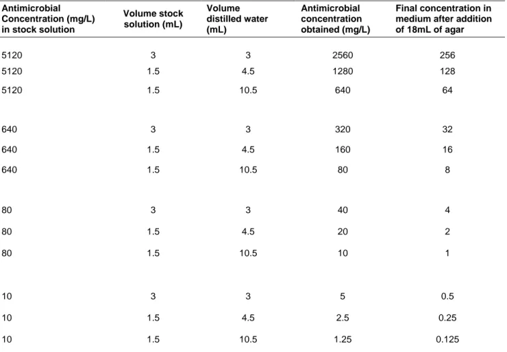
Solutions containing the antimicrobials were diluted in distilled water in 1:1, 1:4 and 1:8 concentrations, in order to obtain an antimicrobial concentration from 2,560 µg/ml to 1.25 µg/ml. In a log2 doubling dilution scheme, 2 mL of each stock solution was mixed in 18 mL of
liquid Muller-Hinton II agar (1:10) in petri dishes, achieving the final MIC range concentration from 256 µg/ml to 0.125 µg/ml (Table 1, see appendix). Final concentrations in SXT were from 1,280 to 5 µg/ml, and the MIC ranged from 0.5/9.5 µg/ml to 64/1,216 µg/ml (Table 2, see appendix). Petri dishes were left at room temperature until agar solidification.
Preparation of inoculum
Inoculums were prepared from a pure overnight culture. Two to four colonies were selected with a 1 µl loop and added to test tubes containing 2 mL of 0.85% normal saline solution (NSS). Turbidity was adjusted to an equivalent of 0.5 McFarland in order to obtain an approximate suspension of 1 to 2x10⁸ CFU/mL.
Inoculation of plates
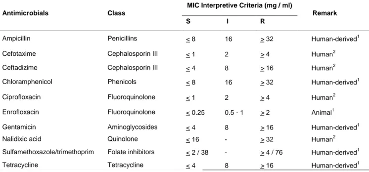
Petri dishes containing the different dilutions of antimicrobials were left to dry at 42ᵒC for 1 h before inoculation. In a biological safety cabinet (Telstar), 100 µl of the inoculum was transferred to eppendorf tubes containing 900 µl of 0.85% NSS. An automatic inoculum-replicator device with 27 micropipettes inserted approximately 2 µL of the prepared inoculum into the surface of each plate with the different concentrations of antimicrobial. Inoculation started from the lowest to the highest concentration. Control plates were used before and after the inoculation to discard contamination. Following the inoculation, plates were allowed to dry at room temperature and then incubated at 37ᵒC for 16-20 h. MIC results were interpreted according to the CLSI (2013) guidelines (Table 3) and registered as the lowest concentration without visible growth of the bacteria. Faint hazes, pinpoints colonies and single colonies were not considered as growth bacteria. SXT results were recorded when a growth reduction of 80% was observed. E. coli ATCC 25922 served as a quality control strain.
PCR
Preparation of DNA template
Salmonella isolates stored at 4ᵒC were recovered and allowed to grow in Muller-Hinton agar (BBL) in overnight incubation at 37ᵒC. One to two colonies were selected by 1 µl loop and transferred to test tubes containing 100 µl of 0.85% NSS. Centrifugation at 1,200
9
rpm for 5 min was preceded. The supernatant was discharged and the pellet was suspended with 100 µl of distilled water. Samples were then boiled in a thermo-shaker (Biosan) for 10 min at 100ᵒC. Centrifugation at 1,200 rpm for 5 min was followed. Samples containing the pellet were cooled down and stored at -30ᵒC until PCR amplification.
Int1 gene amplification
PCR reactions were performed in total volume of 20 µl, including 1.5 mM MgCl2, 50
mM KCl, 10 mM Tris-HCl (pH 8.7), 200 µM of each dNTP (HotStarTaq), 0.25 µM of each primer and 2 µl of DNA. The intI1-specific primers were intI1-F (5‟-AAGGATCGGGCCTTGATGTT-3‟) and intI1-R (5‟-CAGCGCATCAAGCGGTGAGC-3‟). A Pseudomonas aeruginosa strain from Thailand (GenBank accession no. AY553333) was used as positive control.
The PCR for intI1 was as follows: pre-denaturation at 95ᵒC for 15 min, followed by 30 cycles of denaturation at 94ᵒC for 30 s, annealing at 56ᵒC for 30 s, extension at 72ᵒC for 1 min, and final extension at 72ᵒC for 10 min. Amplification reactions were carried out using a DNA thermo-cycler (TProfessional Basic Gradient, Biometra).
Gel electrophoresis
An amount of 3 µl PCR product was mixed with 1 µl of DNA loading dye (Thermo Scientific) and analyzed by electrophoresis in 1.5% agarose gel stained with 1 µl of ethidium bromide (10 mg/mL). The buffer used for the gel preparation and electrophoresis was 0.5X TBE solution. A molecular weight marker with 100 bp increments (Thermo Scientific) was used as a size standard. The gel ran for 30 min/180 mV in an electrophoresis power supply (Enduro, 300V). DNA fluorescence imaging was processed under UV light in a G:BOX XR5 imaging system (Syngene).
10
Results
Serogroup test
Salmonella isolates were serogrouped using specific antisera test, according to the White-Kauffmann-Le Minor scheme (Grimont et al., 2007). All isolates presented a negative reaction with 0.85% NSS implying the presence of somatic (O) antigen. The agglutination test for serogroup A, B, C, D, E, F, G, H and L was preceded and revealed positive results for five different serogroups (Figure 1). Serogroup C had the highest percentage with 35% of samples, followed by B (28%), E (26%), D (10%), and G (1%).
Figure 1. Distribution of five serogroups among the Salmonella isolates (n=100) in poultry
farms from Thailand in 2013-2014.
Antimicrobial susceptibility
Many of the isolates displayed resistance to naxilidic acid (80%), ampicillin (72%), sulfomethoxazole-trimethropim (55%), tetracycline and ceftazidime (38%) and enrofloxacin (31%). Resistance was also observed, but to a lesser extent, to gentamicin (15%) and ciprofloxacin (6%). No resistance was found for ceftotaxime and chloramphenicol. Only two isolates demonstrated susceptibility to all classes of antimicrobials. Susceptibility results and MIC range for each antimicrobial tested are shown in Table 4. Maximum levels of MIC (≥256 µg/ml) were registered in ampicillin, nalidixic acid and tetracycline in 66, 47 and 22 isolates, respectively. Also, 16 isolates reached its maximum level of MIC (64/1216 µg/ml) for sulfomethoxazole-trimethropim. A considerable proportion of the isolates exhibited intermediate resistance to enrofloxacin (42%) and ceftadizime (32%).
28% 35% 10% 26% 1% B C D E G Serogroup
11
Table 4. Antimicrobial sensitivity test and MIC range of Salmonella spp. against the tested
agents. Class Antimicrobial Number of isolates MIC range (µg/ml) S1 I2 R3 Aminoglycosides GEN 83 2 15 <0.125 – 128 ß-lactams AMP 9 19 72 32 – ≥256 CTX 97 3 0 <0.125 – 2 CAZ 30 32 38 1 – 32 Chloramphenicol CHL 100 0 0 0.5 – 2 Quinolones CIP 90 4 6 <0.125 – 8 ENR 27 42 31 <0.125 – 32 NAL 20 - 80 32 – ≥256 Sulfonamides SXT 45 - 55 4/76 – 64/1216 Tetracycline TET 61 1 38 64 – ≥256 1
=susceptible; 2=intermediate; 3=resistant.
GEN=gentamicin; AMP=ampicilin; CTX=cefotaxime; CAZ=ceftadizime; CHL=chloramphenicol; CIP=ciprofloxaxin; ENR=enrofloxacin; GEN=gentamicin; NAL=naxilidic acid; SXT=sulfamethoxazole-trimethropim; TET=tetracycline
Multidrug-resistant (MDR) organisms were considered as being resistant to at least one agent in three or more different antimicrobial categories (Magiorakos et al., 2012). Temporal analysis revealed that MDR strains isolated in 2013 were more frequent than in 2014, with 62% of the total isolates compared to 56%, respectively. The majority of MDR isolates were simultaneously resistant to 3 different antimicrobial categories (Figure 2).
Altogether, different drug resistance profiles were found. Simultaneous resistance to AMP+NAL+SXT was the most common AMR phenotypic profile (9 isolates), followed by AMP+CAZ+NAL+SXT in 6 isolates. AMP+GEN+NAL, AMP+CAZ+SXT+TET and AMP+ENR+NAL+SXT+TET were also typically observed AMR patterns with 4 isolates in each profile. Three isolates depicted simultaneous resistances to 7 of the 10 antimicrobials that were tested: AMP+CAZ+CIP+ENR+NAL+SXT+TET.
12
No. of simultaneous resistances in the MDR isolates
Figure 2. Multi-drug resistant Salmonella isolates (n=59) from poultry farms showing
simultaneous resistance in 3 to 5 antimicrobial categories.
PCR - Class I integrons
The integrase gene (intI) from class I integrons was detected in 36 isolates. PCR results are shown in Figure 3. Isolates harboring the int1 gene showed to be resistant to SXT, AMP, ENR, CAZ, CIP, GEN, NAL and TE with rates of 62%, 44%, 43% 34%, 16%, 33%, 32% and 37%, respectively. The majority of isolates (86%) which carried the integrase gene were multi-drug resistant organisms. Resistant isolates harboring int1 gene and their respective MIC range levels are shown in Table 5.
N o. isol ates 24 6 1 16 9 3 0 5 10 15 20 25 30 3 4 5 2013 2014
13
Figure 3. Amplification of int1 gene in Salmonella isolates. Lane 1, 100 bp DNA Ladder.
Lane 2-11, isolates carrying int1 (471 bp). Lane 12, P. aeruginosa AY553333, positive control.
Table 5. Number of Salmonella spp. isolates positive for int1 gene within each resistant
phenotype
AMP=ampicilin; CAZ=ceftadizime; CTX=cefotaxime; CIP=ciprofloxaxin; ENR=enrofloxacin; GEN=gentamicin; NAL=naxilidic acid; SXT=sulfamethoxazole-trimethropim; TET=tetracycline
AMR Phenotype (N) Rates of resistant isolates
with int1 gene (N)
MIC range for organisms with int1 (µg/mL) AMP (72) 44% (32) 64 - >256 CAZ (38) 34% (14) 16 CIP (6) 16% (1) 8 ENR (31) 43% (13) 2 - 32 GEN (15) 33% (5) 16 - 64 NAL (80) 32% (27) 32 - >256 SXT (55) 62% (34) 4 – 64 TET (38) 37% (14) 64 - >256
14
Discussion
Selective pressure in Salmonella and variations in each serotype may implicate different outcomes of the disease (Jones et al., 2008). Serotyping may be important to understand zoonotic and pathogenic risks posed to humans (Vikash Singh, 2013). In the present study, serotyping was not conducted but we found that the majority of isolates belonged to serogroup C (35%), followed by B (28%), E (26%), D (10%) and G (1%). Previous studies in Thailand (Angkititrakul et al., 2005, Padungtod et al., 2006, Bodhidatta et al., 2013, Lertworapreecha et al., 2013) reported that S. Corvalis, S. Anatum, S. Emek and S. Albany were, respectively, the most common serovars isolated amongst chickens. According to the WhiteKauffaman-LeMinor scheme (Grimont et al., 2007) all the mentioned serovars are representative of serogroup C. Also, a study by Chiu et al. (2010) found that serogroup B and C were the most frequently isolated among chicken isolates. Additionally, S. Weltevreden, was reported to be highly predominant in chicken samples (23/48) and in healthy humans (22/98) in Thailand (Padungtod et al., 2006). This serovar belongs to serogroup E (Grimont et al., 2007) which had also a significant presence in our study. Similarly to our findings, Boodhidatta et al. (2013) and Angkititrakul et al. (2005) observed that serogroup G had the lower rates in their isolates. Serogroups and serovars prevalence in chickens can be age-related and differ between chicken lines and geographic areas (Chiu et al., 2010). The high rates of serogroup B and C in previous studies taken together with our data, may suggest that these serogroups may be more adapted to chickens in Thailand.
This investigation documents the level of antimicrobial resistance among Salmonella spp. isolates obtained in poultry farms in Thailand. High resistance rates to nalidixic acid, ampicillin and sulfamethoxazole-trimethoprim were coherent with the use of antimicrobials in the poultry industry as referred in previous studies (Adeisiji et al., 2014). These antimicrobials are representative of older generation compounds since they were the first to be developed in the quinolones, β-lactams and sulfonamides group, respectively.
Despite the spreading of resistance genes may happen without the direct interaction of antimicrobials by passive or co-selective events, the selective pressure in bacteria due to an active, repeated or intermittently use of antimicrobials in food producing animals may lead to higher prevalence of antimicrobial resistances among animals and humans (Bauer-Garland et al., 2006; Kolár M. et al., 2001). Thus, information such as: daily dose, duration of treatment, number of animals treated and consumption data may be useful to relate the use of antimicrobials to the simultaneous existence of antimicrobial resistances (EFSA, 2015). Unfortunately, in the present study, complementary information regarding antimicrobial usage was not available; therefore it was not possible to associate the observed resistances to an active, passive or co-selective mechanism.
15
Resistance to enrofloxacin was observed in 38 isolates in which six of them showed the highest MIC level of 32 mg/ml. Since quinolones have been commonly used for bacterial disease control in poultry farms (Kang et al., 2005), a correlation may be apparent between the use of quinolones such as nalidixic acid and enrofloxacin. In our study, all six resistant isolates to ciprofloxacin showed a MIC of 8 mg/ml and were simultaneously resistant to enrofloxacin and nalidixic acid. Ciprofloxacin is considered a critically important antimicrobial by the WHO (2014) and the OIE (2007b) given its vast potency against gram-negative bacteria. Also, due to the widespread of resistances to chloramphenicol and ampicillin, fluoroquinolones have been commonly used to treat invasive human salmonellosis (Adesiji et al., 2014). Accordingly to Emmerson et al. (2003), there is an association between the use of enrofloxacin in animal food additives and the incidence of antimicrobial resistances in ciprofloxacin. Additionally, other studies (Gyles et al., 2013; Poppe et al., 2001) stated that organisms resistant to nalidixic acid usually show reduced susceptibility to ciprofloxacin and are easily converted into fluoroquinolones resistant strains.
Resistances in β-lactams, mostly in third and fourth generation cephalosporins, have been a public health concern in the last few years. In 2007, the European Medicine Agency recommended the use of these substances in food-producing animals only in cases of poorly respond to narrower spectrum antimicrobials (EFSA, 2015). In our study, the number of detected antimicrobial resistant isolates was higher in ceftadizime than in cefotaxime. Cefotaxime is one of the most important antimicrobials for the treatment of Salmonella spp. in humans. Children with meningitis caused by invasive Salmonella spp. are also treated with cefotaxime (Price et al., 2000). Due to the possible relation between the use of antimicrobials and the existence of antimicrobial resistances, the low amount of resistant isolates to cefotaxime in this study may suggest that this compound is probably not frequently administrated in the tested farms and that the currently used antimicrobials may not co-select for resistances to cefotaxime.
Chloramphenicol administration in food animals was forbidden by the U.S. Food and Drug Administration in 1997 due to its toxicity in humans. Nevertheless, resistant bacteria to chloramphenicol can still be found in food products including poultry. The illegal use of chloramphenicol and remaining residues from past administrations may be responsible for the maintenance of resistant bacteria in farms. Other conditions such as the use of topical medical preparations containing chloramphenicol by farmworkers and the natural existence of the substance in soil and in plants materials may also contribute for the permanence of resistance traits (Berendsen et al., 2010). Also, in 1997, the European Commission approved the ban of avoparcin due to the high increase of vancomycin-resistant enterococci (VRE) organisms in veterinary and human medicine. In spite of the prohibition, prospective studies in Denmark (Heuer et al., 2002) and in Norway (Borgen et al., 2000), after 5 and 3
16
years, respectively, found that VRE were still extensively present in poultry farms. These studies enhance that antimicrobial resistant organisms may still be disseminated among poultry disregarding the absence of selective pressure. In our study, 100% of the isolates were susceptible to chloramphenicol suggesting that an effective control and monitoring of this compound is probably being conducted in Thailand, and also, that in this case, its interdiction is associated with full susceptibility.
In our study, resistances to tetracycline were lower compared to previous similar studies. Angkititrakul et al., (2005) and Padungtod et al., (2006) conducted their studies in the north of Thailand between 2000-2003 and 2003, respectively, and registered that 100% of Salmonella isolates from chicken meat were resistant to tetracycline. However, a more recent study by Lertworapreecha et al., (2013) in south of Thailand showed lower (60%) resistance rates to tetracycline in their isolates. A report by EFSA showed that tetracycline was the most frequent antimicrobial administrated to food-producing animals in Europe with 2,942.6 tonnes of the active ingredient in 2012 (EFSA, 2015). Newly emerged Salmonella serovars in Europe such as S.Typhimurium DTs 193 / 190 and S. Typhimurium DT104 have been observed to be typically of R-type ASSuT and ACSSuT, respectively, showing resistance to ampicillin (A), streptomycin (S), sulfamethoxazole (Su), tetracycline (T) and chloramphenicol (C) (EFSA, 2015; Mandilara et al., 2013). The prevalence of these multi-resistant organisms may be the consequence of a dominant clone that spreads major determinants of a resistance pattern (Gyles, 2008). In Asia, ACSSuT strains are less dispersed than in Europe (Yu et al., 2008), nevertheless, they have been identified in Korea (Yang et al., 2002) and Japan (Sameshima et al., 2000). In Thailand scarce information is available related to the prevalence of ACSSuT phenotype, thus, in the present study, the absence of resistances in chloramphenicol and the low existence of tetracycline resistances may suggest that this R-type may be less prevalent in the country when compared to European countries.
The presence of int1 gene is related to the existence of integron class I and often considered as responsible for the spreading of MDR organisms. In our study, among MDR isolates, 53% harbored the int1 gene and showed the same phenotypic common resistance profiles: AMP+NAL+SXT in 6 isolates and AMP+CAZ+NAL+SXT in 4 isolates. A correlation between integron class I and the MDR isolates may be assumed, nevertheless only a fraction of the resistant isolates showed as being linked to integron class I. Other genetic mechanisms and elements such as plasmids and transposons should be considered as involved in the transmission of resistances. Previous studies (Miko et al., 2005; Peirano et al., 2006; Ribeiro et al., 2011 and Mahero et al., 2013) mentioned that integron class I may be responsible for a sizeable portion of Salmonella spp. resistances among foodstuffs, animals and humans. Although, the same authors pointed that most resistance genes were
17
located outside of integrons and that not all MDR isolates carried class I integron which is consistent to our results.
Also, isolates carrying int1 gene showed as being highly resistant to sulfamethoxazole-trimethoprim (62%). The sul1, a sulphonamide-resistance gene, found in most class I integrons on their 3‟-conserved segment may be responsible for resistances in sulfamethoxazole-trimethoprim (Recchia and Hall, 1995). A study in Portuguese Salmonella enterica strains from clinical and food samples, found that a significant proportion of isolates resistant to sulfonamides carried class 1 integrons in which the presence of sul1 gene was a consistent marker for sulfonamide resistance (Antunes et al., 2004). In the same study, the authors added that the sul2 and sul3 gene can be also present in integron class 1 but with a lower incidence than sul1. Additionally, Antunes et al. (2004) stated that sul1 gene creates a selective pressure by sulfonamides that can be useful to the maintenance and spreading of resistances to other antimicrobials.
18
Conclusions
The frequency of resistances in Salmonella spp. isolated in parent stock and broiler farms highlight that both populations can be responsible for the dissemination of MDR Salmonella among the poultry industry. Breeders are the top of the pyramid therefore transference of Salmonella spp. and resistance genes they harbor to the subsequent levels should be expected. Surveillance, public policies and guidelines in each sector of the production chain should be implemented actively. The spread of resistances among food producing animals should not be considered only as a domestic public health issue, but also as an international one. Complementary data concerning the use of antimicrobials and information related to possible vehicles and sources associated with the antimicrobial resistances in each tested farm would add value to the present study.
In conclusion, even though the tested compounds in this study are considered as classic, awareness should be given to the fact that selective pressure in poultry stock can eventually lead to the integration of genes conferring resistance to antimicrobials of higher generations. Class I integron can be related to the accumulation of resistance genes and to the emergence of MDR in Salmonella spp. Further studies of genes cassettes inserted in each integron could be useful to follow the evolution of this mobile genetic element and its role in the dissemination of antimicrobial resistances.
19
REFERENCES Literature review
Akbar, A. and Anal, A. (2013). Prevalence and antibiogram study of Salmonella and Staphylococcus aureus in poultry meat. Asian Pacific Journal of Tropical Biomedicine, 3(2), pp 163-168
Angkititrakul S, Chomvarin C, Chaita T, Kanistanon K, Waethewutajarn S. (2005), Epidemiology of antimicrobial resistance in Salmonella isolated from pork, chicken meat and humans in Thailand., Southeast Asian Journal
Tropical Medicine Public Health, 36(6), pp 1510-1515
Archawakulathep, A., Ta Thi Kim, C., Meunsene, D., Handijatno, D., Hassim, H., Rovira, H., Myint, K., Baldrias, L., Sothy, M., Aung, M., Wahyu, N., Chea, R., Boonmasawai, S., Vannamahaxay, S., Angkititrakul, S., Collantes, T., Nguyen Van, T., Punyapornwithaya, V., Zakaria, Z., & Chuanchuen, R. (2014). Perspectives on antimicrobial resistance in livestock and livestock products in ASEAN countries. The Thai Journal of Veterinary Medicine, 44(1) pp 5-13
Bangtrakulnonth, A., Pornreongwong, S., Pulsrikarn, C., Sawanpanyalert, P., Hendriksen, R., Wong, D. and Aarestrup, F. (2004). Salmonella serovars from humans and other Sources in Thailand, 1993–2002. Emerging
Infectious Disease, 10(1), pp 131-136
Bennett, P. (1999). Integrons and gene cassettes: a genetic construction kit for bacteria. Journal of Antimicrobial
Chemotherapy, 43(1), pp 1-4.
Boonprasert, N. Nuanualsuwan S., Pulsrikarn C., Pornaem S., Chokesajjawatee N. (2014), Sources and disseminatios of Salmonella spp. in an integrated broiler meat production. Thai Journal of Veterinary Medicine, 44(1), pp 117-124
Chaisatit, C., Tribuddharat, C., Pulsrikarn, C. and Dejsirilert, S. (2012). Molecular characterization of antibiotic-resistant bacteria in contaminated chicken meat sold at supermarkets in Bangkok, Thailand. Japanese Journal of
Infectious Diseases, 65(6), pp 527-534
Chuanchuen, R. and Pandungtod, P. (2009). Antimicrobial resistance genes in Salmonella enterica isolates from poultry and swine in Thailand, Journal of Veterinary Medical Science, 71(10), pp 1349-1355
Dzidic S., Suskovic J. and Kos B. (2008). Antibiotic resistance in bacteria: biochemical and genetic aspects. Food
Technology and Biotechnology. 46 (1), pp 11–21
EFSA (2014). EFSA explains zoonotic diseases – Salmonella. European Food Safety Authority, consulted in 9th July 2015.
[Available online]
http://www.efsa.europa.eu/sites/default/files/corporate_publications/files/factsheetsalmonella.pdf
FAO (2008), Supply chain auditing for poultry production in Thailand, PPLPI research report, consulted in 12th June 2015
[Available online] http://www.fao.org/ag/againfo/programmes/en/pplpi/docarc/rep-0809_thaipoultrychain.pdf Gebre B. A. (2012) Qualitative screening of antibiotic residues and identification of antibiotic resistant Salmonella from raw and ready to eat meat in Thailand. International Journal of Advanced Life Sciences, 5(2), pp 51-64 Ibrahim M.A., Emeash H.H., Ghoneim N.H. and Abdel-Halim M.A. (2013). Seroepidemiological studies on poultry salmonellosis and its public health importance. Journal of World's Poultry Research 3(1), pp 18-23
Lertworapreecha, M., Sutthimusik, S. and Tontikapong, K. (2012). Antimicrobial resistance in Salmonella enterica isolated from pork, chicken, and vegetables in Southern Thailand. Jundishapur Journal of Microbiology, 6(1), pp 36-41
Mahero M, Byarugaba D.K., Doetkott D.K., Olet S, Khaitsa M.L. (2013). Antimicrobial resistance and presence of class 1 integrons in Salmonella serovars isolated from clinical cases of animals and humans in North Dakota and
Uganda. Clinical Microbiology, 2(6), pp 128
Majowicz, S., Musto, J., Scallan, E., Angulo, F., Kirk, M., O‟Brien, S., Jones, T., Fazil, A. and Hoekstra, R. (2010). The global burden of nontyphoidal Salmonella gastroenteritis. Clinical Infectious Diseases, 50(6), pp 882-889
20
Miko, A. (2005). Molecular mechanisms of resistance in multidrug-resistant serovars of Salmonella enterica isolated from foods in Germany. Journal of Antimicrobial Chemotherapy, 56(6), pp 1025-1033 Momtaz H., Rahimi E., Moshkelani S. (2012). Molecular detection of antimicrobial resistance genes in E. coli isolated from slaughtered commercial chickens in Iran, Veterinarni Medicina, 57 (4), pp 193–197
NaRanong V. (2007) Structural changes in Thailand‟s poultry sector and its social implications, Proceedings of the FAO Conference „Poultry in the 21st Century, Avian Influenza and Beyond consulted on 19th
July 2015 [available online] http://www.fao.org/ag/againfo/home/events/bangkok2007/docs/part1/1_4.pdf
OIE (2007a), Development of on-farm food safety in Thailand‟s poultry industry, Conference OIE 2007, pp 191-195 consulted on 14th July 2015 [available online] http://www.oie.int/doc/ged/D4548.PDF
OIE (2007b) OIE List of antimicrobials of veterinary importance, OIE 75th General Session. Consulted in 20th June 2015 [available online] http://www.oie.int/doc/ged/D9840.PDF
Padungtod, P. and kaneene, J. (2006). Salmonella in food animals and humans in northern Thailand.
International Journal of Food Microbiology, 108(3), pp 346-354
Peirano G, Agersø Y, Aarestrup FM, Reis E., Rodrigues D. (2006) Occurrence of integrons and antimicrobial resistance genes among Salmonella enterica from Brazil. Journal of Antimicrobial Chemotherapy, 58, pp. 305-309
Persoons, D., Haesebrouck, F., Smet, A., Herman, L., Heyndrickx, M., Martel, A., Catry, B., Berge, A., Butaye, P. And Dewulf, J. (2010). Risk factors for ceftiofur resistance in Escherichia coli from belgian broilers.
Epidemiology and Infection, 139(5), pp 765-771
Pui, C. F., Wong, W. C., Chai, L. C., Tunung, R., Jeyaletchumi, P., Noor Hidayah, M. S., Ubong, A., Farinazleen, M. G., Cheah, Y. K. and Son, R. (2011). Salmonella: A foodborne pathogen. International Food Research
Journal, 18, pp 465-473
Randall, L. (2004) Antibiotic resistance genes, integrons and multiple antibiotic resistance in thirty-five serotypes of Salmonella enterica isolated from humans and animals in the UK. Journal of Antimicrobial Chemotherapy, 53(2), pp 208-216
Recchia, G. and Hall, R. (1995). Gene cassettes: a new class of mobile element. Microbiology, 141(12), pp 3015-3027
Ribeiro, V., Lincopan, N., Landgraf, M., Franco, B. and Destro, M. (2011) Characterization of class 1 integrons and antibiotic resistance genes in multidrug-resistant Salmonella enterica isolates from foodstuff and related sources. Brazilian Journal of Microbiology, 42(2), pp 685-692
Sameshima,T., M. Akiba, H. Izumiya, J. Terajima, K. Tamura, H. Watanabe, and M. Nakazawa. 2000. Salmonella
typhimurium DT104 from livestock in Japan. Japonese Journal of Infectious Disease. 53 (1), pp15-16
Sasipreeyajan, J., Jerngklinchan, J., Koowatananukul, C. and Saitanu, K. (1996). Prevalence of Salmonellae in broiler, layer and breeder flocks in Thailand. Tropical Animal Health Production, 28(1), pp 174-180
Sirichote P, Bangtrakulnonth A, Tianmanee K, Unahalekhaka A, Oulai A, Chittaphithakchai P, Kheowrod W, Hendriksen RS. (2010), Serotypes and antimicrobial resistance of Salmonella enterica ssp in central Thailand, 2001-2006, Southeast Asian Journal Tropical Medicine Public Health, 41(6) pp 1405-1415
WHO (2014), Antimicrobial resistances, global report on surveillance, World Health Organization, consulted in 8th July 2015. [Available online] http://www.who.int/drugresistance/documents/surveillancereport/en/
21
Manuscript
Adesiji, Y., Deekshit, V. and Karunasagar, I. (2014). Antimicrobial-resistant genes associated with Salmonella spp. isolated from human, poultry, and seafood sources. Food Science and Nutrition, 2(4), pp 436-442
Angkititrakul S., Chomvarin C., Chaita T., Kanistanon K., Waethewutajarn S. (2005), Epidemiology of antimicrobial resistance in Salmonella isolated from pork, chicken meat and humans in Thailand., Southeast
Asian Journal Tropical Medicine Public Health, 36(6), pp 1510-1515
Angulo, F. and Molbak, K. (2005). Human health consequences of antimicrobial drug--resistant Salmonella and other foodborne pathogens. Clinical Infectious Diseases, 41(11), pp 1613-1620
Antunes, P., Machado, J., Sousa, J. and Peixe, L. (2005). Dissemination of sulfonamide resistance genes (sul1,
sul2, and sul3) in Portuguese Salmonella enterica strains and relation with integrons. Antimicrobial Agents and Chemotherapy, 49(2), pp.836-839.
Bauer-Garland, J., Frye, J., Gray, J., Berrang, M., Harrison, M. and Fedorka-Cray, P. (2006). Transmission of
Salmonella enterica serotype Typhimurium in poultry with and without antimicrobial selective pressure. Journal of Applied Microbiology, 101(6), pp 1301-1308
Bennett, P. (1999). Integrons and gene cassettes: a genetic construction kit for bacteria. Journal of Antimicrobial
Chemotherapy, 43(1), pp.1-4
Berendsen, B., Stolker, L., de Jong, J., Nielen, M., Tserendorj, E., Sodnomdarjaa, R., Cannavan, A. and Elliott, C. (2010). Evidence of natural occurrence of the banned antibiotic chloramphenicol in herbs and grass.
Analytical and Bioanalytical Chemistry, 397(5), pp.1955-1963
Bodhidatta L., Srijan A., Serichantalergs O., Bangtrakulnonth A., Wongstitwilairung B., McDaniel P., Mason C.J., (2013) Bacterial pathogens isolated from raw meat and poultry compared with pathogens isolated from children in the same area of rural Thailand. Southeast Asian Journal of Tropical Medicine and Public Health 44 (2), pp 259–272
Borgen, K., Simonsen, G., Sundsfjord, A., Wasteson, Y., Olsvik, Ø. and Kruse, H. (2000). Continuing high prevalence of VanA-type vancomycin-resistant enterococci on Norwegian poultry farms three years after avoparcin was banned. Journal of Applied Microbiology, 89(3), pp 478-485
Castiglioni Tessari, E., Iba Kanashiro, A., Z. Stoppa, G., L., R., De Castro, A. and P. Cardoso, A. (2012). Important Aspects of Salmonella in the Poultry Industry and in Public Health, Salmonella - A Dangerous
Foodborne Pathogen, pp 182-206
Chiu, L., Chiu, C., Horn, Y., Chiou, C., Lee, C., Yeh, C., Yu, C., Wu, C., Chang, C. and Chu, C. (2010). Characterization of 13 multi-drug resistant Salmonella serovars from different broiler chickens associated with those of human isolates. BMC Microbiology, 10(1), p 86
Clinical and Laboratory Standards Institute (CLSI), (2012). Performance standards for antimicrobial susceptibility testing: twenty-second informational supplement M100-S22
Clinical and Laboratory Standards Institute (CLSI), (2013). Performance standards for antimicrobial disk and dilution susceptibility testing for bacteria isolated from animals: second informational supplement VET01-S2 Clinical and Laboratory Standards Institute (CLSI), (2014). Performance standards for antimicrobial susceptibility testing: twenty-four informational supplement M100-S24
Dzidic S., Suskovic J. and Kos B. (2008). Antibiotic resistance in bacteria: biochemical and genetic aspects. Food
Technology and Biotechnology. 46 (1) 11–21
EFSA (European Food Safety Authority) and ECDC (European Centre for Disease Prevention and Control), (2015). EU Summary Report on antimicrobial resistance in zoonotic and indicator bacteria from humans, animals and food in 2013. EFSA Journal, 13(2)
EFSA (European Food Safety Authority), (2010). Scientific Opinion on monitoring and assessment of the public health risk of “Salmonella Typhimurium-like” strains EFSA Journal, 8(10)
Emmerson, A. (2003). The quinolones: decades of development and use. Journal of Antimicrobial
22
Grimont, P.A.D., Weill, F.-X. (2007) Antigenic formulae of the Salmonella serovars, (9th ed.), WHO Collaborating Center for Reference and Research on Salmonella, Institut Pasteur. Consulted in 13th May 2015 [available online] http://www.pasteur.fr/sante/clre/cadrecnr/salmoms/WKLM_En.pdf
Gyles, C. (2008). Antimicrobial resistance in selected bacteria from poultry. Animal. Health. Research. Reviews., 9(02), pp 149-158
Heuer, O., Pedersen, K., Andersen, J. and Madsen, M. (2002). Vancomycin-Resistant Enterococci (VRE) in Broiler Flocks 5 Years after the Avoparcin Ban. Microbial Drug Resistance, 8(2), pp.133-138
Ibrahim M.A., Emeash H.H., Ghoneim N.H. and Abdel-Halim M.A. (2013). Seroepidemiological studies on poultry salmonellosis and its public health importance, Journal of World's Poultry Research, 3(1) pp 18-23
Ingram, P., Rogers, B., Sidjabat, H., Gibson, J. and Inglis, T. (2013). Co-selection may explain high rates of ciprofloxacin non-susceptible Escherichia coli from retail poultry reared without prior fluoroquinolone exposure.
Journal of Medical Microbiology, 62(11), pp.1743-1746
Jones, T., Ingram, L., Cieslak, P., Vugia, D., Tobin‐D‟Angelo, M., Hurd, S., Medus, C., Cronquist, A. and Angulo, F. (2008). Salmonellosis outcomes differ substantially by serotype. The Journal of Infectious Diseases, 198(1), pp 109-114
Kang, H. (2005). Characterization of antimicrobial resistance and class 1 integrons found in Escherichia coli isolates from humans and animals in Korea. Journal of Antimicrobial Chemotherapy, 55(5), pp 639-644 Kolář, M., Urbánek, K. and Látal, T. (2001). Antibiotic selective pressure and development of bacterial resistance.
International Journal of Antimicrobial Agents, 17(5), pp 357-363
Lertworapreecha, M., Sutthimusik, S. and Tontikapong, K. (2012). Antimicrobial resistance in Salmonella enterica isolated from pork, chicken, and vegetables in Southern Thailand. Jundishapur Journal of Microbiology, 6(1), pp 36-41
Mahero M, Byarugaba D.K., Doetkott D.K., Olet S, Khaitsa M.L. (2013). Antimicrobial resistance and presence of class 1 integrons in Salmonella serovars isolated from clinical cases of animals and humans in North Dakota and
Uganda. Clinical Microbiology, 2(6), pp 128
Mandilara G, Lambiri M, Polemis M, Passiotou M, Vatopoulos A. (2013) Phenotypic and molecular characterisation of multiresistant monophasic Salmonella Typhimurium (1,4,[5],12:i:-) in Greece, 2006 to 2011.
Eurosurveillance, 18(22) consulted on 23th August 2015 [Available online]
http://www.eurosurveillance.org/ViewArticle.aspx?ArticleId=20496
Magiorakos, A., Srinivasan, A., Carey, R., Carmeli, Y., Falagas, M., Giske, C., Harbarth, S., Hindler, J., Kahlmeter, G., Olsson-Liljequist, B., Paterson, D., Rice, L., Stelling, J., Struelens, M., Vatopoulos, A., Weber, J. and Monnet, D. (2012). Multidrug-resistant, extensively drug-resistant and pandrug-resistant bacteria: an international expert proposal for interim standard definitions for acquired resistance. Clinical Microbiology and
Infection, 18(3), pp 268-281
Martins da Costa, P., Loureiro, L., Matos, A. J. F. (2013). Transfer of multidrug-resistant bacteria between intermingled ecological niches: the interface between humans, animals and the environment. International
Journal of Environmental Research and Public Health, 10(1), pp 278–294.
Miko, A. (2005). Molecular mechanisms of resistance in multidrug-resistant serovars of Salmonella enterica isolated from foods in Germany. Journal of Antimicrobial Chemotherapy, 56(6), pp 1025-1033 Padungtod, P. and kaneene, J. (2006). Salmonella in food animals and humans in northern Thailand.
InternationalJournal of Food Microbiology, 108(3) pp 346-354
Peirano G, Agersø Y, Aarestrup FM, Reis E., Rodrigues D. (2006) Occurrence of integrons and antimicrobial resistance genes among Salmonella enterica from Brazil. Journal of Antimicrobial Chemotherapy, 58, pp.
305-309
Price, E. (2000). Antibiotics for Salmonella meningitis in children. Journal of Antimicrobial Chemotherapy, 46(5), pp 653-655
Poppe C., Ayroud M., Ollis G., Chirino-Trejo M., Smart N., Quessy S. and Michel P. (2001). Trends in antimicrobial resistance of Salmonella isolated from animals, foods of animal origin, and the environment of animal production in Canada, 1994–1997. Microbial Drug Resistance, 7 (2), pp 197–212
23
Randall, L. (2004) Antibiotic resistance genes, integrons and multiple antibiotic resistances in thirty-five serotypes of Salmonella enterica isolated from humans and animals in the UK. Journal of Antimicrobial Chemotherapy, 53(2), pp. 208-216.
Recchia, G. and Hall, R. (1995). Gene cassettes: a new class of mobile element. Microbiology, 141(12), pp 3015-3027.
Ribeiro, V., Lincopan, N., Landgraf, M., Franco, B. and Destro, M. (2011). Characterization of class 1 integrons and antibiotic resistance genes in multidrug-resistant Salmonella enterica isolates from foodstuff and related sources. Brazilian Journal of Microbiology, 42(2), pp 685-692.
Vikash Singh (2013) Salmonella serovars and their host specificity. Journal Veterinary Science & Animal
Husbandry 1(3), pp 301
Yang,S. J., K. Y. Park, S. H. Kim, K. M. No, T. E. Besser, H. S. Yoo, S. H. Kim, B. K. Lee, and Y. H. Park. (2002). Antimicrobial resistance in Salmonella enterica serovars Enteritidis and Typhimurium isolated from animals in Korea: comparison of phenotypic and genotypic resistance characterization. Veterinary Microbiology. 86 (4), pp 295-301
Yu, C., Chou, S., Yeh, C., Chao, M., Huang, K., Chang, Y., Chiou, C., Weill, F., Chiu, C., Chu, C. and Chu, C. (2007). Prevalence and characterization of multidrug-resistant (Type ACSSuT) Salmonella enterica serovar Typhimurium strains in isolates from four gosling Farms and a hatchery farm. Journal of Clinical Microbiology, 46(2), pp 522-526
24
25 Antimicrobial Concentration (mg/L) in stock solution Volume stock solution (mL) Volume distilled water (mL) Antimicrobial concentration obtained (mg/L) Final concentration in medium after addition of 18mL of agar 5120 3 3 2560 256 5120 1.5 4.5 1280 128 5120 1.5 10.5 640 64 640 3 3 320 32 640 1.5 4.5 160 16 640 1.5 10.5 80 8 80 3 3 40 4 80 1.5 4.5 20 2 80 1.5 10.5 10 1 10 3 3 5 0.5 10 1.5 4.5 2.5 0.25 10 1.5 10.5 1.25 0.125
Agents used with this dilution method: ampilcin, cefotaxime, ceftadizime, ciprofloxacin, chloramphenicol, enrofloxacin, gentamicin, nalidixic acid and tetracycline
26 Antimicrobial Concentration (mg/L) in stock solution Volume stock solution (mL) Volume distilled water (mL) Antimicrobial concentration obtained (mg/L) Final concentration in medium after addition of 18mL of agar 10240 1.5 10.5 1280 128 1280 3 3 640 64 1280 1.5 4.5 320 32 1280 1.5 10.5 160 16 160 3 3 80 8 160 1.5 4.5 40 4 160 1.5 10.5 20 2 20 3 3 10 1 20 1.5 4.5 5 0.5
Table 2. Preparation of dilutions of Sulfamethoxazole-Trimethropim (SXT) for agar dilution susceptibility test. CLSI (2012)
27
Table 3. MIC interpretive standards for Salmonella according to CLSI guidelines.
Antimicrobials Class
MIC Interpretive Criteria (mg / ml)
Remark
S I R
Ampicillin Penicillins < 8 16 > 32 Human-derived1
Cefotaxime Cephalosporin III < 1 2 > 4 Human2
Ceftadizime Cephalosporin III < 4 8 > 16 Human2
Chloramphenicol Phenicols < 8 16 > 32 Human-derived1
Ciprofloxacin Fluoroquinolone < 1 2 > 4 Human2
Enrofloxacin Fluoroquinolone < 0.25 0.5 - 1 > 2 Animal1
Gentamicin Aminoglycosides < 4 8 > 16 Human-derived1
Nalidixic acid Quinolone < 16 - > 32 Human2
Sulfamethoxazole/trimethoprim Folate inhibitors < 2 / 38 - > 4 / 76 Human-derived1
Tetracycline Tetracycline < 4 8 > 16 Human-derived1
1) CLSI (2013) guidelines, VET01-S2 supplement. 2) CLSI (2014) guidelines.