This Accepted Author Manuscript is copyrighted and published by Elsevier. It is posted here by agreement between Elsevier and University of Brasilia. Changes resulting from the publishing process - such as editing, corrections, structural formatting, and other quality control
mechanisms - may not be reflected in this version of the text. The definitive version of the text was subsequently published in [Theriogenology, Volume 62, Issues 1–2, July 2004, Pages 65–80, doi:10.1016/j.theriogenology.2003.07.025].You may download, copy and otherwise use the AAM for non-commercial purposes provided that your license is limited by the following restrictions:
(1) You may use this AAM for non-commercial purposes only under the terms of the CC-BY-NC-ND license.
(2) The integrity of the work and identification of the author, copyright owner, and publisher must be preserved in any copy.
(3) You must attribute this AAM in the following format: [agreed attribution language, including link to CC BY-NC-ND license + Digital Object Identifier link to the published journal article on Elsevier’s ScienceDirect® platform].
________________________________________________________________________
Este Manuscrito do Autor Aceito para Publicação (AAM) é protegido por direitos autorais e publicado pela Elsevier. Ele esta disponível neste Repositório, por acordo entre a Elsevier e a Universidade de Brasília. As alterações decorrentes do processo de publicação - como a edição, correção, formatação estrutural, e outros mecanismos de controle de qualidade - não estão refletidas nesta versão do texto. A versão definitiva do texto foi posteriormente publicado em [Theriogenology, Volume 62, Número 1–2, Julho de 2004, Páginas 65–80,
doi:10.1016/j.theriogenology.2003.07.025]. Você pode baixar, copiar e utilizar de outra forma o AAM para fins não comerciais , desde que sua licença seja limitada pelas seguintes restrições:
(1) Você pode usar este AAM para fins não comerciais apenas sob os termos da licença CC- BY- NC-ND.
(2) A integridade do trabalho e identificação do autor, detentor dos direitos autorais e editor deve ser preservado em qualquer cópia.
Morphological and ultrastructural analysis of sheep primordial follicles preserved in
0.9% saline solution and TCM 199
M.H.T Matos E.R Andrade C.M Lucci S.N Báo J.R.V Silva R.R Santos M.A.L Ferreira S.H.F Costa J.J.H Celestino J.R Figueiredo Abstract
The objective was to determine the morphological and ultrastructural features of sheep primordial follicles preserved in either 0.9% saline solution or TCM 199 at different temperatures. Soon after death, the ovarian pair of each ewe (n=5) was divided into 25 fragments. One fragment was immediately fixed for morphological evaluation (control). The other 24 fragments were randomly distributed in tubes containing 2 ml of 0.9% saline solution or TCM 199 and maintained at 4, 20 or 39 °C for 2, 4, 12, or 24 h. Based on histological assessment, storage of ovarian fragments in 0.9% saline solution at 20 °C for up to 24 h and in both solutions at 39 °C for 4, 12 or 24 h increased (P<0.01) the percentage of degenerate primordial follicles compared with controls. In contrast, preservation at 4 °C in both solutions, kept the percentage of morphologically normal primordial follicles similar to control values. Although histological integrity of primordial follicles was maintained in fragments stored at 20 °C for up to 24 h in TCM 199, these results were not confirmed by ultrastructural analysis. Based on transmission electron microscopy, only primordial follicles stored at 4 °C for up to 24 h, at 20 °C for up to 12 h and at 39 °C for up to 2 h in both solutions were ultrastructurally normal. In conclusion, sheep primordial follicles were successfully preserved at 4 °C for up to 24 h, at 20 °C for up to 12 h and at 39 °C for 2 h in 0.9% saline solution or TCM 199.
Keywords: Sheep; Primordial follicle; Preservation; Morphology; Ultrastructural
1. Introduction
The mammalian ovary contains a follicular reserve that is determined before birth and
cannot be replenished. This reserve contains thousands of primordial follicles, corresponding
to 95% of the preantral follicle population [1]. Since the vast majority of primordial follicles
become atretic during growth and maturation in vivo, these follicles provide a valuable source
for studies on in vitro development of oocytes and in vitro production of embryos [2].
preantral follicles strive to prevent follicular atresia by rescuing preantral follicles from ovaries
and by maturing these follicles during in vitro culture.
Since oocytes enclosed in primordial follicles are small and less metabolically active,
they may be more tolerant to the preservation process than metaphase II oocytes [8].
Preservation of primordial follicles requires not only the survival of oocyte and granulosa cells,
but also maintenance of gap junctions and metabolic cooperation that are essential for oocyte
growth and development [9]. In addition, maintenance of follicular quality after preservation is
extremely important and should be investigated, because structural or ultrastructural damages
can significantly affect further in vitro growth and maturation of oocytes within primordial
follicles [10]. Some authors have emphasized the importance of ultrastructural evaluation after
in vitro preservation of preantral follicles, since histologically normal follicles may have
degenerative changes detected only with electron microscopy [11] and [12].
Recently, some studies have demonstrated that the quality of the oocytes enclosed in
preantral follicles depends on the preservation medium, temperature and incubation time
during ovary transportation from the field to the laboratory. Saline solution (0.9%) is largely
cited as a short-duration preservation medium for ovaries (cattle [13]; goat [14]) and for
preservation of goat [12] and sheep [15] preantral follicles in situ. However, TCM 199 has been
successfully used in the preservation of goat [16] and sheep [17] preantral follicles. Carvalho et
al. [12] showed that after preservation in Braun–Collins and 0.9% saline solutions, the
degeneration rate of goat secondary follicles was higher than that observed in primordial
follicles. However, studies performed in sheep indicated only the overall incidence of follicular
degeneration after preservation, considering all classes of preantral follicles (primordial,
primary and secondary follicles). In these studies, a high rate of follicular degeneration was
observed after storage at 39 °C for all incubation periods (even for 4 h). On the other hand, the
effect of 0.9% saline solution and TCM 199 for a shorter incubation period (e.g. 2 h), was not
investigated. In addition, ultrastructural analysis of sheep primordial follicles preserved in vitro
has not been reported.
The objectives of the present study were to evaluate the effect of 0.9% saline solution
and TCM 199 on the preservation of sheep primordial follicles, at different temperatures and
incubation times, and to investigate the histologic and ultrastructural morphology of the
2. Materials and methods
2.1. Source of ovaries
Ovaries (n=10) from five adult non-pregnant mixed-breed ewes were collected at a
local abbatoir. The animals were cycling and in good body condition. Under aseptic conditions,
the ovaries were stripped of surrounding fat tissue and ligaments, washed once in a 70%
alcohol solution, and then twice in 0.9% saline solution.
2.2. Media
The media used were: (1) sterile 0.9% saline solution (0.9% NaCl; osmolarity 300
mOsmol/l; pH 7.2) and (2) TCM 199 (osmolarity 280 mOsmol/l; pH7.2; Cultilab, SP, Brazil).
2.3. Experimental protocol
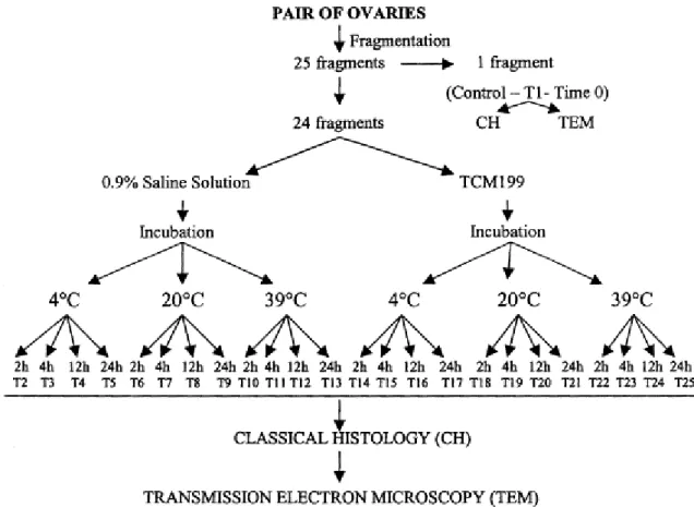
At the abbatoir, the pair of ovaries from each animal was divided into 25 fragments (25
pieces from both ovaries combined). For control purposes (control, treatment 1, time 0), a
small piece of a randomly selected ovarian fragment was removed for transmission electron
microscopy (TEM) with the remainder immediately fixed in Carnoy for histological
examination. The other 24 fragments were randomly distributed into 15 ml tubes (Corning
Glass Works, Corning, NY, USA) containing 2 ml of 0.9% saline solution or TCM 199 at 4, 20 or
39 °C and stored for 2, 4, 12, or 24 h (treatments 2–25) as shown in Fig. 1. The temperatures
were maintained using thermoflasks filled with water at 4, 20 or 39 °C and were monitored at
Fig. 1. General experimental protocol for preservation of ovine primordial follicles.
2.4. Light microscopy
To evaluate the morphology of ovine primordial follicles, at the end of each treatment,
ovarian fragments were processed as follows. Following the removal of a small piece from
each fragment for TEM, the remainder was fixed individually in Carnoy for 12 h. Then, they
were dehydrated in a graded series of ethanol, clarified with xylene and embedded in paraffin
wax. Sections 7 μm thick were stained by a standard protocol using periodic acid schiff (PAS)– hematoxilyn and examined by light microscopy (Zeiss, Germany) under 400× magnification. To
avoid counting a follicle more than once, a primordial follicles was counted only in a sections
where the oocyte nucleus was visible.
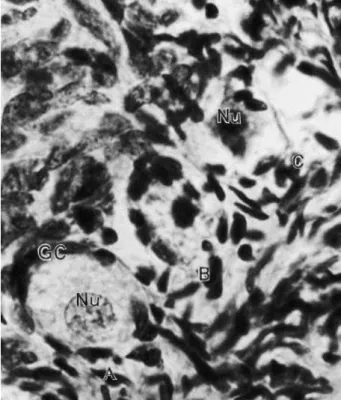
Follicular morphology was evaluated based on the integrity of the basement membrane,
cellular density, presence or absence of pycnotic bodies, and oocyte integrity. Based
on these variables, primordial follicles were classified as morphologically normal (follicles had a
healthy spherical oocyte with uniform cytoplasm, and granulosa cells, well-organized in layers,
without pycnotic nuclei, were observed surrounding the oocyte); Type 1 degenerate follicles
(follicles had an oocyte that was sometimes retracted, with a pycnotic nucleus and
(degeneration of both oocyte and granulosa cells; follicles had a retracted oocyte, with or
without a pycnotic nucleus, and disorganized low-density, swollen granulosa cells) (Fig. 2).
Fig. 2. Histological section of a sheep ovarian fragment, showing normal (A), degenerate Type 1 (B), degenerate Type 2 (C) primordial follicles. GC: granulosa cells, Nu: oocyte nucleus. PAS–hematoxilin stained (400×).
2.5. Transmission electron microscopy
To better evaluate follicular morphology, ultrastructural analysis was performed on
primordial follicles from the control treatment, as well as on treatments that did not differ
from control. Only primordial follicles classified as morphologically normal in semi-thin
sections were evaluated. Briefly, small pieces of ovarian cortex were fixed in a solution
containing 2% paraformaldehyde and 2.5% glutaraldehyde in 0.1 M sodium cacodylate buffer
(pH 7.2). After fixation, specimens were rinsed in buffer and post-fixed in 1% osmium
tetroxide, 0.8% potassium ferricyanide and 5 mM CaCl2 in 0.1 M sodium cacodylate buffer.
Subsequently, samples were dehydrated in acetone and embedded in Spurr’s epoxy resin. Thin sections (80 nm) were prepared when the oocyte nucleus was present in the semi-thin
sections. Semi-thin sections (3 μm) were stained with toluidine blue, while thin sections were
contrasted with uranyl acetate and lead citrate, and examined using a Jeol 100 C (Jeol, Tokyo,
2.6. Statistical analysis
The GLM procedure of the SAS (SAS, Inc., Cary, NC, USA) was used for the analysis of
variance (ANOVA) of data. The factors used in the model for analysis of normal and degenerate
primordial follicles included medium (0.9% saline solution, TCM), temperature (4, 20 or 39 °C),
incubation time (2, 4, 12, or 24 h) and interactions. Differences between the control and other
treatments were performed by Fisher’s PLSD-test. Before statistical analysis, frequency of follicular types was transformed to arcsin. Values were considered statistically significant when
P<0.05.
3. Results
3.1. Storage of sheep primordial follicles in situ in 0.9% saline solution or in TCM 199
A total of 3638 primordial follicles were examined histologically. The number of
follicles ranged from 115 to 152 in each treatment. There was an effect of medium (P<0.05),
temperature (P<0.01) and incubation time (P<0.01), as well as an interaction between
temperature and incubation time (P<0.01).
The effect of temperature and storage time on the percentage of morphologically
normal primordial follides (MNPF) stored in 0.9% saline solution or in TCM 199 is shown in Fig.
3. The percentage of normal follicles stored in 0.9% saline solution at 4 °C for up to 24 h, at 20
°C for 12 h and at 39 °C for 2 h was similar (P>0.01) to control (time zero). A decrease (P<0.01)
in the number of MNPF was observed when primordial follicles were stored in saline solution
at 20 °C for 24 h and at 39 °C for 4, 12 or 24 h when compared to control. Similar results were
obtained when TCM 199 was used, except for the treatment in which the ovarian fragment
was stored at 20 °C for 24 h; in this treatment, the percentage of MNPF was similar (P>0.01) to
Fig. 3. Effect of temperature and storage time on the percentage of morphologically normal primordial follicles preserved in saline solution 0.9% and in TCM 199. (∗) Differ from control (P<0.01); (a, b, c) different letters at the same preservation temperature differ (P<0.01); (A, B, C) different letters in the same preservation period differ (P<0.01); (D, E) different letters among the solution in the same preservation period and temperature differ (P<0.05).
The effect of incubation time within each temperature was analyzed, separately, in
each solution. At 4 °C the percentage of MNPF was not affected by the incubation time, in both
solutions. Similar results were obtained in fragments preserved in TCM 199 at 20 °C. However,
in the fragments stored at 20 °C in 0.9% saline solution, there was a decrease (P<0.01) in the
percentage of MNPF when stored for 24 h compared to 2, 4 or 12 h. A decrease (P<0.01) in the
number of MNPF occurred with the increase of incubation time from 2 to 4 h, 12 and 24 h, in
both solutions at 39 °C.
With respect to the effect of temperature for the same period of incubation, there was
a decrease (P<0.01) in the percentage of MNPF in both solutions at all incubation periods
tested when the fragments were stored at 39 °C, when compared to 4 and 20 °C. However,
fragments stored for 2 h at 39 °C did similar follicular viability when compared with storage at
4 and 20 °C.
The comparison between 0.9% saline solution and TCM 199 at the same temperature
and incubation period showed that the percentage of MNPF at 4 °C in all incubation periods
tested was not different (P>0.05). However, a higher (P<0.05) percentage of MNPF was
observed in TCM 199 at 20 °C for 24 h and at 39 °C for 12 h.
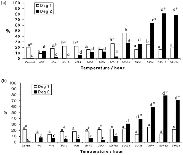
3.2. Distribution of follicular degeneration types in control and other treatments
Fig. 4 shows the distribution of Types 1 and 2 degenerate primordial follicles, in control
and after storage in the different treatments, in 0.9% saline solution (Fig. 4a) and in TCM 199
after storage in 0.9% saline solution at 4 °C for 4, 12 and 24 h, at 20 °C for 12 h, and in TCM 199
at 4 °C for 12 h and at 20 °C for 2 and 4 h. In contrast, an increase (P<0.01) of degenerate Type
2 follicles was observed after storage in both solutions at 39 °C at all incubation times, except
in the fragments stored in saline solution 0.9% for 2 h. Compared to control, a higher (P<0.01)
percentage of Type 1 degenerate follicles was observed in 0.9% saline solution at 4 and 20 °C
for 12 and 24 h and at 39 °C for 4 h. A greater (P<0.01) percentage of degenerate Type 2
follicles compared to control was observed in follicles preserved in 0.9% saline solution at 4 °C
for 2 and 24 h, at 20 °C for 2, 4 and 24 h, at 39 °C for 4, 12 and 24 h, and in TCM 199 at 20 °C
for 24 h and at 39 °C at all incubation periods.
3.3. Ultrastructural analysis of sheep primordial follicles in control and after
preservation
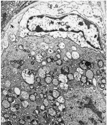
On average, eight primordial follicles per treatment were evaluated by ultrastructural
analysis. Normal primordial follicles exhibited a highly variable number of vesicles spread
throughout the ooplasm. The cytoplasm also contained numerous large pleiomorphic
mitochondria, with irregular cristae and continuous mitochondrial membranes, including
elongated forms with parallel cristae. Well-developed Golgi complexes were observed. Both
smooth and rough endoplasmic reticulum were present, either as isolated aggregations or as
complex associations with mitochondria and vesicles. In normal primordial follicles, there were
sometimes a few microvilli and, occasionally, small amounts of zona pellucida material were
visible, depending on the plane of section. Granulosa cells had irregularly-shaped nuclei, with a
high nucleus-to-cytoplasm ratio. The cytoplasm contained a great number of mitochondria and
well-developed rough endoplasmic reticulum. These features were observed in primordial
follicles from control group (Fig. 5), as well as in follicles preserved in 0.9% saline solution and
in TCM 199 at 4 °C for up to 24 h (Fig. 6), at 20 °C for up to 12 h (Fig. 7), and at 39 °C for up to 2
h (Fig. 8; control).
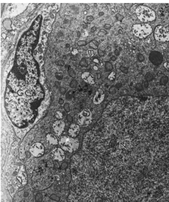
Fig. 6. Electron micrograph of a follicle preserved in 0.9% saline solution at 4 °C for 24 h. GC: granulosa cells, Nu: oocyte nucleus, m: mitochondria, v: vesicles (3700×).
Fig. 8. Electron micrograph of a follicle preserved in TCM 199 at 39 °C for 2 h. GC: granulosa cells, Nu: oocyte nucleus, m: mitochondria, ser: smooth endoplasmic reticulum, v: vesicles (3700×).
When stored in TCM 199 at 20 °C for 24 h, follicles seemed to be well preserved in
semi-thin sections stained with toluidine blue, however, transmission electron microscopy
revealed some discreet changes in their ultrastructure (Fig. 9). In primordial follicles, oocytes
had signs of degeneration before the primitive granulosa cells. The ooplasm of these follicles
was extremely vacated, with the vacuoles often fusing to produce a greater vacated area. In
addition, initial signs of damage to mitochondrial membranes and cristae were observed.
Granulosa cells were slightly swollen, with a low density of organelles present in their
cytoplasm. Some granulosa cells disappeared, leaving a vacated space. The follicles contained
a retracted oocyte and substantial irregularity of the follicular, oocyte and nuclear outlines
(Fig. 9). However, ultrastructural analysis of primordial follicles stored in TCM 199 at 20 °C for
up to 12 h confirmed the integrity of the oocyte, the granulosa cells, and the basement
Fig. 9. Electron micrograph of a follicle preserved in TCM 199 at 20 °C for 24 h. Note the degenerate appearance of the oocyte and granulosa cells. O: oocyte, GC: granulosa cells (4600×).
4. Discussion
In the present study, the storage of ovarian fragments in 0.9% saline solution and in
TCM 199 at 4 °C sustained the number of morphologically normal primordial follicles
compared to control (time zero). In contrast, preservation in either media at 20 or 39 °C,
depending on the incubation period, increased the degeneration rate of sheep primordial
follicles in vitro.
Preservation of ovarian fragments at 4 °C for up to 24 h, in either solution, maintained
the percentage of normal primordial follicles similar to that found in control. Although studies
available did not specifically evaluate the effect of media and temperature on the preservation
of primordial follicles (they usually considered all classes of preantral follicles together), they
demonstrated that a temperature of 4 °C has been successfully used in the follicular
preservation for 24 h in solutions poor (saline [12] and [15]) and rich (TCM 199 [17]) in
nutrients or hyperosmotic (Braun–Collins solution [18]). Therefore, at 4 °C the composition of
the medium is not a limiting factor. In addition, Wood et al. [19] successfully preserved
domestic cat ovarian follicles at 4 °C for 48 h. Moreover, Jewgenow et al. [6] cooled isolated
feline preantral follicles at 4 °C without decreasing the percentage of healthy follicles. Roy and
Treacy [20] observed that a lower metabolic rate at low temperatures may be beneficial for
maintaining viable human preantral follicles in vitro after isolation; preservation at low
temperatures (4 °C) can minimize metabolic requirements and increase follicular resistance to
reduced nutrients and oxygen. Pickering et al. [21] showed that the meiotic spindle of oocytes
was temperature-sensitive during cooling. However, the quality of primordial follicles was not
evaluated. Oocytes enclosed in primordial follicles may be less susceptible to microtubular
disruptions, because most of the microtubular systems remain unorganized, and the
chromatin is in a condensed form protected by the nuclear membrane [22]. In spite of the
good results obtained in this study (as well as in others) in preservation of primordial follicles
at 4 °C, it is unknown if the temperature reduction could affect the culture of primordial
follicles in vitro.
In the present study, when primordial follicles were stored at 20 °C for 12 and 24 h in
0.9% saline solution and in TCM 199, respectively, and at 39 °C for 2 h in both solutions, the
percentage of normal primordial follicles was not significantly different from control. Similar
results were observed with the preservation of preantral follicles (without specific follicular
type) at 20 and 39 °C for 12 and 24 h in 0.9% saline solution (goat [12]; sheep [15]), TCM 199
(goat [16]; sheep [17]) as well as in Braun–Collins or coconut water solution (goat [18]; sheep
The increase of cellular metabolism at the upper limit of temperature (39 °C) or close to it (20
°C) and oxygen consumption could have caused depletion of intracellular energy sources,
followed by consumption of the nutrients and oxygen available in the preservation medium,
resulting in the higher degeneration rates found in these treatments. It is important to note
that in all studies performed with the preservation of goat and sheep preantral follicles, a high
rate of follicular degeneration was observed after storage at 39 °C for all incubation periods.
On the other hand, the best results obtained in our study with preservation at 39 °C (normal
metabolism) may be due to the short incubation period (2 h). Smitz et al. [24] reported that
preantral follicles are able to survive for a short interval under oxygen deficiency, and that
glycolysis can sustain follicle viability for a limited interval.
In this study, TCM 199 was more effective than 0.9% saline solution in the preservation
of sheep primordial follicles at 20 °C for up to 24 h. The primordial follicles had two nutrient
sources, endogenous stores and nutrients from the preservation medium. Although these
follicles were small, quiescent and had a low metabolic rate, they were also sensitive to
adverse conditions in vitro, such as deficiences of oxygen and nutrients. We infer that at 4 °C
(for all storage periods) and at 20 °C (for up to 12 h), sheep primordial follicles were able to
survive on their own energetic sources, since both media had similar efficacy for follicle
preservation. However, with increasing storage time and temperature, the composition of the
medium became an important factor in the maintenance of follicular viability. The better
results observed at higher temperatures and longer storage periods using TCM 199 were
probably due to the nutrient composition of this medium, which is rich in inorganic salts,
glucose, vitamins and amino acids [25]. This effect has already been demonstrated in the
preservation of sheep preantral follicles (all classes taken together), where normal follicles
were found after storage at 20 °C in 0.9% saline solution and in TCM 199 for up to 4 and 12 h,
respectively, suggesting that the TCM 199 is an effective medium for preserving preantral
follicles for longer periods [17]. In contrast to our results, Ferreira et al. [16] observed that
preservation of goat preantral follicles in TCM 199 at 20 °C increased the percentage of
degenerated follicles. However, comparisons between our study results and those of other
studies are tenuous due to species differences, variation in types of follicles included in the
final analysis and different experimental conditions.
Regarding the type of degeneration present in primordial follicles, histological analysis
showed that in control as well as in fragments stored at 4 and 20 °C, the most common type of
degeneration was Type 1 (only in the oocyte). Similar results were also observed with fresh
(cow [1]; rat [26]; goat [14] and [27]; sheep [28]) and stored preantral follicles at 4 °C (cat [19];
[30]. According to Jorio et al. [28], degeneration of oocytes is the mode of atresia most
frequently observed in preantral follicles. Silva et al. [11] reported that the oocyte of goat
primordial follicles were more sensitive to degeneration than granulosa cells. Moreover,
limited morphological evidence of biosynthetic activity in the granulosa cells of primordial
follicles makes these cells less sensitive to degeneration [31]. In contrast, in treatments where
the ovarian fragments were preserved at 39 °C, the most common type of degeneration was
Type 2, where in addition to oocyte degeneration, the granulosa cells were disorganized and
enlarged in volume. Similar results were observed by Silva et al. [18] in the preservation of
goat preantral follicles in Braun–Collins and coconut water solutions at 39 °C for up to 24 h.
Granulosa cell pycnosis was commonly found in follicles preserved for more than 48 h [19] and
may be used as the first histological indicator of atresia in antral follicles [32] and [33]. Barros
et al. [34] suggested that the exposure of cells to a death signal, such as hypoxia, increased the
influx of Na+ to the cytosol, which activated the Na/K ATPase, resulting in expenditure of ATP,
cell swelling and consequently cellular degeneration. Another study has demonstrated that
metabolic depletion in glial cells under chemical anoxia is followed by cell swelling [35].
In the present study, primordial follicles preserved in 0.9% saline solution at 4 °C for up
to 24 h were ultrastructurally normal. In contrast, Carvalho et al. [12] reported that goat
preantral follicles could be stored in 0.9% saline solution at 4 °C for only up to 12 h. These
results suggest that sheep primordial follicles are more resistant to preservation conditions
than goat follicles. Primordial follicles stored in TCM 199 at 20 °C for 24 h were histologically
normal, but on ultrastructural analysis exhibited an ooplasm that was extremely vacated.
Some authors have emphasized that normal sheep oocytes contain a great number of vacuoles
[36] and [37], but in oocytes showing signs of degeneration, they become progressively more
numerous [37]. Cytoplasmic vacuoles are also a characteristic sign of degeneration in
granulosa [38] and cumulus cells [39] during degeneration in vivo and may represent
endoplasmic reticulum swelling [37]. On the other hand, these vacuoles may be altered
mitochondria, as observed by Fuku et al. [40] in cryopreserved bovine oocytes. The initial signs
of damage to the mitochondria were probably induced by preservation. Silva et al. [11]
reported that mitochondria with extensive swelling and disappearance of their cristae, as well
as endoplasmic reticulum with increased volume, were the first signs of degeneration in goat
preantral follicles. In contrast, the transmission electron microscopy analysis of the primordial
follicles stored in TCM 199 at 20 °C for 12 h revealed the ultrastructural integrity of these
follicles.
In conclusion, sheep primordial follicles were stored successfully in 0.9% saline solution
results should be beneficial to the culture of primordial follicles in vitro, which are performed
at temperatures close to 39 °C, due to the maintenance of follicular quality at 39 °C
(physiological temperature) during ovary transportation to the laboratories (approximately 2
h).
Acknowledgements
This study was partially sponsored by CAPES, Brazil. The authors thank Dr. Vicente José de
Figueirêdo Freitas (Laboratório de Fisiologia e Controle da Reprodução of State University of
Ceará, Brazil) for the logistical support and Dr. Davide Rondina for the statistical analyses of
data.
References
[1] Erickson GF. An analysis of follicle development and ovum maturation. Semin Reprod Endocrinol 1986;4:233–54.
[2] Carroll J, Whittingham DG, Wood MJ, Telfer E, Gosden RG. Extra-ovarian production of matures viable mouse oocytes from frozen primary follicles. J Reprod Fertil 1990;90:321–7.
[3] Figueiredo JR, Hulshof SCJ, van den Hurk R, Ectors FJ, Fontes RS, Nusgens B, et al. Development of a combined new mechanical and enzymatic method for isolation of intact preantral follicles from fetal, calf and adult bovine ovaries. Theriogenology 1993;40:789–99.
[4] Amorim CA, Rodrigues APR, Lucci CM, Figueiredo JR, Gonc¸alves PBD. Effect of sectioning on the number of isolated ovine preantral follicles. Small Rumin Res 2000;37:269–77.
[5] Lucci CM, Rumpf R, Figueiredo JR, Ba´o SN. Zebu (Bos indicus) ovarian preantral follicles: morphological characterization and development of an efficient isolation method. Theriogenology 2002;57: 1467–83.
[6] Jewgenow K, Penfold LM, Meyer HHD, Wildt DE. Viability of small preantral ovarian follicles from domestic cats after cryoprotectant exposure and cryopreservation. J Reprod Fertil 1998;112:39–47.
[7] Liu HC, He Z, Rosenwaks Z. In vitro culture and in vitro maturation of mouse preantral follicles with recombinant gonadotropins. Fertil Steril 2002;77:373–83.
[8] Oktay K, Karlikaya GG, Aydin BA. Ovarian cryopreservation and transplantation: basic aspects. Mol Cell Endocr 2000;169:105–8.
[9] Eppig JA. Comparison between oocyte growth in co-culture with granulosa cells and oocytes with granulosa cell-oocyte junctional contact maintained in vitro. J Exp Zool 1979;209:345–53.
[11] Silva JRV, Ba´o SN, Lucci CM, Carvalho FCA, Andrade ER, Ferreira MAL, et al. Morphological and ultrastructural changes occuring during degeneration of goat preantral follicles preserved in vitro. Anim Reprod Sci 2001;66:209–23.
[12] Carvalho FCA, Lucci CM, Silva JRV, Andrade ER, Ba´o SN, Figueiredo JR. Effect of Braun– Collins and saline solutions at different temperatures and incubation times on the quality of goat preantral follicles preserved in situ. Anim Reprod Sci 2001;66:195–208.
[13] Solano R, Armas R, Pupo CA, Castro FO. Short term preservation of intrafollicular oocytes at 4 8C. Theriogenology 1994;41:299.
[14] Lucci CM, Amorim CA, Ba´o SN, Figueiredo JR, Rodrigues APR, Silva JR, et al. Effect of the interval of serial sections of ovarian in the tissue chopper on the number of isolated caprine preantral follicles. Anim Reprod Sci 1999;56:39–49.
[15] Andrade ER, Rodrigues APR, Amorim CA, Carvalho FCA, Dode MAN, Figueiredo JR. Short term maintenance of sheep preantral follicles in situ in 0.9% saline and Braun–Collins solution. Small Rumin Res 2001;41:141–9.
[16] Ferreira MAL, Brasil AF, Silva JRV, Andrade ER, Rodrigues APR, Figueiredo JR. Effects of storage time and temperature on atresia of goat ovarian preantral follicles held in M199 with or without indole-3-acetic acid supplementation. Theriogenology 2001;55:1607–17.
[17] Andrade ER, Amorim CA, Costa SHF, Ferreira MAL, Rodrigues APR, Dode MAN, et al. Preliminary study of short-term preservation of ovine ovarian tissue containing preantral follicles in saline solution or TCM 199. Vet Rec 2002;151:452–3.
[18] Silva JRV, Lucci CM, Carvalho FCA, Ba´o SN, Costa SHF, Santos RR, et al. Effect of coconut water and Braun–Collins solutions at different temperatures and incubation times on the morphology of goat preantral follicles preserved in vitro. Theriogenology 2000;54:809–22.
[19] Wood TC, Montali RJ, Wildt DE. Follicle-oocyte atresia and temporal taphonomy in cold-stored domestic cat ovaries. Mol Reprod Dev 1997;46:190–200.
[20] Roy SK, Treacy BJ. Isolation and long-term culture of human preantral follicles. Fertil Steril 1993;59: 783–90.
[21] Pickering SJ, Braude PR, Johnson MH, Cant A, Currie J. Transient cooling to room temperature can cause irreversible disruption of the meiotic spindle in the human oocyte. Fertil Steril 1990;54:102–7.
[22] Matson BA, Albertini DF. Oogenesis: chromatin and microtubule dynamics during meiotic prophase. Mol Reprod Dev 1990;25:374–83.
[23] Andrade ER, Amorim CA, Matos MHT, Rodrigues APR, Silva JRV, Dode MAN, et al. Evaluation of saline and coconut water solutions in the preservation of sheep preantral follicles in situ. Small Rumin Res 2002;43:235–43.
[24] Smitz J, Cortvrindt R, Van Steirteghem AC. Normal oxygen atmosphere is essential for the solitary longterm culture of early preantral mouse follicles. Mol Reprod Dev 1996;45:466–75.
[26] Hirshfield AN. Size frequency analysis of atresia in cycling rats. Biol Reprod 1988;38:1181– 8.
[27] Bezerra MB, Rondina D, Lima AKF, Oliveira LC, Cecchi R, Lucci CM, et al. Aspectos quantitativos e qualitativos da foliculogeˆnese na fase pre´-natal na espe´cie caprina. Cieˆncia Anim 1998;8:47–56.
[28] Jorio A, Mariana JC, Lahlou-Kassi A. Development of the population of ovarian follicles during the prepubertal period in D’man and Timahdite sheep. Anim Reprod Sci 1991;26:239– 50.
[29] Figueiredo JR, Hulshof SCJ, van den Hurk R, Nusgens B, Bevers MM, Ectors FJ, et al. Preservation of oocyte and granulosa cell morphology in bovine preantral follicles cultured in vitro. Theriogenology 1994;41:1333–46.
[30] Braw-Tal R, Yossefi S. Studies in vivo and in vitro on the initiation of follicle growth in the bovine ovary. J Reprod Fertil 1997;109:165–71.
[31] Hirshfield AN. Development of follicles in the mammalian ovary. Int Rev Cytol 1991;124:43–101.
[32] Grimes RW, Matton P, Ireland JJ. A comparison of histological and non histological indices of atresia and follicular function. Biol Reprod 1987;37:82–8.
[33] Blodin P, Dufour M, Sirard MA. Analysis of atresia in bovine follicles using different methods: flow citometry, enzime-linked immunosorbent assay, and classic histology. Biol Reprod 1996;54:631–7.
[34] Barros LF, Hermosilla T, Castro J. Necrotic volume increase and the early physiology of necrosis. Comp Biochem Physiol 2001;130:401–9.
[35] Jurkowitz-Alexander MS, Altschuld RA, Hohl CM, et al. Cell swelling, blebbing, and death are dependent on ATP depletion and independent of calcium during chemical hypoxia in a glial cell line (ROC-1). J Neurochem 1992;59:344–52.
[36] Cran DG, Moor RM, Hay MF. Fine structure of the sheep oocyte during antral follicles development. J Reprod Fertil 1980;59:125–32.
[37] Tassel R, Kennedy JP. Early follicular development and atretic changes in the ovary of the lamb—fine structure and histochemistry. Aust J Biol Sci 1980;33:675–87.
[38] Hay MF, Cran DG, Moor RM. Structural changes occuring during atresia in sheep ovarian follicles. Cell Tissue Res 1976;169:515–29.
[39] Assey RJ, Hyttel P, Kanuya N. Oocyte structure in dominant and subordinate follicles in zebu cattle (Bosindicus). Anat Embryol 1994;190:461–8.