Alexandra Isabel Cardoso Nunes
Genomic and Transcriptomic Features of
Chlamydia trachomatis
: Tracking the
Basis for the Ecological Success
Lisboa
Author Edition
Copyright © 2010 by A. I. Nunes, Lisbon, Portugal
The work described in this Ph.D. thesis was carried out at the National Reference Laboratory of Bacterial Sexually Transmitted Infections of the Infectious Diseases Department from the National Institute of Health, Lisbon, Portugal.
All rights reserved.
No part of this publication may be reproduced, stored in a retrieval system, or transmitted in any form or by any means, electronic, mechanical, photocopying, recording, scanning or otherwise, without the prior written permission of the author.
ALEXANDRA ISABEL CARDOSO NUNES
Genomic and Transcriptomic Features of
Chlamydia trachomatis
: Tracking the
Basis for the Ecological Success
Dissertation presented to obtain a Ph.D. degree in Biology, speciality in Molecular Genetics, by Universidade Nova de Lisboa, Faculdade de Ciências e Tecnologia.
Supervisors: Doctor João P. Gomes & Doctor Maria J. Borrego Instituto Nacional de Saúde Dr. Ricardo Jorge Co-supervisor: Prof. Doctor Ana Madalena Ludovice
Faculdade de Ciências e Tecnologia, Universidade Nova de Lisboa
Lisboa
Não há ninguém com quem deseje mais partilhar o encerrar desta difícil etapa na minha vida, senão com o meu querido maninho. Ricardo (que saudades em escrever o teu nome…), mesmo estando longe, sinto que tens estado sempre presente. A promessa que te fiz tem-me acompanhado constantemente e, é nela em que me apoio quando preciso de força e coragem. Tens sido o meu modelo ao longo destes anos. Foste, és e sempre serás a pessoa especial da minha vida, a minha inspiração, a minha rocha e, sobretudo, o meu incansável protector… Não houve ninguém que tivesse acreditado em mim como tu… Este ‘triunfo’ é teu também! Não me posso sentir mais orgulhosa, honrada e privilegiada por saber que continuas presente no coração das pessoas com quem conviveste e, no quanto elas ainda recordam a tua humildade, integridade, bondade e excelente sentido de humor. Não foste esquecido, maninho… e, ao fim destes anos, eu sei que a tua passagem aqui será eternamente lembrada com uma saudade imensa, não só por mim e pelos pais, mas por todos. Isso é a maior dádiva que alguém pode ter… e, para nós, tua família, é um conforto mínimo por a vida nos ter sido tão madrasta. Como mana, é um orgulho enorme ter-te como irmão!!
Sinto falta daqueles teus abraços, maninho… Fazes falta… muita falta…
Em primeiro lugar, gostaria de agradecer aos meus orientadores, JP e Zézita, sem os quais nada disto teria sido possível. Obrigada, não só pela excelente orientação, mas também pela paciência, disponibilidade e constante incentivo ao longo destes seis anos. Obrigada por terem acreditado em mim…
Não poderia deixar de agradecer à Professora Ana Madalena pela total disponibilidade e apoio sempre demonstrados ao longo destes anos, mesmo durante a faculdade. Embora nunca lho tenha dito, a Prof.ª foi, sem dúvida, um dos motivos do meu interesse pelo mundo da investigação; a emoção que transmitia nas suas aulas, era simplesmente fascinante…
Gostaria de agradecer à D. Albertina e à D. Arminda pela simpatia com que me acolheram e, total disponibilidade, paciência e sobretudo gentileza demonstradas. Foram as minhas ‘mães’ substitutas… O meu sincero obrigado por terem cuidado de mim!
Ao Vítor e à Rita, pela disponibilidade nos últimos tempos, sobretudo por me terem permitido dedicar exclusivamente à escrita da tese ao assegurarem por completo os trabalhos em execução. Ao meu colega Carlitos, pela sua constante boa disposição!
À Silvita pelas conversas longas e motivação!
Ao Instituto Nacional de Saúde Dr. Ricardo Jorge, em particular, a todas as pessoas do Departamento de Doenças Infecciosas pela simpatia e apoio prestados ao longo destes anos. Em especial, não podia deixar de agradecer à minha querida D. Goretti pela preocupação e amizade, ao Sr. Belém, Sr. Ezequiel, D. Jacinta e D. Alice por me terem tratado sempre bem, ao Nuno Verdasca pela ajuda fundamental na formatação da tese e, a toda a equipa de segurança pela companhia, simpatia e apoio durante as minhas múltiplas noitadas no Instituto.
À Fundação para a Ciência e Tecnologia, pela bolsa de doutoramento atribuída (SFRH/BD/25651/2005) e restante apoio financeiro.
torcer por mim. Obrigada pelas longas conversas às 2h da manhã… Tens sido um verdadeiro amigo! És lindo!!!
A todos os meus amigos, pelo incentivo constante e apoio demonstrados… principalmente àqueles (Martinha e Ruizinho, João e Nês, Carlitos…) que têm impressora e me oferecem cházinho, caipiroscas, ginginhas ou jantares!!! :D Obrigada por terem compreendido a minha ausência, sobretudo nesta última fase, sem nunca se terem esquecido de mim ou deixado de se preocupar comigo. A todos, o meu sincero obrigado!!
Aos meus segundos pais, Titi e Gracias, por se terem sempre preocupado comigo…
Aos meus queridos avós, ‘Janeta’ e ‘Merrau’, que desejaram tanto assistir ao meu ‘sucesso’ mas, com as voltas da vida, infelizmente já não estão entre nós…
- The work reported in this thesis encompasses several studies performed throughout the author’s Ph.D. project, constituting the subject of six independent publications that are presented here as individual chapters. For normalization purposes, chapters were formatted in a unique style since almost all of the described studies are reproductions of published refereed papers with different layouts. Also, to eliminate any confusion arising from the differential use in these publications of various terms, such as ‘serovar’ or ‘genotype’ to designate different types of Chlamydia trachomatis (C. trachomatis) strains, or disparate abbreviations of the same term, each chapter was modified accordingly.
- The order of presentation of these chapters does not necessarily reflect a chronological order, as some of the studies described bellow were performed simultaneously, with the results obtained during one particular study influencing the progress of the others and vice-versa. Also, the time between the submission of an article and its publication date
largely depends on the journal as well as on possible necessary revisions. As a consequence, the prototype strains used as baselines to represent C. trachomatis genotypes may slightly differ among studies/chapters.
- As each chapter contains a specific background and a detailed discussion of the results, this dissertation involves only a first brief overview of the unique C. trachomatis biology and a final global discussion of the major conclusions with regards to recent findings, in order to put into context and emphasize all the performed studies of this thesis.
Chlamydia trachomatis is an obligate intracellular pathogen, comprehending 18 genotypes
responsible for ocular, urogenital, and inguinal lymph node infections worldwide. Genotypes present an intriguing biological uniqueness given their colossal genomic similarity, and the molecular basis underlying those disparities is still elusive.
The scope of this thesis was to search for genomic and transcriptomic features that distinguish C. trachomatis genotypes by analyzing several polymorphic loci potentially important in the chlamydial biology.
We started by validating normalization strategies for real-time expression data in C. trachomatis. Then, we performed transcriptomic and immunoreactivity analyses of the
nine-member polymorphic membrane protein gene (pmp) family for prototype and clinical strains. Both differential immunoreactivity and expression inter-pmps and inter-strains suggest a Pmp variable surface expression according to strain-specific needs, which may promote an important phenotypic diversity in terms of antigenicity, virulence, tissue tropism, and ecological success.
To better understand the impact of the host pressure on C. trachomatis, we evaluated the evolutionary mutational dynamics of the gene encoding the chlamydial key antigen (MOMP). We found that MOMP variability emerges from intrinsic trends likely driven by its complex pathogenesis-related functions. Both the rampant B-cell antigenic variation and the high conservation of T-cell epitope clusters evidence the existence of distinct adaptive evolutionary antigenic scenarios that may benefit the pathogen. Moreover, the apparent MOMP conservation among strains from the two most prevalent genotypes worldwide (E and F) suggests the existence of more fitted antigenic profiles.
Finally, we performed a high-scale phylogenomic analysis to study the evolution of C. trachomatis genotypes. We found that their genetic variability reflects an evolutionary
adaptation to each infected tissue, and also an independent co-evolutionary pathway for E and F. We showed that radiation of genotypes sharing the same cell appetence involved primarily the accumulation of mutations on the whole chromosome, where, beyond surface-exposed protein genes (like pmps), several hypothethical protein genes appear to be important.
the host.
Key-words: Chlamydia trachomatis, evolution, polymorphism, antigen, tissue tropism,
Chlamydia trachomatis é um patogénio intracelular obrigatório, compreendendo 18 genótipos
responsáveis por infecções oculares, urogenitais e dos glânglios linfáticos inguinais. Os genótipos apresentam uma singularidade fenotípica enigmática, dada a elevada similaridade genómica, sendo ainda desconhecida a base molecular responsável por tal disparidade.
O âmago desta tese consistiu na pesquisa de características genómicas e transcriptómicas que diferenciam os diversos genótipos, através da análise de múltiplos loci polimórficos potencialmente importantes para a biologia de clamídia.
Inicialmente, foram validadas estratégias de normalização de dados de expressão genética por PCR em tempo real em C. trachomatis. Em seguida, foram efectuadas análises transcriptómicas e de imunoreactividade para os nove genes (pmps) que codificam proteínas membranares polimórficas, em estirpes protótipo e clínicas. Tanto as diferenças de imunoreactividade como de expressão inter-pmps e inter-estirpes sugerem uma expressão variável das Pmps à superfície da bactéria consoante as necessidades específicas de cada estirpe, podendo promover uma diversidade fenotípica importante.
Para uma melhor compreensão do impacto da pressão imunitária do hospedeiro em C. trachomatis, foi avaliada a dinâmica mutacional do gene que codifica o principal antigénio
(MOMP). Foi possível verificar que a variabilidade na MOMP resulta de pressões selectivas específicas, provavelmente associadas às suas complexas funções patogénicas. A extensa variabilidade nos epitopos das células B e a elevada conservação dos epitopos das células T sugerem a existência de cenários evolutivos distintos potencialmente benéficos para o patogénio. Além disso, a aparente conservação da MOMP entre estirpes pertencentes aos dois genótipos mundialmente mais prevalentes (E e F) sugere que os respectivos perfis antigénios estejam mais adaptados.
singularidade genómica dos dois genótipos com maior sucesso ecológico sugere a emergência de clones dominantes com um make-up genómico favorável à interacção com o hospedeiro.
Palavras-chave: Chlamydia trachomatis, evolução, polimorfismo, antigénio, tropismo celular,
sucesso ecológico, genómica, transcriptómica.
This Ph.D. dissertation is divided into four main sections that encompass:
i) a general introduction (CHAPTER 1) that intend to briefly describe the unique biology
of C. trachomatis, in particular the current knowledge about the molecular factors underlying the distinct tissue tropism, pathogenesis, and ecological success of the 18 C. trachomatis serovars. The issues focused on this chapter aim to put into context and to
emphasize the relevance of the studies described throughout the thesis. Ultimately, the detailed aims of the present Ph.D. thesis are presented.
ii) a set of six genomic and transcriptomic studies over several polymorphic loci (CHAPTERS 2 to 7) in order to get further insights for deciphering the molecular basis of C. trachomatis pathobiologic diversity and ecological success. This section constitutes
the main body of this thesis, and reproduces the contents of the following publications:
- Borges V, Ferreira R, Nunes A, Nogueira PJ, Borrego MJ, Gomes JP (2010) Normalization strategies for real-time expression data in Chlamydia trachomatis. J Microbiol Methods 82: 256-264. (CHAPTER 2)
- Nunes A, Gomes JP, Mead S, Florindo C, Correia H, Borrego MJ, Dean D (2007) Comparative expression profiling of the Chlamydia trachomatis pmp gene family for clinical and reference strains. PLoS ONE 2: e878. (CHAPTER 3)
- Gomes JP, Nunes A, Florindo C, Ferreira MA, Santo I, Azevedo J, Borrego MJ (2009) Lymphogranuloma venereum in Portugal: unusual events and new variants during 2007. Sex Transm Dis 36: 88-91. (CHAPTER 4)
- Nunes A, Borrego MJ, Nunes B, Florindo C, Gomes JP (2009) Evolutionary dynamics of ompA, the gene encoding the Chlamydia trachomatis key antigen. J Bacteriol 191: 7182-7192. (CHAPTER 5)
- Nunes A, Nogueira PJ, Borrego MJ, Gomes JP (2010) Adaptive evolution of the Chlamydia trachomatis dominant antigen reveals distinct evolutionary scenarios for
B- and T-cell epitopes: worldwide survey. PLoS ONE 5: e13171. (CHAPTER 6) - Nunes A, Nogueira PJ, Borrego MJ, Gomes JP (2008) Chlamydia trachomatis
each performed study are highlighted and discussed in a more general context, taking into account the scope of this thesis.
iv) future perspectives (CHAPTER 9) that intend to briefly describe the subsequent research steps that emerge from the findings obtained as well as from the questions raised during the course of the thesis.
General abbreviations
Ab Antibody ANOVA Analysis of variance BEB Bayes empirical Bayes Biovar Biological variant
bp Base pair
CD Constant domain
CEP Cell envelope protein gene CPAF Protease-like activity factor
Ct Threshold cycle
CTL Cytotoxic T lymphocyte
dN Nonsynonymous substitution rate dS Synonymous substitution rate
EB Elementary body
EEA1 Human endosomal antigen 1
ESSTI European Surveillance of Sexually Transmitted Infections FD Fold-difference
gDNA Genomic DNA
HGT Horizontal gene transfer
HIV Human immunodeficiency virus
HK Housekeeping gene
HLA Human leukocyte antigen
HP Hypothetical/unclassified protein gene HPV Human papilloma virus
IAP Inhibitor of apoptosis protein
IDO Indoleamine-2,3-dioxygenase
IFN-γ Interferon-gamma
IGR Intergenomic region
Inc Inclusion membrane protein Indel Insertion/deletion event
IPTG Isopropyl β-D-1-thiogalactopyranoside
LD Lipid droplet
LGV Lymphogranuloma venereum
LPS Lipopolysaccharide M Reference gene stability measure MHC Major histocompatibility complex MOMP Major outer membrane protein
MP Maximum parsimony
MSM Men who have sex with men
MVB Multivesicular body
NF Nuclear factor
NIH National Institute of Health NJ Neighbor-joining
nMOMP Native MOMP
nt Nucleotide
OD Optical density
ORF Open reading frame PBS Phosphate buffered saline PCR Polymerase chain reaction PDI Protein disulfide isomerise pi Post-infection
PID Pelvic inflammatory disease
Pmp Polymorphic membrane protein pt Post-treatment r Recombinant
RB Reticulate body
RBS Ribosome binding sequence
RT Reverse transcription
RT-qPCR Real-time quantitative reverse transcription PCR
SD Standard deviation
SE Standard error
SNP Single nucleotide polymorphism SPG Sucrose-phosphate-glutamate
STD Sexually transmitted disease
Th T helper TN Tamura-Nei
Ts Transition rate
T3S Type III secretion
T3SS Type III secretion system
Tv Transversion rate
VD Variable domain
ω Distribution of the dN/dS ratio WHO World Health Organization
Amino acid abbreviations
Acknowledgements / Agradecimentos ... i
Notes of the Author ... iii
Summary ... v
Resumo ... vii
Thesis Outline ... ix
List of Abbreviations ... xi
General abbreviations ... xi Amino acid abbreviations ... xiiiTable of Contents ... xv
List of Figures ... xix
List of Tables ... xxi
CHAPTER 1 ... 1
1. General Introduction ... 3 1.1. Chlamydia trachomatis biology ... 3 1.2. C. trachomatis pathobiologic diversity ... 9 1.3. C. trachomatis vaccine ... 12 1.4. Scope of the thesis ... 12
CHAPTER 2 ... 15
2. Normalization Strategies for Real-Time Expression Data in C. trachomatis ... 17 2.1. Abstract ... 17 2.2. Introduction ... 17 2.3. Materials and Methods ... 19 2.4. Results ... 23 2.5. Discussion ... 31
CHAPTER 3 ... 35
3. Comparative Expression Profiling of the C. trachomatis pmp Gene Family for Clinical and Prototype Strains ... 37
3.4. Results ... 44 3.5. Discussion ... 50
CHAPTER 4 ... 55
4. Lymphogranuloma Venereum in Portugal: Unusual Events and New Variants During 2007 ... 57
4.1. Abstract ... 57 4.2. Introduction ... 57 4.3. Materials and Methods ... 58 4.4. Results ... 59 4.5. Discussion ... 61
CHAPTER 5 ... 65
5. Evolutionary Dynamics of ompA, the Gene Encoding the C. trachomatis Key Antigen ... 67
5.1. Abstract ... 67 5.2. Introduction ... 67 5.3. Materials and Methods ... 69 5.4. Results ... 72 5.5. Discussion ... 83
CHAPTER 6 ... 87
6. Adaptive Evolution of the C. trachomatis Dominant Antigen Reveals Distinct Evolutionary Scenarios for B- and T-cell Epitopes: Worldwide Survey ... 89
6.1. Abstract ... 89 6.2. Introduction ... 90 6.3. Material and Methods ... 91 6.4. Results ... 95 6.5. Discussion ... 104
CHAPTER 7 ... 109
7. C. trachomatis Diversity Viewed as a Tissue-specific Coevolutionary Arms Race . 111 7.1. Abstract ... 111 7.2. Introduction ... 111 7.3. Materials and Methods ... 113 7.4. Results ... 116 7.5. Discussion ... 125
8.1. Concluding Remarks ... 146
CHAPTER 9 ... 147
9. Future Perspectives ... 149Supplemental Material ... 151
References ... 169
Figure 1.1 - Schematic illustration of the C. trachomatis developmental cycle... 6 Figure 2.1 - Growth profile of C/TW3, E/Bour and L2/434 prototype strains ... 25 Figure 2.2 - Global view of the expression patterns of the 10 genes for C. trachomatis. ... 26 Figure 2.3 - Evaluation of expression stability of the 10 genes for C/TW3, E/Bour and L2/434 prototype strains.. ... 27 Figure 2.4 - RT-qPCR results of 16SrRNA gene by using RT target-specific priming ... 30 Figure 3.1 - Expression profile of the nine pmp genes and ompA throughout the development of C. trachomatis ... 45 Figure 3.2 - Expression profile of pmp and ompA genes throughout the development of C. trachomatis clinical strains ... 46
biological niche ... 119 Figure 7.4 - C. trachomatis evolutionary history ... 121 Figure 7.5 - Impact of indel events on C. trachomatis evolution and ecological success... . 124 Figure 8.1 – Schematic diagram presenting the hypothetical evolutionary scenario of C. trachomatis ... 142
Table 2.1. Gene expression stability evaluation throughout normal growth cycle ... 29 Table 2.2. Gene expression stability evaluation throughout growth cycle under D-cycloserine treatment ... 31 Table 2.3. Top-ranked genes according to geNorm and Normfinder applications ... 31 Table 3.1. Oligonucleotide primers used for RT-qPCR ... 41 Table 3.2. Clinical and microbiologic characteristics of female adolescents from whom sera were used for determining the immunoreactivity against rPmpD and rPmpF ... 49
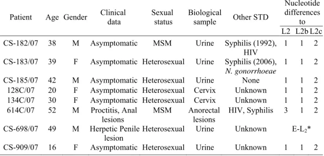
C
C
H
H
A
A
P
P
T
T
E
E
R
R
1
1
G
G
e
e
n
n
e
e
r
r
a
a
l
l
I
I
n
n
t
t
r
r
o
o
d
d
u
u
c
c
t
t
i
i
o
o
n
n
1. General Introduction
1.1.Chlamydia trachomatis (C. trachomatis)biology
Chlamydiae are obligate intracellular bacteria characterized by a highly specialized biphasic
developmental cycle that is unique among prokaryotes. These parasites infect a diverse array of vertebrates (including humans, birds, ruminants and amphibians) and belong to the family Chlamydiaceae, which encompass the genera Chlamydia and Chlamydophila [1]. The later comprises the species C. psittaci, C. abortus, C. caviae, C. felis, C. pecorum, and C. pneumoniae, while the former contains the species, C. trachomatis, C. muridarum, and
C. suis.
As other Chlamydiae, the human pathogen C. trachomatis has undergone massive genome reduction after acquiring an intracellular lifestyle, resulting in the disappearance of several biosynthesis pathways. Consequently, this pathogen has one of the smallest genomes among bacteria [~1.04 million base pairs (bp)], corresponding to just about a quarter of the size of the genome of common free-living bacteria such as Bacillus subtilis or Escherichia coli. Interestingly, of its ~900 open reading frames (ORFs), ~30% have no homology with
other bacterial proteins. C. trachomatis also harbours a 7.5 kbp plasmid, whose function is still unknown [2].
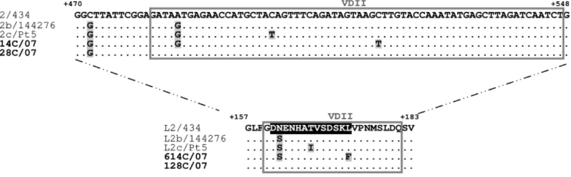
This pathogen can be divided into 18 serovars or genotypes, based on the differential immunoreactivity of its major outer membrane protein (MOMP) or polymorphism of ompA (that encodes MOMP) [3], respectively, which match 100%. Genotypes A-C and Ba usually cause ocular infections, genotypes D-K, Da, Ia and Ja are normally associated with ano-urogenital infections (with E and F representing ~50% of them), while genotypes L1-L3 are the causative agents of lymphogranuloma venereum (LGV). Genotypes can be further grouped into two distinct biological variants (biovars) based on the infected cell-type and on their in vitro infection properties: trachoma biovar (genotypes A-K, Ba, Da, Ia and Ja) is noninvasive, is mucosae-restricted and epitheliotropic, whereas LGV biovar is invasive and infects primarily lymphatic tissue, presenting faster and more vigorous growth in cell culture [4].
1.1.1. Diseases
Infections caused by C. trachomatis genotypes are a serious public health problem worldwide. Ocular infections can result in trachoma, a chronic inflammatory disease that afflicts 300-500 million people and leads to conjunctival scarring and blindness, especially in resource-poor nations [8-12]. Recently, this world’s leading cause of preventable infectious blindness [13] was recognized as one of the seven major neglected diseases of the 21st century [14], and it is currently the target of a World Health Organization (WHO) eradication campaign to the year 2020 (http://www.who.int/pbd/publications/trachoma/en/get_jan1998.pdf). C. trachomatis ano-urogenital infections are the primary cause of bacterial sexually transmitted diseases (STDs) in both industrialized and developing countries, causing cervicitis and urethritis [15]. The asymptomatic character of many of these infections (~75% in women and ~50% in men) often promotes their progression to the upper genital tract, causing epididymitis in men and pelvic inflammatory disease (PID) in women, which can lead to significant long-term sequelae, such as infertility and ectopic pregnancy [16-19]. C. trachomatis is also responsible for more-invasive and systemic diseases, such as LGV, through dissemination via infection of macrophages to regional draining lymph nodes, where it establish a chronic granulomatosis disease [20]. After a recent outbreak in Europe and the United States, LGV has been emerging as a significant problem among men who have sex with men (MSM) [21-23]. Surprisingly, despite aggressive antibacterial control measures over the past decade, the prevalence of C. trachomatis STDs has been increasing [19], with an estimated 92 million new cases
occurring worldwide each year (http://www.who.int/), constituting a enormous morbidity and socioeconomic burden. This gains even greater public health impact as these infections may also increase the risk of acquiring or transmitting human immunodeficiency virus (HIV) [24,25] and hepatitis C [26] as well as of developing invasive cervical carcinoma induced by human papilloma virus (HPV) [27,28].
1.1.2. Morphology
Chlamydiae exist as two morphologically distinct forms: a metabolically inert infectious
peptidoglycan synthesis (such as penicillin and D-cycloserine) and possess a complete set of genes for the peptidoglycan biosynthesis [2,5-7,37] that are expressed in the metabolically active stage of the chlamydial cycle [38] and are thought to be involved in the bacterial cell division [39]. RBs are larger than EBs (~1 μm), structurally flexible and osmotically fragile (due to the paucity of cross-linked membrane proteins), richer in RNA and contain diffuse and fibrillar DNA. These properties enable intracellular replication, uptake and transport of nutrients, protein synthesis as well as several other metabolic activities [32-34].
Both EB and RB cell walls encompass two sets of trilaminar membranes: an inner cytoplasmic membrane and an outer membrane [4,40], whose surface is covered with projections (especially in RBs) that are suspected to be type III secretion system (T3SS) ‘needle’ structures similar to those described for Salmonella spp., Yersinia spp. and Shigella spp. [41-44]. T3SS is a virulence mechanism used by several pathogenic Gram-negative bacteria to translocate effector proteins directly into host cell cytoplasm to manipulate specific cellular functions at distinct stages of infection [45]. It was already reported that Chlamydiae express an active T3SS throughout the infectious cycle, although the identity of the complete set of chlamydial type III secretion (T3S) effectors, which is predicted to encompass >10% of the bacterial chromosome, is still elusive [43,46,47]. Nevertheless, several studies [43,46,48,49] have been showing that this arsenal may be required for efficient cell invasion and inhibition of phagocytosis, establishment of the inclusion, acquisition of nutrients, modulation of intracellular trafficking, early inhibition and late induction of apoptosis, and avoidance of innate immune responses.
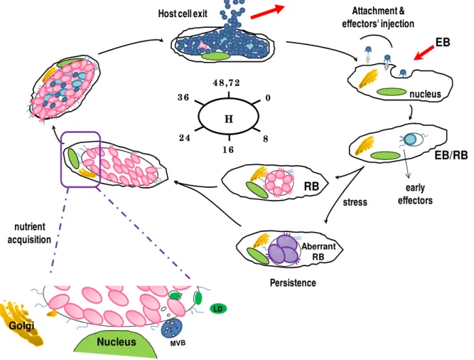
1.1.3. Developmental cycle
EB
1 6
0
8 2 4
3 6
4 8 ,7 2
H
nucleus
RB
EB/RB
Golgi
LD
nutrient acquisition
early effectors Attachment & effectors' injection Host cell exit
Nucleus
Aberrant RB
stress
Persistence
Figure 1.1 - Schematic illustration of the C. trachomatis developmental cycle.
MVB, multivesicular bodies; LD, lipid droplet.
1.1.3.1. Attachment and entry
been proposed that a functional clathrin-mediated pathway may also participate in C. trachomatis entry and may operate together with the Tarp-based mechanism, where Tarp
may alternatively mediate clathrin coat formation [70].
1.1.3.2. Inclusion modification and intracellular growth
Shortly after uptake, EB starts to differentiate into RB, while the RB-containing vacuole is rapidly modified to a membrane-bound inclusion, resulting in trafficking of the inclusion to the host perinuclear region and avoidance of phagolysosomal fusion [71]. RBs then undergo repeated cycles of binary fission, synthesizing their own DNA, RNA and proteins while obtain energy, nutrients and biosynthetic precursors from the host cell. As the chlamydial inclusion membrane is impervious to the diffusion of molecules >520 Da [72], Chlamydiae are adept at acquiring host metabolites through selective interaction with host organelles and subversion of membrane trafficking [49]. For instance, it has been showed that chlamydial recruitment of host-derived lipids, including cholesterol [73], sphingolipids [74,75], and glycerophospholipids [76,77] results from interception with Golgi-derived exocytic vesicles [74,78] and multivesicular bodies (MVBs) [79], or direct uptake of lipid droplets (LDs) [80]. Cumulative evidences indicate that chlamydial T3S-secreted inclusion membrane proteins (Incs) might have a central role in orchestrating inclusion modification through recruitment of key regulators of host cell membrane trafficking to the inclusion membrane, hence rerouting host cell vesicular trafficking and modulating inclusion fusion events [81]. During this process, chlamydial inclusion grows to accommodate the increasing number of RBs. Roughly midway through infection, chlamydial replication becomes asynchronous as RBs begin redifferentiating into EBs (by remodelling bacterial outer membrane and condensation of chromosome), which accumulate within the inclusion while the remainder RBs continue to multiply. Meanwhile, effector proteins required for both bacterial release and infection of new cells are assembled and preloaded onto EBs [43].
1.1.3.3. Persistence
temperature [89], infection of monocytes [90], or concomitant herpes infection [91]. Although the in vitro persistence is considered reversible upon removal of the stressful factor, aggressive inflammatory responses to repeated and persistent infections in vivo are thought to initiate irreversible pathogenic events (including promotion of cellular proliferation, tissue remodelling and scarring) that ultimately lead to debilitating chronic sequelae of blinding trachoma and tubal infertility [92,93].
1.1.3.4. Host cell exit
At about 48-72 h post-infection (pi), the expanded inclusion ultimately occupies almost of the entire host cell cytoplasm, and Chlamydiae are released by two mutually exclusive independent mechanisms: host cell lysis and extrusion. The former is a destructive process that consists of sequential rupture of bacterial inclusion, host nucleous and cell membranes, resulting in host cell death. In contrast, extrusion is a slow process dependent on actin polymerization, in which a portion of the inclusion is released by a membranous protrusion, leaving both the inclusion and host cell intact, although left-over inclusion remnants remain inside the host cell [94], which may contribute for persistent infection. The released multitude chlamydial infectious forms will then infect neighboring host cells or new hosts, initiating subsequent rounds of infection.
1.1.3.5. Evasion of host surveillance
Considering the unique biphasic life-cycle of Chlamydiae, these bacteria must be able to overcome various sets of host immune defence during both the extracellular and intracellular phases of their development in order to survive: i) before invading the host cells, extracellular EBs must be able to avoid attack by host pre-existing humoral and cellular effector mechanisms (such as neutralizing antibodies (Abs) and antigen-presenting cells), and ii) during infection, Chlamydiae have not only to block the early host phagolysosomal action
been showing that the early chlamydial antiapoptotic activity encompass the inhibition of mitochondrial cytochrome c release and downstream caspase activation [100], stabilization of inhibitor of apoptosis proteins (IAPs) [101], sequestration of proapoptotic proteins [102], and inhibition of transcription nuclear factor (NF)-kB activation pathway [103]. These antiapoptotic activities are prolonged during chlamydial persistence [104]. Interestingly, apoptosis also seems to have an immunomodulatory role in chlamydial infections as C. trachomatis appear to induce T-cell apoptosis by paracrine effects [104], which might
suppress host immune responses.
Alternatively, Chlamydiae may also avoid host immune recognition through degradation of specific host transcription factors required for IFN-γ-induced major histocompatibility complex (MHC) class I and II expression and lipid antigen presentation, both by secreting the protease-like activity factor (CPAF) [105,106].
1.2.C. trachomatis pathobiologic diversity
In vivo, C. trachomatis genotypes present biological uniqueness, exhibiting significant
differences in tissue tropism, pathogenesis and ecological success. To date, relatively little is known about the molecular factors underlying pathobiologic specificities of genotypes. However, the colossal genomic similarity (~99%) observed among the few currently fully-sequenced C. trachomatis genomes [2,5-7] suggests that the polymorphism of a relatively small number of loci likely determine C. trachomatis diversity.
Relevant contributions to explain the colonization of a particular tissue (eyes/genitalia) or cell-type (epithelial/lymph cells) came from the putative virulence factors type III effector Tarp [2,5-7], cytotoxin gene [107], and mainly trpRBA operon [108,109]. For instance, the presence of twice more tyrosine-rich tandem repeats within the Tarp N-terminus of LGV genotypes than that of noninvasive genotypes may function as a putative virulence mechanism that influence the colonizing capacity of the former [5]. In fact, as tyrosine phosphorylation of this domain is critical for chlamydial entry into host cells [110], the putative increased LGV Tarp phosphorylation may result in a more efficient entry process, and consequently in an enhanced ability to avoid the early host phagolysosomal action by rapid modification of chlamydial inclusion membrane.
genotypes have both domains entirely deleted [107]. As chlamydial cytotoxic activity appears to be correlated with a high multiplicity of infection, this putative virulence factor may allow epithelial-genital genotypes to circumvent host immune system by inactivating specific IFN-γ-mediated antimicrobial effectors at attachment sites, which may lead to disruption of host cytoskeleton and alteration of early vesicular trafficking [111].
Moreover, it was recently demonstrated that a fully active trpRBA operon is a mandatory condition for any strain to infect the genital but not the ocular tract [108]. Indeed, only genital-infecting strains retain the ability to encode a functional Trp synthase [109] that catalyzes the final steps in the biosynthesis of Trp from endogenous sources such as indole, which is produced by other microorganisms existing solely on genital flora. This ability is particularly important in an IFN-γ-rich environment (that is a result of a anti-chlamydial immune response), where an IFN-γ-induced tryptophan-catabolizing enzyme (indoleamine-2,3-dioxygenase, IDO) depletes intracellular tryptophan pools, thereby starving the pathogen from this essential amino acid [108]. Consequently, both the bacterial replication and secondary differentiation are inhibited, which may induce a persistent phase. Thus, the ability to synthesize tryptophan from endogenous indole, produced in a mixed microbial environment, is therefore a potential virulence factor for genital-infecting strains, allowing them to avoid host IFN-γ-mediated eradication.
However, none of these putative virulence factors fully explain the existence of three major tropism groups (eyes, epithelial-genitalia, and lymph nodes) neither the distinct invasiveness (mucosotropic and lymphotropic) nor the ecological success of genotypes. This becomes even more complex as, in contrast to the LGV strains, the preference for infecting mucosae-epithelial cells is not exclusive, and therefore ocular strains can occasionally be found in the urogenital tract and vice-versa [108]. Moreover, several studies [112-116] have been showing that, despite of its intracellular condition, this pathogen may undergo intragenic recombination and even extensive interstrain genetic exchanges (during mixed infections) involving polymorphic and immunogenic loci, which indicate some pathogen plasticity to deal with host niche-specific environmental changes in order to survive. Some of these loci belong to the nine-member polymorphic membrane protein gene family (pmpA to pmpI).
1.2.1. Pmps
Over the last decade, Pmps have been intense research targets as they are unique to Chlamydiaceae and encompass ~3.2% of the C. trachomatis chromosome (13.6% of the
suggests their importance in the chlamydial biology. Supporting this, a recent comparative genomic analysis between prototype strains from genotypes A and D [5] has shown that 20% of the total single nucleotide polymorphisms (SNPs) observed within coding regions derived from the nine pmp genes.
Although much more work still needs to be done to completely understand the nature, function and processing of Pmps, all the exposed findings suggest that Pmps may be important virulence factors with a dual role of promoting niche-specific pathogenesis-related adhesion function while providing antigenic diversity for chlamydial host immune evasion.
1.3. C. trachomatis vaccine
It is believed that the effective management of human chlamydial diseases will likely require the development of an efficacious prophylactic or therapeutic vaccine. Over the last two decades, there has been a gradual shift from the use of inactivated or attenuated intact pathogens (due to the existence of immunopathogenic components) to peptide or subunit vaccines [131]. However, despite of the intensive efforts, attempts to develop a chlamydial vaccine have been unsuccessful to date, conferring only a partial short-lived genotype-specific protective immunity [132].
Nevertheless, contemporary vaccine research continues to seek not only potent adjuvants and effective delivery vehicles but also a stable optimal antigen cocktail able to strongly elicit, at different stages of cycle, both the humoral and cellular immune responses required for a vaccine to confer long-lasting multi genotype-broad protection. The major chlamydial membrane component MOMP [133] is regarded as one of the leading candidates as it is immunodominant, immunoaccessible, neutralizing, highly variable among genotypes, and possesses species and genotype-specific epitopes that elicit both humoral and cellular immune responses [134-140]. However, it has been suggested that the success of a MOMP-based vaccine may depend on the presence of other antigens with neutralizing effects [132,141,142], such as the promising Pmps.
1.4. Scope of the thesis
It is assumed that a detailed understanding of the molecular factors that determine the differential pathogenesis of C. trachomatis genotypes, in particular their tissue tropism, virulence and immune evasion strategy, is vital for deciphering the basis of their ecological success as well as for defining and evaluating protective antichlamydial immune responses to foster vaccine design.
mirror the chlamydial infection in vivo. Therefore, due to the inapplicability of many of the traditional research methodologies used for other bacteria, genomics, transcriptomics and bioinformatics gain an enhanced relevance in Chlamydiae, being crucial tools for identifying loci, whose genetic and transcriptomic polymorphisms may get further insights in deciphering
the molecular basis of the pathobiologic diversity and ecological success of C. trachomatis. In order to contribute for this knowledge, the scope of the present Ph.D. thesis was to evaluate the genomic and transcriptomic features of several polymorphic loci, given especial relevance to the nine pmp family and the chlamydial key antigen (MOMP) as they are predicted to play an important role in C. trachomatis biological diversity. In particular, this thesis intended to scrutinize the putative biological role of the nine pmp family, to provide a deeper understanding of the impact of the host pressure on MOMP, to study the evolution of the C. trachomatis genotypes by analyzing a panel of heterogeneous loci scattered throughout the chromosome, and ultimately to outline the putative molecular aspects that may underline genotype ecological success. To achieve robust and reliable conclusions, both the traditional prototype strains (‘study-models’) and current clinical isolates were used in parallel whenever it was logical, since the later are not laboratory-adapted and reflect the immune selection that results from the infection of several hots. Therefore, the following detailed objectives were pursued and constituted the subject of six independent chapters that can be read separately:
i) to evaluate several normalization strategies for validation of real-time expression data in C. trachomatis (CHAPTER 2);
ii) to determine the expression profile of the nine-member pmp gene family throughout development for prototype and clinical strains representing the two chlamydial biovars, as well as to evaluate their immunoreactivities (CHAPTER 3);
iii)to perform a genetic characterization of the chlamydial major antigen (MOMP) of the strains isolated in Portugal on behalf of the recent LGV outbreak observed worldwide (CHAPTER 4);
iv) to perform an evolutionary mutational trend analysis of MOMP using all variant strains reported to date in Portugal (CHAPTER 5) as well as in the rest of the globe (CHAPTER 6);
v) and finally, to perform a high-scale evolutionary concatenation-based phylogenomic survey that comprised about one third of all chromosome SNPs of the 15 main C. trachomatis genotypes (CHAPTER 7), in order to understand the molecular
C
C
H
H
A
A
P
P
T
T
E
E
R
R
2
2
N
N
o
o
r
r
m
m
a
a
l
l
i
i
z
z
a
a
t
t
i
i
o
o
n
n
S
S
t
t
r
r
a
a
t
t
e
e
g
g
i
i
e
e
s
s
f
f
o
o
r
r
R
R
e
e
a
a
l
l
-
-
T
T
i
i
m
m
e
e
E
E
x
x
p
p
r
r
e
e
s
s
s
s
i
i
o
o
n
n
D
D
a
a
t
t
a
a
i
i
n
n
C
C
.
.
t
t
r
r
a
a
c
c
h
h
o
o
m
m
a
a
t
t
i
i
s
s
Published in
Borges V, Ferreira R, Nunes A, Nogueira PJ, Borrego MJ, Gomes JP (2010) J Microbiol Methods
82: 256-264.
Author Contributions
MJB and JPG conceived and designed the experiments; VB, RF, AN, MJB and JPG performed the
experiments; PJN performed the statistics; VB, RF, AN and JPG analyzed the data; VB, RF and JPG
2. Normalization Strategies for Real-Time Expression Data in
C. trachomatis
2.1.Abstract
C. trachomatis is a widespread obligate intracellular pathogen genetically nontractable for
which transcriptomics is a fundamental tool to better understand its biology. However, the suitability of endogenous controls for normalization of transcriptomic data in this bacterium still needs validation. We aimed to assess the stability of 10 genes for their potential use as endogenous controls in real-time quantitative polymerase chain reaction (PCR) assays at both normal and stress (D-cycloserine treatment) growth conditions throughout the developmental cycle of three C. trachomatis strains with different tissue tropism. Normalization was performed by real-time absolute quantification of the bacterial genomes. We also tested the applicability of two widely used softwares (geNorm and Normfinder) to our data. For all strains, we found that 16SrRNA was the most stably expressed gene throughout the chlamydial normal developmental cycle, which indicates its potential use as endogenous control in relative expression assays. However, it was highly unstable under D-cycloserine treatment (where oppA_2 was top-ranked), suggesting prudence when using ribosomal genes in expression experiments involving stress conditions. The geNorm and Normfinder algorithms revealed contrasting results and seem inappropriate for the selected pool of genes. Considering the multiplicity of experimental conditions, there should be an in loco validation of endogenous controls, where 16SrRNA gene appears to be in the front line. Alternatively, normalization of expression data against genomic DNA, which is less influenced by experimental constraints that are especially relevant for intracellular organisms, likely constitutes a good option. Moreover, the number of genomes also seems to be less subject to variation than expression of endogenous controls when working under stress conditions. The present study constitutes the first evaluation of putative endogenous controls for real-time expression assays in C. trachomatis.
2.2.Introduction
C. trachomatis is a widespread obligate intracellular bacterium, where genotypes A–C are the
which replicates within a host cell inclusion. The normal developmental cycle can be disturbed by several stress conditions such as nutrient starvation, temperature, host immune response or antibiotic treatment, which are known to affect transcription [143,144].
Gene expression in Chlamydia has been mostly performed using real-time quantitative reverse transcription PCR (RT-qPCR), which presents well-known advantages over traditional mRNA quantification methods [145,146]. However, the requirement of a proper normalization strategy is probably the major problem when performing relative expression assays, where inappropriate methodologies can lead to inaccurate data and incorrect conclusions [147,148]. To date, numerous strategies for RT-qPCR data normalization have been applied, such as the use of the total RNA mass [148], the number of cells determined by quantitative culture [149] and the use of an external reference of known amount added to the cultures prior to sample processing [146,150]. Besides the inherent and well-known disadvantages of these three approaches, additional drawbacks limit their application to obligate intracellular organisms like C. trachomatis: i) the total RNA that is extracted is composed not only by variable proportions of bacterial rRNA, tRNA, mRNA, small non-coding RNA but also by eukaryotic RNA [150]; ii) quantitative cultures have low sensitivity and are difficult to reproduce [145,151]; iii) the amount of external reference that enters or stays out the host cell cannot be controlled, hampering the determination of bacteria-specific mRNA.
Considering the lack of a validated method for selecting genes as endogenous controls in C. trachomatis, we aimed to assess the stability of 10 chromosome-spread genes that revealed an apparent stability during development through microarray analysis [96]. These genes are not likely metabolically related to each other and include a ribosomal RNA gene (16SrRNA), housekeeping genes (yraL, fer, oppA_2, radA, yaeI, hemN_2, tyrP_2 and map), and CT147 that codes for a hypothetical “early endosomal antigen 1 homologue”. To achieve this, the number of chlamydial genomes, determined by cloning-based real-time absolute quantification, was used as the normalizing factor of gene expression. We tested three strains presenting distinct tissue tropism and growth rate. The gene expression stability was assessed in normal growth and under stress condition induced by D-cycloserine, an antibiotic that is known to interfere with bacterial cell wall synthesis, preventing RB differentiation into EB [163,164]. We further evaluated the accuracy of using statistical algorithms to validate endogenous control genes in this intracellular pathogen. Finally, we discuss the reliability of normalizing gene expression against the number of bacterial genomes in opposition to using the experimentally and/or mathematically determined top-ranked endogenous control genes.
2.3.Materials and methods
2.3.1. Preliminary assays
2.3.1.1. Preliminary assay 1: Evaluation of D-cycloserine toxicity
In order to determine the D-cycloserine concentration to be used in the expression assays under stress conditions, we first evaluated the cytotoxic effect of this antibiotic on cell monolayers and C. trachomatis. Therefore, the old enriched medium in the T25 cm2 flasks
containing HeLa 229 cells not infected and infected with this bacterium (see below details of chlamydial culture) was replaced by fresh enriched medium with a concentration range of 0.001 μg/ml to 200 μg/ml of D-cycloserine at 18 h (pi). The morphologic alterations of HeLa 229 cells and C. trachomatis inclusions were then examined by phase-contrast microscopy every hour post-treatment (pt), in order to select the antibiotic concentration that induces the abnormal RB development without interfering with normal growth of HeLa 229 cells.
independent DNA and RNA extraction using QIAamp® DNA Mini Kit (Qiagen) and RNeasy Mini Kit (Qiagen), respectively, according to manufacturer's instructions. After elution, the DNA and RNA yields were determined by optical density (OD) measured at A260 nm. The
reproducibility of both extraction methods was statistically evaluated by calculating the variation coefficient within each group of five samples, and then by the determination of the mean value of the three independent assays.
2.3.1.3. Preliminary assay 3: Evaluation of RT efficiency using random hexamers versus target-specific primer
In order to evaluate if different RT methodologies influence gene expression data, we assessed if the levels of cDNA obtained by using random hexamers or a target-specific primer reflect the initial quantities of RNA. This assay was performed solely for 16SrRNA as it was the most stably expressed gene (see results for details). cDNA was generated from four-fold serial dilutions of the same RNA, ranging from about 50 pg to 50 ng of total RNA. Subsequently, 5 μl of each cDNA were used in qPCR reaction. Both RT and qPCR reaction conditions are described below. To evaluate the performance of both assays we calculated the respective sum of the square residuals calculated for each concentration. Assuming that each residual approximates to the standard normal distribution, their sum was considered to follow a chi-square distribution enabling the probability calculation of the observed deviation values being equal to zero.
2.3.2. Chlamydial culture and harvesting
The C. trachomatis prototype strains used in the present study represent the three disease groups: C/TW3 (ocular), E/Bour (urogenital) and L2/434 (LGV). Cell culture of each strain was performed as previously described [165]. Briefly, eight T25 cm2 flasks of confluent HeLa
229 cell monolayer were inoculated with each strain by centrifuging for 1 h at 34 °C and subsequent incubation for 1 h at 37 °C in 5% CO2. Cell medium was then replaced by
enriched medium (MEM with 10% fetal bovine serum, vitamins, nonessential amino acids, glucose and 0.5 μg/ml cycloheximide) and cultures were allowed to grow at 37 °C in 5% CO2.
For C. trachomatis growth under normal conditions, five T25 cm2 flasks were used and the
same cell harvesting protocol, as previously described [166]: medium was removed and cells were scraped with 1400 μl of phosphate buffered saline (PBS) at 4 °C; for disruption of eukaryotic cells and bacterial release, the suspension was sonicated for 30 s and the harvested cells were submitted to low-speed centrifugation (1500 rpm for 2 min) at 4 °C; finally, the supernatant was collected, homogenized and rigorously divided into two identical aliquots. One of these was stored at −20 °C until DNA extraction and the other aliquot was subjected to immediate RNA extraction due to its very short half-life. The use of two aliquots is a mandatory step as RNA quantification demands exclusion of any contaminant DNA and, on the other hand, DNA extraction procedure does not protect RNA. To ensure accuracy, three biological replicates of each strain were used.
2.3.3. Nucleic acid extraction
From each time-point, extraction of total RNA was carried out as previously described [127]. Basically, lysates were centrifuged at 14,000 rpm for 10 min (at 4 °C) to collect the Chlamydia-containing pellet which was subsequently resuspended in lysozyme-containing TE buffer. Then, RNeasy Mini Kit (Qiagen) was used according to the package inserts. Since double-stranded DNA is more effectively amplifiable than the single stranded cDNA that is generated from RNA, an on-column DNA digestion using 30 U of RNase-free DNase (Qiagen) was performed to ensure the complete removal of residual contaminant DNA. In fact, DNase treatment has proven to be highly effective by removing >98% of DNA [149]. RNA was eluted in 50 μl of RNase-free water and stored at −80 °C in RNase-free tubes after A260 nm quantification.
For DNA extraction, the respective −20 °C aliquot was centrifuged at 14,000 rpm for 10 min (at 4 °C), the pellet was resuspended in 200 μl PBS and the QIAamp® DNA Mini Kit (Qiagen) was used, according to manufacturer's instructions. DNA was eluted in 50 μl of AE buffer, quantified at A260 nm and stored at −80 °C until use.
2.3.4. RT assays
cDNA was generated from 2 μl of RNA sample from each time-point using TaqMan RT Reagents (Applied Biosystems), as previously described [127,166]. Briefly, 2.5 μM of random hexamers, 5.5 mM MgCl2, 500 μM of each dNTP, 1×RT Buffer, 0.8 U/μl RNase
random hexamers or target-specific primer (see results), we decided to evaluate if the expression profile throughout development for the most stable gene (16SrRNA) would be similar using both methodologies. Thus, we have also generated cDNA for 16SrRNA by using 0.75 μM of 16SrRNA-10 primer (Table S2.1) and keeping the remaining RT conditions. cDNA were stored in DNase-free tubes at −80 °C, until use.
2.3.5. Generation of standard curves
For quantification of the number of bacterial genomes, a plasmid standard curve was generated as previously described [167]. Briefly, an amplified fragment of the single-copy of C. trachomatis ompA (Table S2.1) was cloned into the TOPO vector using the TOPO TA
technology for PCR products prior to transformation into DH5α competent cells (Invitrogen), according to manufacturer's instructions. Plasmid DNA was purified using the QIAprep Spin Miniprep Kit Protocol (Qiagen), according to package inserts. To ensure elimination of any contaminating RNA that can interfere with plasmid quantification, RNase A was used. Confirmation of the cloning and transformation success was performed by EcoRI restriction digestion of the plasmids as well as by PCR amplification (using the same primers used to generate the fragments for cloning) and sequencing of each cloned fragment. The plasmid copy number was determined by A260 nm measurement of the plasmid concentration,
according to the formula: No. plasmid/μl = (Avogadro's No. × Plasmid conc.(g/μl)) / MW of 1 mol of plasmids (g). Finally, eight-serial plasmid dilutions (concentration ranging from about 30 to 1×10⁶ plasmid copies/μl) were prepared in order to generate the external standard curve for qPCR.
For transcripts quantification, a 48 h (pi) chlamydial culture was used for chromosomal DNA extraction. Subsequently, this stock DNA was subjected to eight two-fold serial dilutions in DNase-free water to generate the standard curves. The use of DNA standard curves allowed us to simultaneously compare the expression levels among different genes at each time-point, as DNA represents equal amounts of each single copy gene. This cross-comparison could not be achieved using cDNA standard curves.
2.3.6. Real-time quantitative PCR
The real-time quantification was achieved using the ABI7000 SDS, SYBR Green chemistry and optical plates (Applied Biosystems), as previously described [166]. The qPCR reagents consisted of 1× SYBR Green PCR Master Mix (Applied Biosystems), 400 nM of each primer and 5 μl of sample DNA or cDNA, in a final volume of 25 μl.
For each strain, plates included a plasmid standard curve and duplicates of DNA extracted at each time-point (for absolute quantification of bacterial genomes), as well as DNA standard curves and duplicates of cDNA obtained at each time-point (for quantification of transcripts). “No template” and “no RT” controls were also included in every qPCR assays. The thermocycling profile was: 10 min/95 °C followed by 40 cycles of 15 s/95 °C and 1 min/60 °C. The dissociation melting curves of the PCR products, generated by stepwise increase of the temperature from 60 °C to 95 °C, were used to verify the specificity of the amplified products. Finally, raw qPCR data at each time-point was normalized against the number of chlamydial genomes determined for the corresponding T25 cm2 flask. The final
qPCR results were based on three independent experiments for all strains.
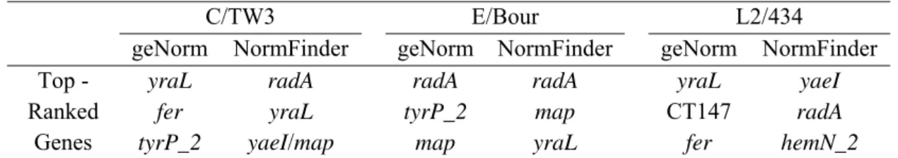
2.3.7. Software applications (geNorm and Normfinder)
Gene expression stability was evaluated using two widely used MS Excel applications, geNorm version 3.5 [160] and Normfinder version 0.953 [161]. Briefly, the geNorm application assumes that the expression ratio of two stable genes will remain identical in all samples, independently of experimental conditions and cell-type, and computes a reference gene stability measure (M) for all genes included in a study. It also allows the stepwise elimination of the gene with the highest M value (less stable) and the recalculation of new M values for the remaining genes, and so on, until the two most stable are left. geNorm further determines the minimal number of genes required for normalization, taking into account a normalization factor based on the geometric mean of these best-scoring reference genes. With respect to Normfinder analysis, it assumes groups of samples and intra/intergroup variations to calculate the stability value for all genes tested, allowing the identification of the best endogenous control candidate and also the selection of the most stable pair of reference genes.
2.4.Results
2.4.1. Preliminary assays
the subsequent experiments, we used a D-cycloserine concentration of 30 μg/ml, which is in agreement with previous studies [168,169]. During D-cycloserine treatment no apparent motion inside inclusions was observed at 6 h (pt) for all strains and both inclusions and RB showed abnormal size and shape (data not shown), without visible cytotoxic effects on HeLa cells. Contrarily, typical chlamydial inclusions and bacterial Brownian movements were detected (noticeably from 20 h pi) during the normal growth cycle.
The reproducibility of the nucleic acids extraction is a crucial aspect of the protocol that could bias the final normalized expression results. According to the normalization strategy adopted in this study, the number of chlamydial genomes found for each T25 cm2
flask and the corresponding transcript amounts are obtained independently from one another, since both DNA and RNA were subjected to different extraction methods. In our preliminary assay, a mean variation coefficient of 22.1% (standard deviation (SD)±1.0%) and 26.9% (SD±2.1%) was obtained for DNA and RNA, respectively. Thus, it is reasonable to expect that the final normalized gene expression data may be slightly biased.
Evaluation of RT efficiency using random hexamers versus target-specific primer (for 16SrRNA) showed that the final proportions of cDNA obtained mirrored the initial amounts of RNA used in RT reaction. Indeed, qPCR assays revealed differences of about two threshold cycle (Ct) values between every two consecutive four-fold dilutions, where the probability of the deviation between the theoretical and the experimental obtained Ct being similar to zero was found to be 0.999 for both assays (data not shown). Curiously, the A260 nm
determined for all cDNA samples was similar instead of reflecting their four-fold difference between consecutive dilutions. Thus, the A260 nm determination is not a reliable method for
cDNA quantification because the nonconsumed RT reagents (nucleotides, primers, and enzyme) are the major responsible for the A260 nm measurements rather than the generated
cDNA.
2.4.2. Real-time quantitative PCR
2.4.2.1. Chlamydial growth
The growth profile for C/TW3, E/Bour and L2/434 (Fig. 2.1) revealed an overall increase in chlamydial genomes of about 43-fold, 22-fold and 188-fold (from 4 h to 42 h pi), respectively. However, one E/Bour replicate showed less bacterial load for 12 h (pi) than for 4 h (pi), which could be due to the ~22% variation coefficient obtained in the DNA extraction procedure and/or to the inherent losses during cell harvesting. Surprisingly, C/TW3, which is assumed to have slow growth characteristics as any ocular strain [170], presented a doubling time of 2.87 h (SD±0.25) and the exponential phase starting at 12 h, although this period ended up at 20 h (pi). On the other hand, for E/Bour and L2/434, an extended exponential period (12–30 h pi) was observed with doubling times of 4.14 h (SD±1.05) and 2.36 h (SD±1.65), respectively.
Figure 2.1 - Growth profile of C/TW3, E/Bour and L2/434 prototype strains. For each strain,
the results at each time-point are represented as a mean±SD of the logarithmic fold-difference of
the genome copy number (in each T25 cm2 flask) compared to 4 h (pi). Black bars refer to
normal growth conditions (4, 12, 20, 30 and 42 h pi), while grey bars represent bacterial growth
under D-cycloserine treatment. Antibiotic was added at 18 h (pi) and harvesting was done at
18+6, 18+16 and 18+36 h (pi). Results are based on three biological replicates of each strain.
2.4.2.2. Gene expression
Figure 2.2 illustrates a global view of the expression pattern of each gene based on the log2 value of the mean expression at each time-point. In a rough estimation, genes of L2/434
For instance, for C/TW3, five genes (yraL, fer, CT147, radA and yaeI) were simultaneously expressed at both the early- and mid-stages (4 h to 20 h pi) of chlamydial development. However, this chart does not allow an evaluation of the expression stability, as variations of about eight-fold can be represented by the same color.
Figure 2.2 - Global view of the expression patterns of the 10 genes for
C. trachomatis. Strains C/TW3, E/Bour and L2/434 were grown under normal conditions (4, 12, 20, 30 and 42 h pi). Expression values were normalized against the
number of bacterial genomes. Each interval represents the log2 values of the mean
expression levels at each time-point, where the same color can represent variations
up to eight-fold. Results are based on three biological replicates of each strain.
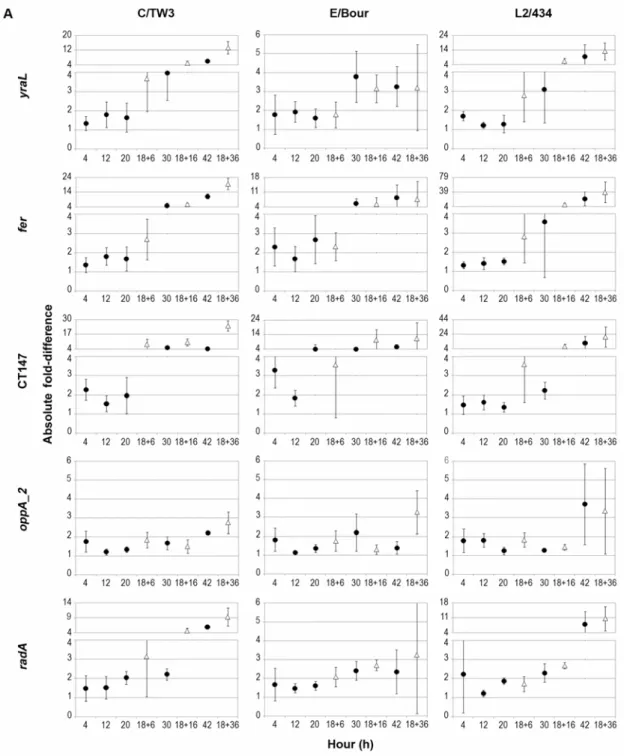
Thus, in order to accurately assess their expression stability, we carried out another strategy by calculating the absolute fold-difference (FD) between each time-point and the mean expression (determined for the entire cycle under normal growth conditions) (Fig. 2.3). Globally, most genes showed variation above four-fold to the respective mean. Exceptions were observed for: oppA_2, hemN_2 and 16SrRNA for C/TW3, oppA_2 and 16SrRNA for L2/434, and all genes except fer and CT147 for E/Bour. Moreover, 16SrRNA was the only gene for which expression variations never exceeded two-fold for all strains. However, oppA_2 and hemN_2 also exhibited variations below two-fold except for one time-point for
Figure 2.3 - Evaluation of expression stability of the 10 genes for C/TW3, E/Bour and
L2/434 prototype strains. (A) Includes yraL, fer, CT147, oppA_2 and radA, whereas (B) includes yaeI, hemN_2, tyrP_2, map and 16SrRNA. Each graph represents the mean±SD absolute FD of expression values of each time-point compared to the mean expression value
calculated for the entire developmental cycle under normal growth conditions. Expression
values were normalized against the number of bacterial genomes. Black circles represent
normal growth (4, 12, 20, 30 and 42 h pi), and white triangles refer to D-cycloserine
Figure 2.3 (continued)
Table 2.1. Gene expression stability evaluation throughout normal growth cycle
a Sum of the absolute FD between each time-point and the mean calculated for
the whole cycle.
b
Sum of the corresponding SD.
Since 16SrRNA was the most stable gene in the expression study using random hexamers in the RT reaction, we determined its expression profile throughout development of all strains using a target-specific primer. Matching results were obtained, where 16SrRNA gene did not exceed two-fold variation relatively to the mean expression value for all time-points (Figs. 2.3B & 2.4A).
2.4.2.3. D-cycloserine assays
As expected, the influence of D-cycloserine treatment on bacterial growth did not seem to affect the normal increase of genome copy number (Fig. 2.1). In fact, this antibiotic is known to block the peptidoglycan synthesis rather than interfering with chromosomal replication [163,164,168].
The effect of D-cycloserine in gene expression was also assessed (Fig. 2.3). For most genes, the largest expression variation occurred at 36 h (pt), when compared to the mean expression calculated from mRNA levels at time-points with no antibiotic treatment. In general, for all strains, the prolonged exposure to D-cycloserine resulted in a gradual decrease of gene expression (not shown in Fig. 2.3). For E/Bour, all genes except fer, CT147 and yaeI showed no expression variations above four-fold to the mean value, suggesting a lower influence of this antibiotic on the gene expression of this strain compared to that of C/TW3 and L2/434 where greater expression fluctuations were observed for all genes. Globally, oppA_2 and hemN_2 were the least variable genes under this stress condition with no
variations larger than four-fold to the mean value, for all strains (except hemN_2 for L2/434).
C/TW3 E/Bour L2/434
Genes Rank Σ FDa Σ SDb Rank Σ FDa Σ SDb Rank Σ FDa Σ SDb 16SrRNA 1 6.27 0.94 1 7.46 1.11 1 7.46 2.15
oppA_2 2 8.13 1.27 2 7.92 2.21 2 9.85 3.39
hemN_2 3 8.81 2.31 4 8.86 2.67 3 14.17 6.91
map 4 11.56 2.51 3 8.77 1.69 7 17.51 2.43
yaeI 5 11.97 2.49 7 10.95 4.38 5 16.02 8.16
radA 6 13.28 2.38 5 9.55 2.99 4 15.54 8.99
yraL 7 14.87 3.98 8 12.39 4.47 6 16.87 10.93 CT147 8 15.03 4.16 9 18.93 7.73 8 18.50 10.90
fer 9 20.93 5.07 10 20.61 11.18 10 30.29 20.16