Abstract
Submitted: April 24, 2017 Modification: August 7, 2017 Accepted: September 26, 2017
Dynamics of the seasonal airborne
propagation of
Staphylococcus aureus
in academic dental clinics
Objective: Staphylococcus aureus strains can be disseminated during dental treatments and occasionally lead to the contamination and infection of patients and dentists, which is an important public health problem. The dynamics of the airborne propagation and the genetic diversity of S. aureus
isolated in an academic dental clinic environment were investigated using isoenzyme typing. Material and Methods: The isoenzymes of 44 previously reported isolates were obtained from fresh cultures and extracted using glass beads. Nine isoenzymes were investigated using multilocus enzyme electrophoresis (MLEE). The genetic diversity and relationship among the strains (electrophoretic type – ET) were determined using statistics previously described by Nei25 (1972) and the SAHN grouping method (UPGMA algorithm). Results: Clonal pattern analyses indicated a high level of genetic polymorphism occurring among the 33 ETs, which were grouped into five taxa. Each taxon presented one or more clusters that were moderately related and that contained two or more identical/highly related isolates, revealing seasonal airborne propagation in these dental clinic environments. Conclusions: These data suggest the occurrence of active microevolutionary processes in S. aureus as well as the possibility of environmental propagation during a 14-month time span. Such findings are important to show that multi-user academic dental clinics can retain certain strains that are spreadable to different niches.
Keywords: Staphylococcus aureus. Dentistry. Environment. Molecular biology. Genetic diversity.
Wagner Luiz de Carvalho
BERNARDO1
Jeferson Júnior da SILVA1,2 José Francisco HÖFLING1 Edvaldo Antônio Ribeiro ROSA3 Marcelo Fabiano Gomes BORIOLLO1,2
1Universidade Estadual de Campinas, Faculdade de Odontologia de Piracicaba, Departamento de
Diagnóstico Oral, Laboratório de Microbiologia e Imunologia, Piracicaba, São Paulo, Brasil.
2Universidade José do Rosário Vellano, Faculdade de Medicina, Laboratório de Farmacogenética e
Biologia Molecular, Alfenas, Minas Gerais, Brasil.
3Pontifícia Universidade Católica do Paraná, Escola de Ciências da Vida, Unidade de Pesquisa com
Xenobióticos, Curitiba, Paraná, Brasil. Corresponding address:
Marcelo F. G. Boriollo Universidade Estadual de Campinas -Faculdade de Odontologia de Piracicaba -
Introduction
The dissemination of S. aureus is an important public health problem because its resistant strains are involved in severe infections, predominantly in children and hospitalized patients21. Dental practitioners treat
a wide range of patients. Therefore, it is likely that they will have contact with people colonized or infected with drug-resistant microorganisms4. High resistance
rates against the antibiotics used for prophylaxis in Dentistry have been detected for pathogens associated with bacterial endocarditis such as S. aureus19. S.
aureus strains can be disseminated during dental treatments and occasionally lead to the contamination and infection of patients and dentists24. Certain
aspects of practicing Dentistry may contribute to the transmission of microorganisms. Skin, environment, and instruments can be contaminated with saliva, blood, or organic debris during routine dental treatment29. Several investigators have observed an
increase in the amount of microorganisms during clinical procedures in a dental environment, suggesting contamination from aerosols and especially when high-speed handpieces or ultrasonic scalers are used1,20.
Among the species identified by microbiological studies, Streptococcus viridans and Staphylococcus
spp. are the most prevalent microorganisms found on the surfaces of dental equipment12, which includes
the methicillin-resistant S. aureus that has been detected on the surfaces of dental operatories, air-water syringes, and reclining chairs18.
Phenotype- and genotype-based methods have allowed researchers to classify microbial isolates in systematic, taxonomic, evolutionary, phylogenetic, and epidemiological studies2,28. MLEE has been used
for several decades as a standard method in eukaryotic population genetics and systematics26 as well as in
large-scale studies to estimate the genetic diversity and structure in natural populations of a variety of bacterial23, fungal, and yeast3 species. Its special
advantage is that mobility variants (electromorphs or allozymes) of an enzyme can be directly equated with alleles at the corresponding structural gene loci. Another attractive feature is the likelihood that much of the electrophoretically demonstrable polymorphic variation in enzymes is selectively neutral or nearly so and, therefore, minimally subjected to evolutionary convergence2,28. The genetic interpretation of
patterns depends on the ploidy of the organism, and
different rules have to be applied for haploid and diploid organisms2,28. Based on these rules, the allelic
makeup of each isolate is determined over the set of different enzymes studied (between 10 and 30). The enzymes examined in MLEE typically participate in the basic metabolism of the cell and are less likely than other loci to be under selective pressure from the environment or to be subjected to convergence2.
Therefore, MLEE data are considered representative of the whole genome of an organism and form a good basis for population genetic studies of bacteria, fungi, and protozoa2. The genetic data obtained by MLEE
can be used for a variety of purposes in the medical
microbiology field: (i) to infer the degree of genetic
recombination occurring within a natural population, (ii) to assess the degree of genetic isolation of natural populations, either for geographical and ecological reasons or because of biological barriers, (iii) to assess the degree of genetic relatedness between organisms or populations, and thus, provide systematic and taxonomic implications, (iv) to identify bacterial clones within species that can be associated with particular clinical patterns, with a particular frequency in clinical diseases or with a higher level of pathogenicity, and (v) epidemiological tracing of microorganisms. This method represents an invaluable complement to the most recent molecular typing methods, particularly for large-scale epidemiological studies2. In addition,
MLEE possesses an excellent typeability (i.e., the percentage of distinct strains obtained), excellent reproducibility (i.e., the percentage of strains that show the same results in repeated assays), and good discriminatory power (i.e., its ability to differentiate unrelated strains)2,3.
Here, we evaluated the genetic diversity and the distribution of airborne S. aureus in a multi-user academic clinic by MLEE and a clustering analysis in an attempt to understand the air-spreading behavior of this bacterium.
Material and methods
Bacteria
A total of forty-four S. aureus isolates (predominantly
ampicillin-resistant and β-lactamase positive) were employed. The bacteria were isolated, identified, and had their resistance profiles performed as described
passively harvested from a clinical environment (the air) in the academic clinic (Dental School). Open plates containing MSA (mannitol salt phenol-red agar) selective medium (Merck; Darmstadt, Germany) were placed in 12 dental clinic environments for two hours during intense and periodical multi-activities. This procedure was conducted for 12 months twice a month with intervals of ±15 days between one harvest and another, totaling 24 collections (from September to mid-December, 2000; from March to June, 2001; from August to mid-December, 2001). The plates were then incubated at 37°C for 48 h. Colonies indicating mannitol fermentation by staphylococci were selected, and the characterization of S. aureus was performed by Gram staining; growth in a chromogenic medium CHROMagar™ Staph aureus (Probac do Brasil Produtos Bacteriológicos Ltda.; São Paulo, SP, Brazil); and
catalase, coagulase, and β-lactamase tests using CefinaseTM discs (Becton, Dickinson and Company;
Franklin Lakes, NJ, EUA). Antimicrobial susceptibility testing was also performed1.
Enzyme extraction
Bacterial cultures were grown in flasks containing
200 mL BHI (Brain Heart Infusion) culture medium (HiMedia Laboratories GmbH; Einhausen, Germany) at 37°C for 24 h under constant shaking at 150 rpm. After growth, the cells were centrifuged at 5,000xg
for 3 min and washed in sterile 40 mM PBS (pH 7.5) three times. Pellets (~250 mg) were transferred to 2 mL microtubes containing 250 mL PBS and ~250 mg glass beads (0.45-0.55 mm). These mixtures were kept on ice (4°C) for 5 min and then agitated 4 times in a BeadBeater® (Biospec Products, Inc.; Bartlesville,
OK, USA) at 4,200 rpm for 30 s with one-minute intervals. Cell fragments were centrifuged at 5,000xg
for 5 min at 4°C using a centrifuge 5403, rotor 16F24-11 (Eppendorf AG; Hamburg, Germany). The resulting upper aqueous phase was applied to Whatman #3
filter papers wicks (12x5 mm) and maintained at
-70°C until use3,28.
Electrophoresis and specific enzyme stainings
The enzymes were separated in 13% (wt/vol) Penetrose 30® starch gels (dimensions 200x120x10 mm) (Refinações de Milho Brasil Ltda.; Mogi-Guaçu,
SP, Brazil) in Tris-citrate buffer pH 8.0 (electrode buffer diluted 1:29)28. The wicks were then immediately
soaked in 5 mL [0.02% (wt/vol)] bromophenol-blue solution and then perpendicularly applied to a
longitudinally cut gel (20 mm). Electrophoresis was performed in a horizontal and continuous system at 130 volts at 4°C overnight (bromophenol-blue migration equivalent to 80 mm) using Tris-citrate electrode buffer (pH 8.0). To assure reproducible results, S. aureus
ATCC® 29213 enzymes were included in each gel. After
the electrophoresis, the gels were put on acrylic base and sliced into 1.5-mm sections with the aid of rulers and #15 nylon strings. The sections were carefully placed inside white porcelain containers and stained for 9 enzymes (14 enzymatic loci)28. The enzymatic
activities that were analyzed included: alcohol dehydrogenase (EC 1.1.1.1), mannitol-1-phosphate dehydrogenase (EC 1.1.1.17), malate dehydrogenase (EC 1.1.1.37), glucose dehydrogenase (EC 1.1.1.47), D-galactose dehydrogenase (EC 1.1.1.48), glucose-6-phosphate dehydrogenase (EC 1.1.1.49), catalase (EC 1.11.1.6), and a- and b-esterase (EC 3.1.1.1.) (Figure 1). The enzymatic expression levels of alcohol dehydrogenase (Adh-1 and Adh-2), mannitol-1-phosphate dehydrogenase (M1p-1 and M1p-2), malate dehydrogenase (Mdh-1 and Mdh-2), and a- and b-esterase (a-Est-1, a-Est-2, b-Est-1 and b-Est-2)
identified two genetically interpretative loci.
Genetic interpretation of the MLEE patterns
Pattern interpretation was performed following the general rules commonly accepted in the deduction of allelic composition for haploid organisms. The bands on the gels were numbered in order of decreasing mobility, and their corresponding alleles were numbered by using the same nomenclature. A lack of demonstrable activity for an enzyme was scored as one null allele at the corresponding gene locus. Each unique combination of alleles over the 14 enzyme loci examined resulted in an electrophoretic type (ET)
–
subtype or strain28.Discriminatory power
The discriminatory power of the MLEE method, which is based on the numeric interpretation of electrophoretic patterns, was established using the numerical index of discrimination (D), which is according to the probability that two unrelated isolates
sampled from the test population will be classified into
following equation: , where N is the total number of isolates in the sample population,
S is the total number of types (strains) described, and
nj is the number of isolates belonging to the jth type
(strain). This equation was derived as follows. The probability that two isolates sampled consecutively will belong to that type (strain) is: .
These probabilities can be summed for all the described types (strains) to determine the probability that any two consecutively sampled isolates will be the same type (strain). This summation can be subtracted from 1 to obtain the equation above. This equation can be applied both to a direct comparison of the discriminating power of typing methods and to an analysis of the discriminating power of the combined typing schemes. To interpret the typing results with
confidence, an index greater than 0.90 is desirable3.
Cluster analysis
The statistics of Nei25 (1972) were used to estimate
the genetic distance among the S. aureus isolates: dij
= – In(I) or ,
where I is the normalized identity of genes between two populations (ranging from 0 to infinity), a measure of genetic distance based on the identity of genes (frequency of alleles for all loci, including monomorphic loci) among populations. This genetic distance measures the accumulated allelic differences
per locus, which can also be estimated from the amino acid sequences of proteins and even for a distantly related species. Therefore, if enough data Enzyme Compound
for staining
EC number Name Symbol Substrate Buffer Salt Coenzyme Dye and Catalyser
1.1.1.1. alcohol
dehydrogenase ADH Ethanol (3 mL) Isopropanol (2 mL)
200 mM Tris-HCl pH 8.0 (q.s.p. 50 mL)a
1% NAD
(2 mL) 1.25% MTT (1 mL)1% PMS (500 mL)
1.1.1.17 mannitol-1-phosphate dehydrogenase M1P Mannitol 1-phosphate (5 mg)
200 mM Tris-HCl pH 8.0 (q.s.p. 50 mL)a
1% NAD
(2 mL) 1.25% MTT (1 mL)1% PMS (500 mL)
1.1.1.37. malate
dehydrogenase MDH 2M Malic acid (6 mL)b
200 mM Tris-HCl pH 8.0 (q.s.p. 50 mL)a
1% NAD
(2 mL) 1.25% MTT (1 mL)1% PMS (500 mL)
1.1.1.47 glucose
dehydrogenase GDH D-glucose(500 mg) 200 mM Tris-HCl pH 8.0 (q.s.p. 50 mL)a
1% NAD
(2 mL) 1.25% MTT (1 mL)1% PMS (500 mL)
1.1.1.48 D-galactose
dehydrogenase GLDH Galactose(450 mL) Tris-HCl 100mM pH 8.4 (q.s.p. 50 mL)c
1% NAD
(1 mL) 1.25% MTT (1 mL)1% PMS (500 mL)
1.1.1.49 glucose-6-phosphate dehydrogenase G6PDH Glicose-6-phosphate disodium salt (100 mg)
200 mM Tris-HCl pH 8.0 (q.s.p. 50 mL)a
100 mM MgCl2 (1 mL)d
1% NADP
(1 mL) 1% PMS (500 mL) 1.25% MTT(1 mL)
1.11.1.6 catalase CATe
3.1.1.1. a- and b-
esterase EST a- and b- Naphthyl acetate (1%
solution in acetone)
(1.5 ml)
50 mM Sodium phosphate pH 6.0
(q.s.p. 50 mL)f
Fast Blue RR salt (25 mg)
Electrode buffer: Tris–citrate pH 8.0 [83.2 g of C4H11NO3 (Tris), 33.09 g of C6H8O7.H2O (Citric acid), 1L of H2O]; Gel buffer: Electrode buffer
diluted 1:29.
a 24.2 g of C
4H11NO3 (Tris), 1L of H2O (pH adjusted with HCl); b 26.8 g of C
4H6O5 (DL-malic acid) and 16 g of NaOH in 0.1L of H2O (caution: potentially explosive reaction);
c 12.1 g of C
4H11NO3 (Tris), 1L of H2O (pH adjusted with HCl); d 2.03 g of MgCl
2.6HCl (Magnesium chloride) in 0.1 L of H2O;
e Incubate gel slice for 30 min at 0oC in 50 mL of 0.1 M sodium phosphate pH 7.0 buffer, then pour off solution, and immerse it in 50 mL of
1.5% potassium iodide solution (KI) for 2 min. Therefore, rinse gel slice with water, and immerse it in 50 mL of 0.03% hydrogen peroxide (H2O2) solution. Mix gently and remove stain solution when white zones appear on dark-blue background;
f Sodium phosphate buffer pH 7.0: mix equal parts of 27.6 g of NaH
2PO4.H2O (monobasic) in 1 L of water and 53.6 g of Na2HPO4.7H2O in
1 L of water, then dilute the mixture 1:25 with water
are available, the genetic distances between any pair of organisms can be measured in terms of dij. In addition, this measure is applicable to any type of organism regardless of ploidy or matting scheme. Its interpretation in terms of enzyme loci infers that, on
average, 0 to an infinite number of allelic substitutions
are detected (by electrophoresis) in every 100 loci, from a common ancestral strain3,25. A tree with two-dimensional classifications (dendrogram), based on
matrix dij, was generated using the SAHN grouping method (Sequential, Agglomerative, Hierarchic, Nonoverlapping Clustering Methods) and UPGMA algorithm (Unweighted Pair-Group Method using an Arithmetic Average)3. Because MLEE provides
all levels of relatedness, which must be resolved by DNA fingerprinting methods (i.e., identifying the same strain in independent isolates, identifying microevolutionary changes in a strain, identifying clusters of moderately related isolates, and identifying completely unrelated isolates), a threshold (average value:dij) was established in the dendrograms to identify clusters of identical isolates and highly related isolates and taxa (singular taxon, i.e., a taxonomic group of any nature or rank) (0£dij< dij)3.
The Pearson product–moment correlation coefficient
(ranging from -1 to +1),
,
[where Xij stands for the character state value of character i in the operational taxonomy unit (OTU)
j, Xj is the mean of all state values for OTU j, and n
is the number of characters sampled], was used as a measure of the agreement between the genetic distance values implied by the UPGMA dendrograms and the ones from the original genetic distance matrices (dij). These agreements were interpreted as follows: r>0.9, very good fit; 0.8≤r<0.9, good
fit; 0.7≤r<0.8, poor fit; r<0.7, very poor fit. All of
these analyses were obtained using the NTSYS-pc 2.1 program3.
Results
The S. aureus isoenzymatic patterns were reproducible in different gels after three repetitions of each electrophoretic run. Based on the haploid nature of S. aureus, such patterns indicate the following characteristics (Figures 2 and 3): 12 (85.7%) enzymatic loci were polymorphic for two, three, four,
and five alleles (2 alleles: Adh-1, Adh-2, M1p-2, Mdh-1, Gldh, and G6pdh; 3 alleles: M1p-Mdh-1, Gdh, β-Est-1; 4 alleles: α-Est-1 and α-Est-2; 5 alleles: β-Est-1).
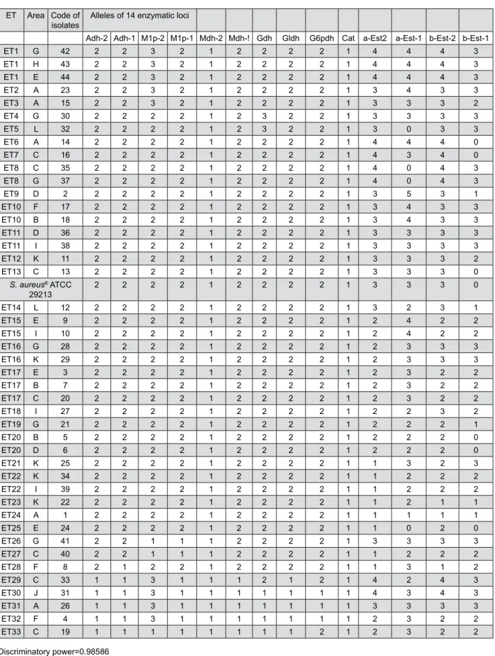
The mean number of alleles per locus and the mean number of alleles per polymorphic locus were equal to 2.57 and 2.83, respectively. The combination of existing alleles in 14 enzymatic loci revealed 33 electrophoretic types (ETs) (75% of the total isolates), Figure 2- Diagram of the multilocus enzyme electrophoresis (MLEE) patterns of the S. aureus isolates. The migration of the enzymes
that is, identical isolates corresponding to the same strain or ET (i.e., dij=0.000). Only two loci were monomorphic for only one allele, Mdh-2 and Cat. The
discriminatory power of the MLEE method, which is based on the numeric interpretation of electrophoretic patterns, was 0.98586, that is, a 98.6% probability
ET Area Code of
isolates Alleles of 14 enzymatic loci
Adh-2 Adh-1 M1p-2 M1p-1 Mdh-2 Mdh-! Gdh Gldh G6pdh Cat a-Est2 a-Est-1 b-Est-2 b-Est-1
ET1 G 42 2 2 3 2 1 2 2 2 2 1 4 4 4 3
ET1 H 43 2 2 3 2 1 2 2 2 2 1 4 4 4 3
ET1 E 44 2 2 3 2 1 2 2 2 2 1 4 4 4 3
ET2 A 23 2 2 3 2 1 2 2 2 2 1 3 4 3 3
ET3 A 15 2 2 3 2 1 2 2 2 2 1 3 3 3 2
ET4 G 30 2 2 2 2 1 2 3 2 2 1 3 3 3 3
ET5 L 32 2 2 2 2 1 2 3 2 2 1 3 0 3 3
ET6 A 14 2 2 2 2 1 2 2 2 2 1 4 4 4 0
ET7 C 16 2 2 2 2 1 2 2 2 2 1 4 3 4 0
ET8 C 35 2 2 2 2 1 2 2 2 2 1 4 0 4 3
ET8 G 37 2 2 2 2 1 2 2 2 2 1 4 0 4 3
ET9 D 2 2 2 2 2 1 2 2 2 2 1 3 5 3 1
ET10 F 17 2 2 2 2 1 2 2 2 2 1 3 4 3 3
ET10 B 18 2 2 2 2 1 2 2 2 2 1 3 4 3 3
ET11 D 36 2 2 2 2 1 2 2 2 2 1 3 3 3 3
ET11 I 38 2 2 2 2 1 2 2 2 2 1 3 3 3 3
ET12 K 11 2 2 2 2 1 2 2 2 2 1 3 3 3 2
ET13 C 13 2 2 2 2 1 2 2 2 2 1 3 3 3 0
S. aureus® ATCC
29213 2 2 2 2 1 2 2 2 2 1 3 3 3 0
ET14 L 12 2 2 2 2 1 2 2 2 2 1 3 2 3 1
ET15 E 9 2 2 2 2 1 2 2 2 2 1 2 4 2 2
ET15 I 10 2 2 2 2 1 2 2 2 2 1 2 4 2 2
ET16 G 28 2 2 2 2 1 2 2 2 2 1 2 3 3 3
ET16 K 29 2 2 2 2 1 2 2 2 2 1 2 3 3 3
ET17 E 3 2 2 2 2 1 2 2 2 2 1 2 3 2 2
ET17 B 7 2 2 2 2 1 2 2 2 2 1 2 3 2 2
ET17 C 20 2 2 2 2 1 2 2 2 2 1 2 3 2 2
ET18 I 27 2 2 2 2 1 2 2 2 2 1 2 2 3 2
ET19 G 21 2 2 2 2 1 2 2 2 2 1 2 2 2 1
ET20 B 5 2 2 2 2 1 2 2 2 2 1 2 2 2 0
ET20 D 6 2 2 2 2 1 2 2 2 2 1 2 2 2 0
ET21 K 25 2 2 2 2 1 2 2 2 2 1 1 3 2 3
ET22 K 34 2 2 2 2 1 2 2 2 2 1 1 2 2 2
ET22 I 39 2 2 2 2 1 2 2 2 2 1 1 2 2 2
ET23 K 22 2 2 2 2 1 2 2 2 2 1 1 2 1 1
ET24 A 1 2 2 2 2 1 2 2 2 2 1 1 1 1 1
ET25 E 24 2 2 2 2 1 2 2 2 2 1 1 0 2 0
ET26 G 41 2 2 1 1 1 2 2 2 2 1 3 3 3 3
ET27 C 40 2 2 1 1 1 2 2 2 2 1 1 2 2 2
ET28 F 8 2 1 2 2 1 2 2 2 2 1 1 3 1 2
ET29 C 33 1 1 3 1 1 1 2 1 2 1 4 2 4 3
ET30 J 31 1 1 3 1 1 1 1 1 1 1 4 3 4 3
ET31 A 26 1 1 3 1 1 1 1 1 1 1 3 3 3 3
ET32 F 4 1 1 3 1 1 1 1 1 1 1 2 3 2 2
ET33 C 19 1 1 1 1 1 1 1 1 2 1 2 3 2 2
Discriminatory power=0.98586
Figure 3- Allelic profiles of the 44 S. aureus isolates (33 S. aureus strains ETs) isolated from clinical environments (air) at the School of
that two unrelated S. aureus isolates sampled from
the test population will be classified as different types
(strains or ETs).
The genetic diversity among the S. aureus isolates/ strains was evaluated through the matrix dij and a UPGMA dendrogram (Figure 4). When considering
the threshold obtained (0≤dij<0.025: highly related
or identical isolates; 0.025≤dij<0.063: moderately
related isolates; 0.063≤dij<0.184: distantly related
isolates), the results showed five major groups or
taxa (i.e., singular taxon – a taxonomic group of any nature or rank), named A, B, C, D, and E. The taxon
A comprised three isolates (2 ET9 – area D, 19 ET33 – area C, 41
ET26 – area G) and four moderately related clusters (from
I to IV and totaling 29 isolates 65.9% or 21 ETs 60.6%):
Cluster I: 18 highly related and/or identical isolates
40.9% (isolates 3 ET17 – area E, 7 ET17 – area B, 9 ET15 – area E, 10 ET15
– area I, 11 ET12 – area K, 15 ET3 – area A, 17 ET10 – area F, 18 ET10 – area B, 20 ET17 – area C, 23 ET2 – area A, 28 ET16 – area G, 29 ET16 – area K,
30 ET4 – area G, 36 ET11 – area D, 38 ET11 – area I, 42 ET1 – area G, 43 ET1 – area H, and 44 ET1 – area E), or 10 highly related ETs 30.3%
obtained from 10 areas 83.3% during ±14 months (from
Sept. 19, 2000 to Nov. 27, 2001).
Cluster II: 3 highly related isolates 6.8% (isolates
13 ET13 – area C, 14 ET6 – area A, and 16 ET7 – area C), or 3 highly
related ETs 9.1%, including S. aureus ATCC 29213,
Figure 4- Genetic diversity of the 44 S. aureus isolates from the dental clinic environments (air) and the S. aureus type strain (ATCC®
29213). UPGMA dendrogram (rjk=0.83459) obtained from the genetic distance matrix dij25. The letters A to L correspond to the sampling
obtained from 2 16.7% areas during ±3 months (from
Dec. 12, 2000 to March 27, 2001).
Cluster III: 2 highly related isolates 4.5% (isolates 12 ET14 – area L and 27 ET18 – area I), or 2 highly related ETs 6.1%
obtained from 2 16.7% areas during ±5 months (from
Dec. 5, 2000 to May 29, 2001).
Cluster IV: 3 highly related isolates 6.8% (isolates
5 ET20 – area B, 6 ET20 – area D, and 21 ET19 – area G), or 2 highly
Figure 5- Design of the Academic Dental Clinic (School of Dentistry at Piracicaba, State University of Campinas, Brazil). The letters A
related ETs 6.1% obtained from 3 25% areas during ±6
months (from Oct. 31, 2000 to Apr. 3, 2001).
Taxon B comprised one isolate (isolate 8 ET28 – area F)
and two moderately related clusters (V and VI; totaling 7 isolates 15.9% or 6 ETs 18.2%):
Cluster V: 4 highly related and/or identical isolates
9.1% (isolates 25 ET21 – area K, 34 ET22 – area K, 39 ET22 – area I, and
40 ET27 – area C), or 3 highly related ETs 9.1% obtained from
3 areas 25% during ±5 months (from May 15, 2001 to
Oct. 19, 2001).
Cluster VI: 2 highly related isolates 4.5% (isolates 1 ET24 – area A and 22 ET23 – area K), or 2 highly related ETs 6%
obtained from 2 areas 16.7% during ±7 months (from
Sept. 19, 2000 to Apr. 3, 2001).
Taxon C comprised two isolates (4 ET32 – area F and
33 ET29 – area C) and one moderately related cluster (VII;
totaling 4 isolates 9.1% or 4 ETs 12.1%):
Cluster VII: 2 highly related isolates 4.5% (31 ET30 – area J and 26 ET31 – area A), or 2 highly related ETs 6.1% obtained
from 2 areas 16.7% during ±1 month (from May 29, 2001
to June 26, 2001).
Taxa D and E comprised only one cluster [Cluster VIII: 3 highly related and/or identical isolates 6.8% (32 ET5 – area L, 35 ET8 – area C, 37 ET8 – area G), or 2 highly related
ETs 6.1% obtained from 3 areas 25% during ±4 months
(from June 26, 2001 to Oct. 9, 2001)] and one isolate (isolate 24 2.3% or ET25 3% obtained from area E),
respectively. These results indicate that highly related or identical S. aureus isolates can emerge or remain for long periods in the same or different areas of dental
office environments (Figures 4 and 5).
Discussion
Strain delineation using multilocus enzyme electrophoresis (MLEE) allows for evaluations of genetic diversity and population structure analyses and provides a high discriminatory power and reproducibility2,3. Considered neutral markers
(i.e., invariable when they suffer environmental selective pressures), metabolic isoenzymes have
demonstrated a significant potential for the taxonomic,
systematic, genetic, evolutionary, and epidemiological characterization of bacteria and yeasts of medical importance2,3,23,26,28.
In this study, the enzyme electrophoretic profiles of
S. aureus samples from different gels were reproducible after three repetitions of each electrophoretic run. The
high discriminatory capacity (i.e., >98% probability that two unrelated S. aureus isolates sampled from
the test population will be classified as different
types or strains) of MLEE, which is based on the interpretation of enzyme electrophoretic patterns, was also observed (i.e., the combination of existing alleles in 14 enzymatic loci revealed 33 ETs). MLEE has again been proven to be a powerful tool for typing S. aureus strains for epidemiological studies. The results related to the reproducibility and discriminatory power of MLEE corroborate with those previously reported by other medically relevant studies24,28; however, the
discriminatory power described here is higher than those reported by other groups7,17.
Genetic polymorphisms have been reported in almost all natural populations, at all levels of genetic organization and from genotypic characteristics to phenotypic traits. The possible reasons for their existence have been the object of a long debate in population genetics and molecular evolution10,11. S.
aureus is a heterogeneous (polymorphic) species8
that has a clonal population structure6. Therefore,
it is believed that S. aureus does not undergo extensive recombination, diversifies largely by nucleotide mutations, and displays a high degree of linkage disequilibrium (nonrandom associations between genetic loci). A certain structural genetic
locus is defined as polymorphic when the frequency
of its most common allele presents a value below 0.99 (99%). Some of the measurements used to quantify this variability in organism populations are the gene and allele frequencies, the polymorphic loci percentage, the average number of alleles per locus, the average number of alleles per polymorphic locus, and the heterozygosity14. In this study, quantitative
and qualitative variations of the polymorphic loci (12
85.7% enzymatic loci polymorphic for two, three, four, and five alleles), the average number of alleles per
locus (2.57) and the average number of alleles per
polymorphic locus (2.83) were observed in S. aureus
population isolated from dental clinic environments (air). These variations have been observed in several genetic diversity studies of S. aureus populations recovered from human and bovine sources7,8,17,22. In
addition, the genetic polymorphisms found among
38 ET11, 28 ET16 and 29 ET16, 5 ET20 and 6 ET20, 35 ET8 and 37 ET8) and for some of the periods of time, ranging from
<1 to ±7 months (isolates 42 ET1, 43 ET1 and 44 ET1 for
<1 month, isolates 17 ET10 and 18 ET10 for <1 month,
isolates 3 ET17, 7 ET17 and 20 ET17 for ±7 months, and
isolates 34 ET22 and 39 ET22 for ±2 months).
The genetic relationship among the S. aureus
strains could be explained using Nei’s distance
dij25 and a UPGMA dendrogram3, according to the Pearson product-moment correlation coefficient value
(rjk=0.83459) [i.e., good concordance between the dij
(genetic distance matrix) and Cjk elements (correlation matrix derived from the UPGMA dendrogram)]. A high
degree of genetic polymorphism (0.000≤dij<0.184) was found among the clinical isolates (i.e., on average, from zero to 18.4 allelic substitutions were detected in every 100 loci from a common ancestral strain). Such
isolates were distributed into five taxa (A to E), which
established a genetic distance of 0.063≤dij<0.184.
Taxon A contained a larger number of isolates, strains or bacterial clusters (29 isolates 65.9%, 20 ETs 60.6%, and
4 clusters I, II, III and VI), followed by taxon B (7 isolates 15.9%, 6 ETs 18.2%, and 2 clusters V and VI), taxon C (4
isolates 9.1%, 4 ETs 12.1%, and 1 cluster VI), taxon D (3
isolates 6.8%, 2 ETs 6.1%, and 1 cluster VIII), and taxon E
(1 isolate 2.3% and 1 ET 3%). Each taxon contained one
or more moderately related clusters and/or isolates
(0.025≤dij<0.063). In turn, these clusters harbored
two or more highly related isolates (0≤dij<0.025). These results reinforce the hypothesis that S. aureus
is dispersed in clinical microenvironments (multi-user and urgency clinics and sterilization sectors) following both monoclonal and polyclonal patterns during a certain period and/or re-emerging temporarily. However, when considering that highly related strains come from a common ancestor [i.e., descendants have undergone microevolution and adaptations as a consequence of recombination (not extensive), nucleotide mutations, and nonrandom associations between genetic loci (linkage disequilibrium)6,8], these
data also suggest the possibility of microevolutionary processes in S. aureus populations, as demonstrated in each cluster (i.e., on average, from zero to 2.5 allelic substitutions were detected in every 100 loci from a common ancestral strain) from ±1 to ±14 months. The existence of transmission methods and the horizontal/vertical spreading of such strains must be further explored.
Several sources might contribute to the appearance
and reappearance of these strains in a clinical environment, which include such avenues as healthy carriers (patients, dental surgeons, and auxiliary staff) to the previous contamination of central air conditioning systems. As a consequence, microorganisms would be maintained in the air of dental environments for prolonged periods of time, which are characterized as spaces that might serve to spread microbial contamination. The high frequency of identical or highly related strains in the air of different
environments might be due to the intense traffic of
carriers such as students, assistants or professors. On the other hand, the relatively low frequency of S. aureus strains can be explained by the admission of new students/patients, which only reside in clinical environments for short periods of time. In addition, no correlation was observed between S. aureus strains (or clusters) and virulence characteristics, such as the
production of β-lactamase and resistance to ampicillin,
erythromycin, and tetracycline.
Certain aspects of practicing Dentistry may contribute to the transmission of microorganisms. Skin, environment, and instruments can be contaminated with saliva, blood or organic debris during routine dental treatment29. In the dental environment,
investigators have observed an increase in the amount of microorganisms during clinical procedures, suggesting contamination by aerosols, especially when high-speed handpieces or ultrasonic scalers are used1,20. Among the species identified in microbiological
studies, Streptococcus viridans and Staphylococcus
spp. are the most prevalent microorganisms found on the surfaces of dental equipment1,12,20. In addition,
the high-speed drills and cavitrons used in dental offices generate aerosols and droplets that are contaminated with blood and bacteria and may be a route for the transmission of diseases, such as SARS (severe acute respiratory syndrome), tuberculosis, and Legionnaires’ disease13,15,16. Methicillin-resistant
S. aureus (MRSA) has frequently been detected on surfaces in dental operatories, including the air-water syringe and reclining chair18. Nosocomial
infections or the colonization of MRSA occurred in eight out of 140 patients who showed no evidence of MRSA upon admission to a clinic. Antibiogram tests revealed that the isolates from the eight patients were of the same strain as those from the surfaces of the dental operatory, suggesting S. aureus
the clinical environment18. Beyond their resistances
to antimicrobial agents, S. aureus strains have the capacity to survive on dry surfaces for an average of 5 days9. Recently, the frequency of S. aureus isolated
from the noses, hands, and tongues of students and patients and from the clinical environment of a pediatric Dentistry clinic before and after dental treatment was determined24. The highest concentration of S. aureus
was found in the noses and on the tongues of children and among the dental students, and the highest level of contamination was observed on gloved hands, which was followed by the tongue and hands without gloves before clinical care. At the end of dental treatment,
S. aureus colonies isolated from the gloved hands of students decreased significantly. Considering the clinical environment, S. aureus dissemination increased at the end of dental procedures, and the most contaminated areas were the auxiliary table and the storeroom, which was located at the center of the clinic. Such results can be explained by the intense circulation of people in the clinic and the use of high-speed dental handpieces. However, it is still speculated that much of the S. aureus contamination detected in the clinical environment came from other sources, such as direct contact, skin exfoliation or the improper handling of plates, and it is concluded that the dental clinic is an appropriate environment for S. aureus cross-transmission.
The survival of S. aureus on white coats over a 24-h period was investigated by van der Reijdena and colleagues30 (2009). Immediately after inoculation,
only 0.16% of the cells survived. The number of viable cells decreased further to 0.046% after 1 h and to 0.014% after 24 h. To explore the capacity of bacteria to transfer from a white coat to another surface, the number of viable bacteria following a transfer to a stainless steel disc was also determined30. The
number of recovered cells after contacting the stainless
steel surface was not significantly different from the
number before the contact, regardless of the dryness of the coat. Even airborne transfers of bacteria by moving contaminated pieces of white coat over agar
plates in a disinfected laminar flow cabinet revealed a
comparably low recovery of pathogenic bacteria after a direct recovery from the fabric30. The use of white
coats in dental and medical clinics has been a common practice to prevent the contamination of healthcare workers by the pathogenic microorganisms found on patients or vice versa. It is well known that aerosols of
oral secretions are frequently produced during dental interventions and that these secretions contain large numbers of microorganisms27.
The persistent presence of microorganisms on patient-derived dental impressions and gypsum casts and preliminary surveys of the practices and attitudes of 59 general dentists in Japan, concerning cross-infection control and their awareness of the possibility of microbial contamination on dental impressions and gypsum casts, were analyzed. The results demonstrated the presence of streptococci (100%), staphylococci (65.4%), Candida (46.2%), methicillin-resistance S. aureus (MRSA) (15.4%), and
Pseudomonas aeruginosa (7.7%) on the impressions and the gypsum casts. Only 54% of the general dentists had a cross-infection policy in their dental clinics, and only 30% to 40% were aware of the possible persistence of MRSA or P. aeruginosa on impressions
and gypsum casts. This knowledge confirmed the
need for dental clinics to use appropriate infection control procedures and to prevent the possibility of cross-contamination, which results in infections by opportunistic pathogens, among patients and dental
office and laboratory personnel5.
In a previous study, the contamination degree and antimicrobial susceptibility of S. aureus isolated from different touchable surfaces before, during, and after clinical procedures revealed an increase in the number of microorganisms during clinical procedures19. In
addition, that study provided evidence that clinical procedures increased the number and proportion of antimicrobial-resistant S. aureus isolates dispersed in a dental clinic environment and highlighted the need to establish strategies to prevent the emergence of drug-resistant bacterial strains in dental settings. In this context, infection control guidelines and published research pertinent to dental infection control principles and practices, such as the one revised by the Centers for Disease Control and Prevention (CDC), must be applied by the dentist as a matter of routine in
academic dental offices16.
Conclusion
the dynamics of the spread and seasonal retention of S. aureus strains resistant to antibiotics and points to the need for containment barriers, use of personal protective equipment, periodic identification and treatment of carriers among clinical staff, and installation of air purifiers. Finally, the MLEE method associated with genetic and clustering analysis allowed to identify a high degree of genetic polymorphism among S. aureus isolates, mono- and polyclonality patterns (bacterial strains dispersion in air environment of the academic dental clinic over time), taxa and clusters displaying variable frequencies of strains and possibly microevolutionary changes.
Acknowledgments
This research was supported by the FAPEMIG
(Fundação de Amparo à Pesquisa do Estado de Minas
Gerais, process no. APQ-3897-4.03/07) and CNPq
(Conselho Nacional de Desenvolvimento Científico e
Tecnológico, process no. 157768/2011–2). We thank the Language Services of Elsevier for helping in the English language editing.
Transparency declaration
The authors declare no potential conflicts of interest
regarding the authorship and/or publication of this article.
References
1- Bernardo WL, Boriolo MF, Gonçalves RB, Höfling JF. Staphylococcus aureus ampicillin-resistant from the odontological clinic environment. Rev Inst Med Trop São Paulo. 2005;47(1):19-24.
2- Boerlin P. Applications of multilocus enzyme electrophoresis in medical microbiology. J Microbiol Methods. 1997;28(3):221-31.
3- Boriollo MF, Rosa EA, Gonçalves RB, Höfling JF. Parity among
interpretation methods of MLEE patterns and disparity among clustering methods in epidemiological typing of Candida albicans. J Microbiol Methods. 2006;64(3):346-65.
4- Dawson MP, Smith AJ. Superbugs and the dentist: an update. Dent Update. 2006;33(4):198-208.
5- Egusa H, Watamoto T, Abe K, Kobayashi M, Kaneda Y, Ashida S, et al. An analysis of the persistent presence of opportunistic pathogens on patient-derived dental impressions and gypsum casts. Int J Prosthodont. 2008;21(1):62-8.
6- Feil EJ, Cooper JE, Grundmann H, Robinson DA, Enright MC, Berendt T, et al. How clonal is Staphylococcus aureus? J Bacteriol. 2003;185(11):3307-16.
7- Fitzgerald JR, Meaney WJ, Hartigan PJ, Smyth CJ, Kapur V. Fine-structure molecular epidemiological analysis of Staphylococcus aureus
recovered from cows. Epidemiol Infect. 1997;119(2):261-9.
8- Fitzgerald JR, Sturdevant DE, Mackie SM, Gill SR, Musser JM. Evolutionary genomics of Staphylococcus aureus: insights into the origin of methicillin-resistant strains and the toxic shock syndrome epidemic. Proc Natl Acad Sci U S A. 2001;98(15):8821-6.
9- Getchell-White SI, Donowitz LG, Gröschel DH. The inanimate
environment of an intensive care unit as a potential source of nosocomial bacteria: evidence for long survival of Acinetobacter calcoaceticus. Infect Control Hosp Epidemiol. 1989;10(9):402-7. 10- Gillespie JH. The causes of molecular evolution. New York: Oxford University Press; 1991.
11- Golding B. Non-neutral evolution: theories and molecular data. London: Chapman and Hall; 1994.
12- Hackney RW Jr, Crawford MA, Tulis JJ. Using a biological indicator to detect potential source of cross-contamination in the dental operatory. J Am Dent Assoc. 1998;129(11):1567-77.
13- Harrel SK, Molinasi J. Aerosols and splatter in dentistry: a brief review of the literature and infection control implications. J Am Dent Assoc. 2004;135(4):429-37.
14- Hartl DL, Clark AG, eds. Principles of population genetics. Sunderland: Sinauer Associates; 1997.
15- Hubar JS, Pelon W. Low-cost screening for microbial contaminants
in aerosols generated in a dental office. Gen Dent. 2005;53(4):270-2.
16- Kohn WG, Collins AM, Cleveland JL, Harte JA, Eklund KJ, Malvitz DM. Guidelines for infection control in dental health-care settings. MMWR Morb Mortal Wkly Rep. 2003;52(RR17):1-61.
17- Koreen L, Ramaswamy SV, Graviss EA, Naidich S, Musser JM, Kreiswirth BN. spa typing method for discriminating among
Staphylococcus aureus isolates: implications for use of a single marker to detect genetic micro- and macrovariation. J Clin Microbiol. 2004;42(2):792-9.
18- Kurita H, Kurashina K, Honda T. Nosocomial transmission of methicillin-resistant Staphylococcus aureus via the surfaces of the dental operatory. Br Dent J. 2006;201(5):297-300.
19- Motta RH, Groppo FC, Bergamaschi CC, Ramacciato JC, Baglie S, Mattos-Filho TR. Isolation and antimicrobial resistance of
Staphylococcus aureus isolates in a dental clinic environment. Infect Control Hosp Epidemiol. 2007;28(2):185-90.
20- Motta RH, Ramacciato JC, Groppo FC, Pacheco AB, Mattos-Filho TR. Environmental contamination before, during and after dental treatment. Am J Dent. 2005;18(5):340-4.
21- Murashita T, Sugiki H, Kamikubo Y, Yasuda K. Surgical results for active endocarditis with prosthetic valve replacement: impact of culture-negative endocarditis on early and late outcomes. Eur J Cardiothorac Surg. 2004;26(6):1104-11.
22- Musser JM, Kapur V. Clonal analysis of methicillin-resistant
Staphylococcus aureus strains from intercontinental sources: association of the mec gene with divergent phylogenetic lineages implies dissemination by horizontal transfer and recombination. J Clin Microbiol. 1992;30(8):2058-63.
23- Napimoga MH, Kamiya RU, Rosa RT, Rosa EA, Höfling JF,
Mattos-Graner RO, et al. Genotypic diversity and virulence traits of
Streptococcus mutans in caries-free and caries-active individuals. J Med Microbiol. 2004;53(Pt 7):697-703.
24- Negrini TC, Duque C, Oliveira ACM, Hebling J, Spolidorio LC, Spolidorio DM. Staphylococcus aureus contamination in a pediatric dental clinic. J Clin Pediatr Dent. 2009;34(1):13-8.
25- Nei M. Genetic distances between populations. Am Nat. 1972;106:283-92.
26- Oxford GS, Rollinson D. Protein polymorphism: adaptive and
taxonomic significance. London: Academic Press; 1983.
Whittam TS. Methods of multilocus enzyme electrophoresis for bacterial population genetics and systematics. Appl Environ Microbiol. 1986;51(5):873-84.
29- Suzuki J, Komatsuzawa H, Kozai K, Nagasaka N. In vitro susceptibility of Staphylococcus aureus including MRSA to four disinfectants. ASDC J Dent Child. 1997;64(4):260-3.