Rosa Helena de Figueiredo ChavesI, Celice Cordeiro de SouzaII, Ismari Perini FurlanetoIII, Renan Kleber
Costa TeixeiraIV, Carolina Pinheiro de OliveiraV, Emanuelle de Matos RodriguesV, Daniel Arthur Santos
dos SantosV, Renata Cunha SilvaVI, Nelson Elias Abrahão da PenhaVII, Ana Rita de LimaVIII
Influence of tramadol on functional recovery of acute
spinal cord injury in rats
1Acta Cir Bras. 2018;33(12):1087-1094 Abstract
Purpose: To evaluate the influence tramadol on functional recovery of acute spinal cord
injury in rats.
Methods: Ten rats were divided into two groups (n = 5). All animals were submitted by a
laminectomy and spinal cord injury at eighth thoracic vertebra. In control group, the rats didn’t receive any analgesic. In tramadol group, the rats received tramadol 4mg/Kg at 12/12h until 5 days by subcutaneous. Animals were following by fourteen days. Was evaluated the Basso, Beattie, Bresnahan scale (locomotor evaluation) and Rat Grimace Scale (pain evaluation) at four periods.
Results: There no difference between the groups in locomotor evaluation in all periods
evaluated (p>0.05) and in both groups there was a partial recover of function. The tramadol group show a lower pain levels at the first, third and seventh postoperatively days when comparing to the control group.
Conclusion: The tramadol as an analgesic agent don’t influence on functional recovery of
acute spinal cord injury in rats
Key words: Neuralgia. Pain. Analgesics. Rats.
DOI: http://dx.doi.org/10.1590/s0102-865020180120000006
IFellow PhD degree, Postgraduate Program in Health and Animal Production in Amazon, Universidade Federal Rural da Amazônia (UFRA), Belem-PA, Brazil. Conception, design, intellectual and scientific content of the study; interpretation of data; manuscript writing.
IIPhD, Associate Professor, School of Medicine, Centro Universitário do Estado do Pará (CESUPA), Belem-PA, Brazil. Acquisition and interpretation data, manuscript writing.
IIIFellow PhD degree, Postgraduate Program in Parasitic Biology at Amazon, Universidade Federal do Pará (UFPA), Belem-PA, Brazil. Interpretation of data.
IVMD, MS, Department of Experimental Surgery, School of Medicine,Universidade do Estado do Pará (UEPA), Belem-PA, Brazil. Interpretation of data, statistical analysis, manuscript writing.
VGraduate student, School of Medicine, CESUPA, Belem-PA, Brazil. Acquisition and interpretation of data.
VIGraduate student, School of Occupational Therapy, UEPA, Belem-PA, Brazil. Acquisition and interpretation of data. VIIPhD, Associate Professor, School of Medicine, CESUPA, Belem-PA, Brazil. Conception, design, intellectual and scientific content of the study; critical revision; final approval.
pain, cancer and postoperative. It acts as a μ-opioid agonist, but also has a variety of other properties that may contribute to its analgesic effect, including inhibition of serotonin and norepinephrine14,15.
Some studies mention the use of opioids for pain control after the neuropathic injury procedure12. However, there is resistance
in the use due to the possibility that these medications may influence the results of the study. Thus, this study proposes to evaluate the effect of tramadol on the functional recovery after injury of the acute spinal cord in rats.
■
Methods
This research followed the rules of the Brazilian Law for Animal Care (Law: 11.794/08) and ARRIVE guideline; and it was approved by the Animal Use and Care Committee at the Centro Universitário do Pará, Protocol Nº 06/17.
Ten Wistar male rats (Rattus norvegicus) weighing between 250-350 grams and aging between 90-120 days provided from the Animal Colony of the Instituto Evandro Chagas were used. The animals were kept in a vivarium of the Experimental Surgery Laboratory at the Pará State University (Brazil) with a controlled environment with a 12h light and 12h dark cycle. Water and food were provided ad libitum. They were randomly divided into two groups (n = 5):
Control Group: Animals’ submitted to laminectomy, without analgesic protocol.
Tramadol Group: Animals’ submitted to laminectomy, treat with tramadol as analgesic.
All surgical procedures were performed in anesthesia (ketamine 60mg/Kg, xylazine 8 mg/ kg and fentanyl 0.03mg/Kg, intraperitoneal). Fentanyl was repeat for each 30 minutes until the end of the surgery. All animals underwent the same surgical procedure, with the two
■
Introduction
Medullar injury is a dramatic event that interference on normal brain function, such as sensory, motor and autonomic functions, and subsequently affects the patient’s physical, psychological and social well-being1,2.
Traumatic spinal cord injury leads not only to motor impairment but also to central chronic pain, making this lesion difficult to treat3,4.
Chronic compression caused by trauma and subsequent fibrosis may result in loss of motor neurons in the anterior horn5, but the detailed
mechanism of this type of neuronal loss is not fully understood6.
Despite the surgical and pharmacological resources available, it is not possible to totally reverse the neurological damage after trauma7. Surgical therapy is
restricted to spinal cord decompression, fragments removal, and spinal stabilization8. In
drug therapy, studies indicate that medullary lesions can be minimized by drugs, provided they are administered in a short time after the trauma9.
The use of experimental models has improved the knowledge of the pathophysiology of these lesions, providing new opportunities for therapeutic strategies in vivo10, using methods of reproduction of acute
and chronic spinal cord injury in small rodents, mainly rats11.
Studies involving neurological injury have become increasingly widespread and frequent in the scientific field, but the precaution and care with the analgesia of the experimental models have not followed the development of the projects, not being noted the adequate application of postoperative analgesic protocol level to the intensity of pain and discomfort caused12,13.
groups differing only in the analgesic protocol used, the surgical procedures were performed by the same researcher. The rats were placed in a horizontal ventral position, then was shaved the thoracic region. Antisepsis was performed with Povidone-iodine.
Microsurgical procedures were performed under a DFVasconcelos©
microscope with 40× magnification. A 3-cm incision was performed in the thoraco-lumbar region above the column. The paravertebral muscles were dissected and then the eighth thoracic vertebra was identified. The spinal cord was exposed through a laminectomy of the eighth thoracic vertebra with a precision surgical pliers. The spinal cord injury was performed with a Scalpel blade Nº12. Was performed a complete right hemisection, based on posterior spinal vein, with total depth. The procedure ended with the suture of muscles with 5-0 nylon and skin using 4-0 nylon.
The animals were followed up by 14 days postoperatively. In both groups, enrofloxacin was administrated by subcutaneous at 10mg/ Kg once a day until 7 days and lidocaine topical was used in the incision at 12/12h until 5 days. On Tramadol group, the animals received tramadol hydrochloride 4mg/Kg at 12/12h until 5 days by subcutaneous. It was not necessary to perform the bladder massage. The animals were housed in isolated cages after the procedure to avoid injuries and pressure ulcers.
The parameters analyzed were Basso, Beattie, Bresnahan (BBB) scale16 and Rat
Grimace Scale17. Both scales were performed
at first, third, seventh and fourth quarter postoperatively days. The BBB scale16 is a
locomotor evaluation performed by the filming of the motricity of each rat during five minutes, three times at least. This test was performed along a 50×20 cm open field cage, lined with ethylene vinyl acetate to avoid slip action, by
only a trained researcher. The BBB scale ranging from 0 – No observable movement of the hindlimbs to 21 - Consistent plantar stepping and coordinated gait, consistent movement of the toes; paw position is predominantly parallel to the body during the whole support stage; consistent trunk stability; consistent tail elevation.
The rat grimace scale17 consists of
four facial “action units” (orbital tightening; nose/cheek flattening; ear changes; and whisker change) scored on a 0-2 scale for their prominence in still photographs taken from digital video of mice in either a baseline or pain condition. For the time-course study, 120 images were collected from 10 rats at each of the 4 time points (24 hour, 3, 7 and 14 days) and were analyzed by an experienced blinded observer.
The software BioEstat© 5.3 was
used. All data were expressed as means ±standard deviation. Student’s t test was used to compare two groups in both scales (intergroup comparisons). The Kruskal-Wallis test, followed by Dunn post hoc test correction when necessary, was used to comparing the intragroup evolution. The Pearson’s correlation test was used to determine the association between BBB scale and rat grimace scale. Statistical significance was assumed at p< 0.05.
■
Results
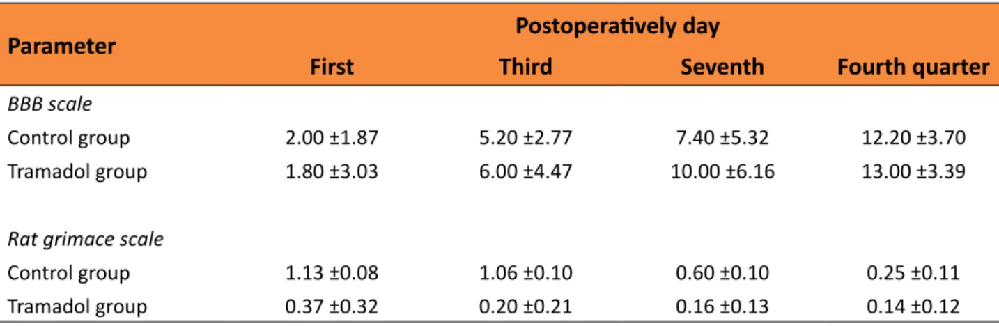
All animals survived during the study period. Table 1 presents the mean and standard error of mean BBB and rat grimace scales. Regarding the BBB scale (Figure 1), in the control group at the 14th postoperatively
Table 1 – Mean scores of BBB and rat grimace scales according groups.
Parameter Postoperatively day
First Third Seventh Fourth quarter
BBB scale
Control group 2.00 ±1.87 5.20 ±2.77 7.40 ±5.32 12.20 ±3.70 Tramadol group 1.80 ±3.03 6.00 ±4.47 10.00 ±6.16 13.00 ±3.39
Rat grimace scale
Control group 1.13 ±0.08 1.06 ±0.10 0.60 ±0.10 0.25 ±0.11 Tramadol group 0.37 ±0.32 0.20 ±0.21 0.16 ±0.13 0.14 ±0.12
Figure 1 – Evolution of score of Basso, Beattie, Bresnahan (BBB) scale according each group.
Regarding the rat grimace scale (Figure 2), there were no significant differences in the tramadol group at the four analyzed periods (p>0.05). In control group, in the first and third postoperatively days have a higher pain level than the 14th postoperatively days (p<0.0001).
There were no significant differences between the groups at the 14th postoperatively days
(p>0.05), however at the first, third and seventh postoperatively days the tramadol
group show a lower score the control group (p<0.0001).
Figure 2 – Evolution of score of rat grimace scale according each group.
The purpose of pain is to alert an individual to withdraw from immediate tissue damaging stimuli and to prevent further damage to the site of injury during the healing process20.
This fact was observed in this study, whereas, both GC and GT groups showed inversely proportional relationships between the rat Grimace score (the pain scale) and the BBB scale (functional scale), suggesting that the lower the pain, the greater the function of the affected member.
Philips et al.21 evaluated the
neuropathic pain in a cervical radiculopathy model in rats and verified the persistence of pain after injury on days 1 and 7, which confirms the verified data in this study, where the GC presented higher score in the rat grimace scale compared to the GT until the 7th postoperative day, not being identified
between the groups on the 14th day.Although
rat grimace scale most sensitively detects pain at acute time points17,22-24, the disappearance
of facial grimace is not necessarily indicative of the resolution of spontaneous pain17, whereas
natural adaptations have led prey species, like rodents, to inhibit facial grimace as soon as possible so that they do not become the target of predators25. By hiding painful expressions,
■
Discussion
Models of nerve damage in animals have been developed over the last 25 years to mimic the clinical manifestation of neuropathic pain. Normally, they have focused on damaging nerves that innervate the hind paw, as this is likely to show changes in sensory function via measurement of withdrawal reflexes in response to mechanical, thermal or chemical stimulation12,18. Regardless of the
method applied, trauma-induced neuropathic models are typically invasive and give rise to perioperative nociceptive signaling of surrounding tissue. Despite this, the use of appropriate post-operative analgesics is not performed as rigorously as it should be, despite being an irrefutable example from an animal welfare perspective and minimizing experimental variance due to stress induced by pain12,13,19. In this study, no difference
statistically significant was identified, considering the functional outcome of the BBB scale in any of the evaluated time, showing no influence of tramadol in this injury protocol.
pain may persist for longer than is detectable by rat grimace scale. This hypothesis has been already suggested previously for visceral, inflammatory, and neuropathic conditions17,24.
Other tests of spontaneous behaviors, like conditioned place preference and activity monitoring, also fail to capture robust pain behaviors after acute time points26.
In the GT, tramadol was administered postoperatively in order to avoid the pain felt by the animal during anesthetic awakening. Previous work showed that inflammatory cytokine levels increase in the spinal cord after injury as early as 1 h after nerve root compression27,28 being indicated preemptive,
postoperative analgesics or even early after injury, and this was verified in the GT group, which presented lower values in the Grimace score in the first 24 hours, in relation to the control group.
Despite the apparent lack of applicability of the rat grimace scale for animal care staff in evaluating pain levels on a day-to-day basis, the use of facial coding to detect pain has clinical utility in refining our understanding of acute pain29-34. This study confirms the importance of applying a pain scale in spinal cord injury procedures reaffirming that cited by Sperry
et al.33, as a useful modality to predict long
periods in clinical pain models. Although the non-invasive collection of rat grimace scale data is simple to execute, its scoring can be a labor-intensive process17. Recent advances in
real-time rat grimace scale32 and convolutional
neural networks for automated facial grimace scoring34 provide methods to streamline its
use in both clinical and pre-clinical research applications.
Some limits of this study must be highlight, we don’t evaluated the effect of tramadol on chronic stage of spinal cord injury, and was not performed inflammatory markets, however it don’t invalided this study because we proved that tramadol don’t affect
the functional recovery, so new studies could evaluate the effect of tramadol or others opioids’ drugs on chronic spinal cord injury.
■
Conclusion
The tramadol attenuates pain in a model of spinal cord injury without altering the functional evaluation of the injured member.
■
References
1- Yang H, Liu CC, Wang CY, Zhang Q, An J, Zhang L, Hao DJ. Therapeutical strategies for spinal cord injury and a promising autologous astrocyte-based therapy using efficient reprogramming techniques. Mol Neurobiol. 2016 Jul;53(5):2826-42. doi: 10.1007/s12035-015-9157-7.
2- He J, Zhao J, Peng X, Shi X, Zong S, Zeng G. Molecular mechanism of MiR-136-5p targeting NF-κB/A20 in the IL-17-mediated inflammatory response after spinal cord injury. Cell Physiol Biochem. 2017;44(3):1224-41. doi: 10.1159/000485452.
3- Hains BC, Waxman SG. Activated microglia contribute to the maintenance of chronic pain after spinal cord injury. J Neurosci. 2006 Apr 19;26(16):4308-17. PMID: 16624951. 4- Zhang D, Ma G, Hou M, Zhang T, Chen
L, Zhao C. The neuroprotective effect of puerarin in acute spinal cord injury rats. Cell Physiol Biochem. 2016;39(3):1152-64. doi: 10.1159/000447822.
5- Liao B, Zhang Y, Sun H, Ma B, Qian J. Ryanodine receptor 2 plays a critical role in spinal cord injury via induction of oxidative stress. Cell Physiol Biochem. 2016;38(3):1129-37. doi: 10.1159/000443063.
6- Kasahara K, Nakagawa T, Kubota T. Neuronal loss and expression of neurotrophic factors in a model of rat chronic compressive spinal cord injury. Spine (Phila Pa 1976). 2006 Aug 15;31(18):2059-66. doi: 10.1097/01. brs.0000231893.21964.f2.
7- Gebrin AS, Cunha A dos S, Da Silva CF, Barros Filho TEP de, Azze RJ. Perspectivas de recuperação do lesado medular. Rev Bras Ortop. 1997;32(2):103-8.
spinal injuries. Spine (Phila Pa 1976). 1990 Jul;15(7):662-6. PMID: 2218712.
9- Gerbrin AS, Cristante AF, Marcon RM, Da-Silva CF. Pharmacological interventions in spinal cord trauma: a therapeutic approach. Acta Ortop Bras.1997;5(3):123-36.
10- Su YF, Lin CL, Lee KS, Tsai TH, Wu SC, Hwang SL, Chen SC, Kwan AL. A modified compression model of spinal cord injury in rats: functional assessment and the expression of nitric oxide synthases. Spinal Cord. 2015 Jun;53(6):432-5. doi: 10.1038/ sc.2014.245.
11- Marques SA, Garcez VF, Del Bel EA, Martinez AM. A simple, inexpensive and easily reproducible model of spinal cord injury in mice: morphological and functional assessment. J Neurosci Methods. 2009 Feb 15;177(1):183-93. doi: 10.1016/j. jneumeth.2008.10.015.
12- Hestehave S, Munro G, Christensen R, Brønnum Pedersen T, Arvastson L, Hougaard P, Abelson KSP. Is there a reasonable excuse for not providing post-operative analgesia when using animal models of peripheral neuropathic pain for research purposes? PLoS One. 2017 Nov 22;12(11):e0188113. doi: 10.1371/journal.pone.0188113.
13- Stokes EL, Flecknell PA, Richardson CA. Reported analgesic and anaesthetic administration to rodents undergoing experimental surgical procedures. Lab Anim. 2009;43(2):149–54. PMID: 19116297. 14- Kaneko K, Umehara M, Homan T, Okamoto
K, Oka M, Oyama T. The analgesic effect of tramadol in animal models of neuropathic pain and fibromyalgia. Neurosci Lett. 2014 Mar 6;562:28-33. doi: 10.1016/j. neulet.2014.01.007.
15- Kimura M, Obata H, Saito S. Antihypersensitivity effects of tramadol hydrochloride in a rat model of postoperative pain. Anesth Analg. 2012 Aug;115(2):443-9. doi: 10.1213/ANE.0b013e31825683c3. 16- Barros Filho TEP, Molina AEIS. Analysis
of the sensitivity and reproducibility of the Basso, Beattie, Bresnahan (BBB) scale in Wistar rats. Clinics (Sao Paulo). 2008 Feb;63(1):103-8. PMID: 18305873.
17- Sotocinal SG, Sorge RE, Zaloum A, Tuttle AH, Martin LJ, Wieskopf JS, Mapplebeck JC, Wei P, Zhan S, Zhang S, McDougall JJ, King OD, Mogil JS. The Rat Grimace Scale: a partially
automated method for quantifying pain in the laboratory rat via facial expressions. Mol Pain. 2011 Jul 29;7:55. doi: 10.1186/1744-8069-7-55.
18- Jurga AM, Rojewska E, Piotrowska A, Makuch W, Pilat D, Przewlocka B, Mika J. Blockade of Toll-Like Receptors (TLR2, TLR4) attenuates pain and potentiates buprenorphine analgesia in a rat neuropathic pain model. Neural Plast. 2016;2016:5238730. doi: 10.1155/2016/5238730.
19- Nagakannan P1, Shivasharan BD, Thippeswamy BS, Veerapur VP. Effect of tramadol on behavioral alterations and lipid peroxidation after transient forebrain ischemia in rats. Toxicol Mech Methods. 2012 Nov;22(9):674-8. doi: 10.3109/15376516.2012.716092.
20- Cox JJ, Reimann F, Nicholas AK, Thornton G, Roberts E, Springell K, Karbani G, Jafri H, Mannan J, Raashid Y, Al-Gazali L, Hamamy H, Valente EM, Gorman S, Williams R, McHale DP, Wood JN, Gribble FM, Woods CG. An SCN9A channelopathy causes congenital inability to experience pain. Nature. 2006 Dec 14;444(7121):894-8. PMID: 17167479. 21- Philips BH, Weisshaar CL, Winkelstein BA.
Use of the rat grimace scale to evaluate neuropathic pain in a model of cervical radiculopathy. Comp Med. 2017 Feb 1;67(1):34-42. PMID: 28222837.
22- Langford DJ, Bailey AL, Chanda ML, Clarke SE, Drummond TE, Echols S, Glick S, Ingrao J, Klassen-Ross T, Lacroix-Fralish ML, Matsumiya L, Sorge RE, Sotocinal SG, Tabaka JM, Wong D, van den Maagdenberg AM, Ferrari MD, Craig KD, Mogil JS. Coding of facial expressions of pain in the laboratory mouse. Nat Methods. 2010 Jun;7(6):447-9. doi: 10.1038/nmeth.1455.
23- De Rantere D, Schuster CJ, Reimer JN, Pang DS. The relationship between the Rat Grimace Scale and mechanical hypersensitivity testing in three experimental pain models. Eur J Pain. 2016 Mar;20(3):417-26. doi: 10.1002/ejp.742.
24- Philips BH, Weisshaar CL, Winkelstein BA. Use of the rat grimace scale to evaluate neuropathic pain in a model of cervical radiculopathy. Comp Med. 2017 Feb 1;67(1):34-42. PMID: 28222837.
of pain in laboratory animals. Contemp Top Lab Anim Sci. 2003 Jul;42(4):13-20. PMID: 12906396.
26- Klinck MP, Mogil JS, Moreau M, Lascelles BDX, Flecknell PA, Poitte T, Troncy E. Translational pain assessment: could natural animal models be the missing link? Pain. 2017 Sep;158(9):1633-46. doi: 10.1097/j. pain.0000000000000978.
27- Rothman SM, Huang Z, Lee KE, Weisshaar CL, Winkelstein BA. Cytokine mRNA expression in painful radiculopathy. J Pain. 2009 Jan;10(1):90-9. doi: 10.1016/j. jpain.2008.07.008.
28- Rothman SM, Winkelstein BA. Cytokine antagonism reduces pain and modulates spinal astrocytic reactivity after cervical nerve root compression. Ann Biomed Eng. 2010 Aug;38(8):2563-76. doi: 10.1007/ s10439-010-0012-8.
29- Fairbanks CA, Goracke-Postle CJ. Neurobiological studies of chronic pain and analgesia: rationale and refinements. Eur J Pharmacol. 2015 Jul 15;759:169-81. doi: 10.1016/j.ejphar.2015.03.049.
30- Faller KM, McAndrew DJ, Schneider JE,
Lygate CA. Refinement of analgesia following thoracotomy and experimental myocardial infarction using the Mouse Grimace Scale. Exp Physiol. 2015 Feb 1;100(2):164-72. doi: 10.1113/expphysiol.2014.083139.
31- Préfontaine L, Hélie P, Vachon P. Postoperative pain in Sprague Dawley rats after liver biopsy by laparotomy versus laparoscopy. Lab Anim (NY). 2015 May;44(5):174-8. doi: 10.1038/laban.731. 32- Leung V, Zhang E, Pang DS. Real-time
application of the Rat Grimace Scale as a welfare refinement in laboratory rats. Sci Rep. 2016 Aug 17;6:31667. doi: 10.1038/ srep31667.
33- Sperry MM, Yu YH, Welch RL, Granquist EJ, Winkelstein BA. Grading facial expression is a sensitive means to detect grimace differences in orofacial pain in a rat model. Sci Rep. 2018 Sep 17;8(1):13894. doi: 10.1038/s41598-018-32297-2.
34- Tuttle AH, Molinaro MJ, Jethwa JF, Sotocinal SG, Prieto JC, Styner MA, Mogil JS, Zylka MJ. A deep neural network to assess spontaneous pain from mouse facial expressions. Mol Pain. 2018 Jan-Dec;14:1744806918763658.
Correspondence:
Rosa Helena de Figueiredo Chaves Travessa do Chaco, 729/802 66083-180 Belém – PA Brasil Tel.: (55 91)98896-1978
rosahelenamedvet@gmail.com
Received: Aug 25, 2018 Review: Oct 21, 2018 Accepted: Nov 23, 2018
Conflict of interest: none Financial source: none
1Research performed at Biotery, Centro