http://dx.doi.org/10.1590/s2175-97902017000300204
A
r
*Correspondence: J. P. Robinson. Department of Biotechnology. K.S.Rangasamy College of Technology. Trichengode, Namakkal District. Tamil Nadu, India. E-mail: [email protected]
Antioxidant and cytotoxic activity of
Tecoma stans
against lung
cancer cell line (A549)
Jayachandran Philip Robinson
*, Kumaresan Suriya, Ramasamy Subbaiya, Ponnusamy
Ponmurugan
Department of Biotechnology, K.S.Rangasamy College of Technology, Trichengode, Tamil Nadu, India
Human have been constantly using plants and plant products to overcome many diseases. The antioxidant property of the plant sources is studied to obtain an eicacious drug against cancer. The objectives of the present study is to evaluate the antioxidant and cytotoxic activity of the Tecoma stans extracts against lung cancer cell line in comparison with vincristine drug. The antioxidant activity was studied using the standard DPPH assay and the cytotoxic activity using MTT assay. DPPH assay results show that methanolic extract of T. stans inhigher concentration show better antioxidant potential than the standard L-ascorbic acid. They exhibited strong antioxidant potential at 20 µg/mL concentration. The absorbance at 517 nm showed that in the range of 0.201-0.0203 compared to that of absorbance of ascorbic acid at 0.023.Cytotoxic activity was studied using MTT assay which showed that the increase in concentration of extract increases the cell death. At 100µg/mL concentration there is an increased cytotoxic activity, i.e., 99% of cell inhibition. The results of antioxidant and anticancerous activity may be positively correlated.
UNITERMS: Tecoma stans/extract/cytotoxicity. Tecoma stans/extract/antioxidant activity. Free radical scavenging. Plant extracts. Lung cancer cell line/study/drugs.
INTRODUCTION
Cancer is one of the disease that occurs in both developed and developing countries and is the leading cause of death. An extensively used treatment for cancer
is chemotherapy and one of the major drawbacks is the
toxicity that is caused to the normal cells due to the
inability of the chemical drugs to diferentiate between
normal and cancerous cells (Balamurugan et al., 2014). Traditional medicines have been tested and researched upon to obtain an effective drug against cancer. Plant derived compounds are widely studied for their holistic value. These plant derived compounds have clinical
signiicance which can be further developed into efective
drugs against cancer. Bio active compounds have received the attention of researchers to overcome the burden of chemotherapy related problems. Oncovin is the brand name for vincristine, which is used as a chemotherapy drug for many types of cancer. Vincristine is also used to
treat neurodegenerative disease (Chun-Fai et al., 2013). The mode of action of vincristine is that it acts on the tubulin protein, thereby inhibiting the metaphase stage, thus leading the cell to apoptosis (Jordan, 2002).
Tecoma Stans is an ornamental tree that has its origin from the Americas belongs to the family Bignoniaceae. Presently the plant is widely cultivated throughout India
for its lowers which bloom throughout the year. Presence
of alkaloids tecomine and tecostamine which are potential hypoglycemic agents present in the leaves of T. stans. In addition to that presence of also anthranilic acid in the roots of plant which is an antidiabetic agent (Khare, 2007). There are a number of compounds from the fruits and
lowers of the Tecoma, which had antioxidant activity and
anti proliferative efect against cancer cell lines (Marzouk et al., 2006). Antioxidants play a major role in deciding the pharmaceutical efect of plants and to make them potent
drugs against the chronic diseases. These antioxidants are taken in our dietary from the plant compounds, which is a rapid and simple method. The DPPH assay is the simple method to evaluate the presence of antioxidants in any source based on the principle of radical scavenging
2-diphenyl-1-picryl-hydrazyl-hydrate) i.e., DPPH is reduced to DPPHH, the odd element is formed is absorbed at 517nm (Tailor, Goyal, 2014). The cytotoxic activity of the plant extract is studied using the
3-(4,5-dimethylthiazol-2-yl)-2,5- diphenyltetrazolium bromide (MTT) which forms formazan (insoluble)
crystals, by interacting with the mitochondria of the
cells and the insoluble formazan that is formed is solubilized using isopropanol solvent which is studied
spectrophotometrically at 540 nm. MTT is reduced only by metabolically active cells, thus in, turn it helps to study the viability of the cell.
MATERIAL AND METHOD
Collection of plant material
The fresh plant of Tecoma stans was collected from Paramathi velur, Namakkal district of Tamil Nadu, India.
The collected plants were further surface sterilized using
tween 80 and it was shade dried for future investigation.
Preparation of plant extract
10 g of both fresh and dried leaves, lowers were
chopped into fine pieces and macerated in 100 mL of methanol. The plant material was extracted at room
temperature for 3 days in a shaker. The extract was iltered and the iltrate was concentrated in a rotary evaporator
under reduced pressure to dryness. The extract obtained was stored at 4 o
C until use.
Antioxidant assay
DPPH assay
The percentage of antioxidant assay was determined using the free radical scavenging activity (2,
2-diphenyl-1-picryl-hydrazyl-hydrate). About 1 mg of fresh and the
dried plant extracts was dissolved in 1ml of methanol. The standard procedure for DPPH assay was performed based on Ochuko et al. (2012). About 10 mL of 0.1 mM of DPPH was prepared in methanol and stored in cool dark condition until use. Accurately, 1 mL of DPPH was
added to diferent concentration (20, 40, 60, 80, 100 µg/ mL) of T.stans extract. The mixture of DPPH and extract was shaken and incubated at room temperature in the dark for 30 minutes, then the absorbance was measured at 517 nm in the UV spectrophotometer (Tailor, Goyal, 2014). Ascorbic acid was used as a reference and DPPH without the extract served as negative control. The IC50 value of
the sample was calculated based on the absorbance. The percentage of inhibition was calculated using the formula,
DPPH scavenging efect (%) or
Percent inhibition = (Absorbance of sample-absorbance of blank)/Absorbance of Control X 100
Lung Cancer (A549) cell line
Lung cancer cell line (A549) cell line was procured from NCCS, Pune and maintained in Dulbecco’s minimal essential medium (DMEM) with 10% FBS, and antibiotic mixture (Penicillin, streptomycin and ampicillin 100 units/
mL) under deined conditions of temperature at 37 o
C, 95% humidity and 5% CO2.
Cell viability assay
The cytotoxic activity of the fresh and dried extracts of leaves and flowers of Tecoma stans was
determined using the 3-(4,5-dimethylthiazol-2-yl)-2,5- diphenyltetrazolium bromide (Terry et al., 1992). Tecoma stans extract concentrations were prepared in the range of 100-20µg/mL from the stock solution by serial dilution using Dimethyl sulphoxide (DMSO). Vin-Cristine wide range anti-cancer drug was used as the standard (0.1 g/ mL). Lung cancer cell line (A549) was trypsinised and the cells were counted using haemocytometer following standard procedure.100µl of the lung cancer cell line at 1 X 10⁴ cells/mL was added to poly L-lysine coated 96
well plate and incubated at 37 oC in a humidiied 5% CO
2
incubator. After 24 hours of incubation, the old medium was replaced with fresh medium and 50 µL of the extract was added and incubated for 48 hours at 37 o
C in a humidified 5% CO2 incubator. 30µl of 0.5% w/v MTT was added and incubated at room temperature for 4 hours. After incubation, 50 µL of acid-isopropanol was added
to dissolve the formazan formed and incubated at room
temperature for 30 minutes. Then absorbance was taken at 554nm using Bio-Rad micro-titer plate reader. The assay was performed in triplicates.
RESULTS AND DISCUSSION
Antioxidant property plays an important role in reducing chronic diseases like cancer and cardiovascular (CAD) diseases. Crude plant extracts are screened on
cell culture, to determine their eiciency as a potential
alternate drug and also to check their efficiency in clinical application as suggested by Balamurugan et al.
(Prajapati, Patel, 2010). A number of lowers and fruits having anti oxidant activity have been identiied in the lowers and fruits of Tecoma their efective anti-cancer activity was studied against cancer cell line (Marzouk et al., 2006).
DPPH assay is performed to study the radical scavenging activity, the scavenging activities in the present investigation were similar to that as reported by Erukainure et al. (2012), Where the scavenging activity follows a dose dependen pattern i.e., increase in activity to increase in concentration. The alcoholic and aqueous extract of T.stans on phytochemical analysis has revealed
the presence of polyphenolic,ß- sitosterol and lavonoids.
In earlier reports by Hamburger and Hostettmann (1991), Beltrame et al. (2002) and Suffredimi et al. (2004), these compounds have exhibited anti-bacterial, antiviral,
immunological and cytotoxic property on diferent cancer
cell lines.
The current study was carried out to report the antioxidant activity of the fresh and dried leaves and flowers of Tecoma stans using the DPPH assay.
1,1-diphenyl-2-picrylhydrazyl reacts with the
anti-oxidants present in the plant extract and accepts the hydrogen atom, thus addition of hydrogen atom converts it
to 1, 1-diphenyl-2-picrylhydrazine (Ochuko et al., 2012).
The chemical 1, 1-diphenyl, 1-2- picrylhydrazyl changes
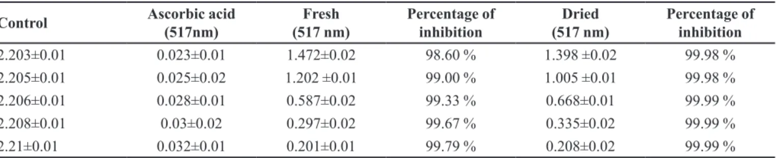
from purple color to yellow color. In case of T stans the antioxidant is higher from 20-100 µg/mL when compare to the standard of L-ascorbic acid at absorbance 517 nm in UV visible spectrophotometer. Table I shows the radical scavenging activity of T.stans against stable DPPH.
The scavenging activity values were compared with that of ascorbic acid (concentrations of 20,40,60,80 and 100 µg/mL) the values were dose dependent and their percentage of inhibition show that at 20 µg/mL concentration the percentage inhibition was 99.98%, which increase as 99% (40 µg/mL), 99.33% (60 µg/mL), 99.67% (80 µg/mL) and 99.79% (100 µg/mL) for dried
leaves and lowers while for fresh lowers and leaves its
99.98% (20, 40 µg/mL) and 99.99% (60,80,100 µg/mL) respectively. These results were compared to standard ascorbic acid favorably.
From Figure 2 it is observed that cancer cells have undergone certain morphological changes, like cellular shrinkage and blebbing, which are characteristic features of apoptosis. These morphological changes are less observed in standard (vincristine) treated cells and these changes seem increasing with increase in concentration of
FIGURE 1 - Antioxidant activity of fresh and dried leaves and lowers of Tecoma stans in comparison with ascorbic acid at 517 nm.
TABLE I - Absorbance and percentage of inhibition of Fresh and Dried extract of Tecoma stans
Control Ascorbic acid
(517nm)
Fresh (517 nm)
Percentage of inhibition
Dried (517 nm)
Percentage of inhibition
2.203±0.01 0.023±0.01 1.472±0.02 98.60 % 1.398 ±0.02 99.98 %
2.205±0.01 0.025±0.02 1.202 ±0.01 99.00 % 1.005 ±0.01 99.98 %
2.206±0.01 0.028±0.01 0.587±0.02 99.33 % 0.668±0.01 99.99 %
2.208±0.01 0.03±0.02 0.297±0.02 99.67 % 0.335±0.02 99.99 %
the extract. At 100 µg/mL the morphological changes are higher, followed by 80µg/ml concentration.
The antioxidant assay results further, directed the study towards cytotoxic assay. The phytochemical and antioxidant assay which showed the expression of carotenes and phytophenols, hence lung cancer cell line was chosen for cytotoxic activity. The results of MTT assay in Figure 3 show that the methanolic extract of
Tecoma stans has high cytotoxic activity against the cancer cell line from 20µg/mL concentration i.e., 99.3%. Earlier
reports conirm the better cytotoxic activity of the plant
(Pusapathi et al., 2015).
Presence of phenolic compounds and its congers have shown to induce a cascade based apoptosis in cancer cells, thus inducing cytotoxicity (Owen et al., 2000). The cell viability of the lung cancer cell line decreased with increase in concentration of the plant extract and it was found to be the highest in 100 µg/mL concentration. The decrease in cell viability with increased concentration of the plant extract of Tecoma stans suggests the ability of the
extract as an efective anti-cancer medicine. Antioxidant
and free radical scavenging activity of the extract may be the reason behind its anti-cancer property.
CONCLUSION
The present investigation revealed that T. stans can act as a potential alternative remedy for lung cancer. The extract of T. stans can be used as an efective ingredient in
drug recipe cancer. Further investigation is undertaken to identify the active compound behind the cytotoxic activity of the plant. The study in the future is to be extended to other cancer cell lines and there is a need to carry out in vivo studies to further authenticate the anti-oxidant potentials of this species.
ACKNOWLEDGEMENT
The authors are thankful to the Department of Science and Technology, New Delhi for the financial
FIGURE 2 - Morphological changes of lung cancer cell lines after plant extract treatment a) Untreated lung cancer cells; b) T. stans
treated lung cancer cells (after 24 hours).
support through DST- FIST grant for the improvement of infrastructure facilities to the laboratories.
REFERENCES
Balamurugan V, Balakrishnan V, Robinson JP, Ramakrishnan M. Anticancer and apoptosis-inducing effects of Moringa concanensis using hepG2 cell lines. Bangladesh J Pharmacol. 2014; 9(4):604-09.
Beltrame FL, Pessini GL, Doro DL, Filho BPD, Bazotte RB, Cortez DAG. Evaluation of the anti diabetic and antibacterial activity of Cissus sicyodes. Braz Arch Biol Technol. 2002;45(1):21-25.
Chun-Fai NG, Chun-Hay KO, Chi-Man K, Jia-Wen X, Ping-Chung L, Kwok-Pui F, Ho YEC, Clara B-SL. The aqueous extract of rhizome of gastrodia elata protected drosophila and PC12 cells against beta-amyloid-induced neurotoxicity. Evid-Based Complem Altern Med. 2013;5(1):1-12.
Erukainure OL, Oke OV, Owolabi FO, Kayode FO, Umanhonlen EE, Aliyu M. Chemical properties of Monodora myristica and its
protective potential against free radicals in vitro. Oxid Antioxid
Med Sci. 2012;1(2):127-132.
Hamburger M, Hostettmann K. Bioactivity in plants: the link between phytochemistry and medicine. Phytochemist ry.1991;30(12):3864-3874.
Jordan MA. Mechanism of action of antitumor drugs that interact with microtubules and tubulin. Anticancer Agents Med
Chem. 2002;2(1):1-17.
Khare CP. Indian medicinal plants and illustrated dictionary. New Delhi: Springer Science Publishers; 2007. 900 p.
Marzouk M, Gamal-Eldeen A, Mohamed M, El-Sayeed M.
Anti-proliferative and antioxidant constituents from Tecoma stans. Z
Naturforsch C. 2006;61(11-12):783-791.
Ochuko LE, Oluwatoyin VO, Folashade OO, Funmi OK, Emmanuel EU, Muhammad A. Chemical properties of
Monodora myristica and its protective potentials against free
radicals in vitro. Oxid AntioxidMed Sci. 2012;1(2):127-132.
Owen RW, Giacosa A, Hull WE, Haubner R, Spiegelhalder B, Bartsch H. The antioxidant/anticancer potential of phenolic compounds isolated from olive oil. Eur J Cancer. 2000; 36(10):1235-47.
Prajapati DK, Patel NM. Pharmacognostic and phytochemical investigations of the leaves of Tecoma Stans Linn. Int J Pharm Sci Rev Res. 2010;3(1):70-72.
Pusapati MR, Nagarani T, Swathi V, Pahni KK, Chowdary YA, Siva Reddy CH, Girijasankar G. Evaluation of phytochemical content and In vitro cytotoxic activity of various ornamental plant lower extracts against MCF-7 Cell lines. Int J Curr Res Life Sci. 2015;4(3):172-176.
Sufredimi IB, Sader HS, Gonçalves AG, Reis AO, Garles AC, Varella AD, Younes RN. Screening of antibacterial extracts from plants nature to the Brazilian amazon rain forest. Braz J Med Biol Res. 2004;37(3):379-384.
Tailor CS, Goyal A. Antioxidant activity By DPPH radical
scavenging method of Ageratum conyzoides Linn. Leaves. Am
J Ethnomed. 2014;1(4):244-249.
Terry BJ, Liu WC, Cianci CW, Proszynski E, Fernandes P, Meyers E. Inhibition of Herpes Simplex virus type I DNA polymerase by the natural product Oosporein. J Antibiot. 1992;45(2):286-288.
Received for publication on 21st November 2016