Yuselis Castaño Guerrero
Lic. Biochemistry
Report of professional activity
Dissertação para obtenção do Grau de Mestre em Bioquímica
L
O
M
B
A
D
A
2
0
1
Yuselis Castaño Guerrero
Lic. Biochemistry
Report of professional activity
Dissertação para obtenção do Grau de Mestre em Bioquímica
Copyright
Title: Report of professional activity.
Eu, Yuselis Castaño Guerrero concedo à Faculdade de Ciências e Tec-nología e à Universidade Nova de Lisboa, nos terrnos dos regulamentos aplicãveis, 0 direito de divulgar e distribuir copias da dissertaçao.
“A Faculdade de Ciências e Tecnología e a Universidade Nova de Lisboa tém 0 direito, perpétuo e sem limites geográficos, de arquivar e publicar esta dissertação através de exemplares impressos reproduzidos em papel ou de forma digital, ou por qualquer outro meio conhecido ou que venha a ser inventado, e de a divulgar atraves de repositôrios científicos e de admitir a sua côpia e distnibuiçao com objectivos educacionais ou de investigacao, não comerciais, desde que seja dado crédito ao autor e editor’.
Acknowledgements
This professional report involved many people that I would like to thank. First and foremost I would like to thank my actual supervisor Prof. Maria Goreti Ferreira Sales for giving me a chance of being part of Biomark and the opportunity of present this report.
I am grateful as well to Rocio García Miniet, for encouraging me to con-tinue in the research and giving the first oppotunity as research, and well to Maria Elena González Fraguela. I would also thank to Dra. María de los Àngeles Ribas Antúnez for being a great supervisor at my first job. I would also thank to Gerardo González Aguilar for giving me the first opportunity to work in Portugal.
Abstract
"Biochemistry has become the foundation for understanding all biological processes. It has provided explanations for the causes of many diseases in humans, animals and plants".
Resumo
"A bioquímica tornou-se a base para a compreensão de todos os processos biológicos. Ele forneceu explicações para as causas de muitas doenças em humanos, animais e plantas".
Contents
1 Abbreviations 9
2 Introduction 11
3 Metabolism disorders in a rats model 15
3.1 Introduction . . . 16
3.2 Materials and methods . . . 16
3.2.1 Experimental subjects . . . 16
3.2.2 Surgical procedure . . . 17
3.2.3 Study of oxidative metabolism . . . 17
3.2.4 Evaluation of cell damage . . . 18
3.2.5 Evaluation of behaviour disorders . . . 19
3.2.6 Statistical method . . . 19
3.3 Results . . . 20
3.3.1 Oxidative metabolism . . . 20
3.3.2 Evaluation of cell damage . . . 20
3.3.3 Behavioural trials . . . 23
3.4 Discussion . . . 25
3.5 Conclusions . . . 28
4 Norovirus and Rotavirus detection 31 4.1 Introduction . . . 31
4.2 Materials and methods . . . 31
4.3 Results . . . 31
4.4 Discussion . . . 32
5 Bacterial threat remote detection using electronic tongues 35
5.1 Introduction . . . 35
5.2 Materials and methods . . . 38
5.2.1 Bacillus Thuringiensis Preparation . . . 38
5.2.2 Electrode Preparation . . . 38
5.2.3 Data Acquisition . . . 38
5.3 Results . . . 39
5.4 Discussion . . . 40
5.5 Conclusions . . . 40
6 Smart nanomaterials for drug-delivery 41 6.1 Introduction . . . 41
6.2 Materials and methods . . . 42
6.2.1 Materials and instruments . . . 42
6.2.2 Preparation of Apo . . . 43
6.2.3 DOXO encapsulation . . . 44
6.2.4 Intrinsic emission fluorescence . . . 44
6.2.5 Cell culture . . . 44
6.2.6 MTT viability Test . . . 44
6.3 Results . . . 45
6.4 Discussion . . . 49
6.5 Conclusions . . . 49
7 Conclusions 51 A Shiga Toxin Detection : A Short Review 55 A.1 Introduction . . . 55
A.1.1 Preliminaries . . . 55
A.2 The Shiga family of toxins . . . 58
A.2.1 Structural Characteristics . . . 58
A.2.2 Genetics Encoding . . . 60
A.2.3 Shiga Receptor . . . 60
A.2.4 Mechanism of action . . . 61
A.3 Medical Applications . . . 62
A.4 Detection . . . 63
A.4.1 Biological Methods . . . 63
A.4.2 Detection by Biosensors . . . 65
A.5 Conclusions . . . 68
References 69
List of Figures
3.1 Activity of superoxide dismutase in the cortex, hippocampus, and striatum of rats subjected to permanent occlusion of com-mon carotid arteries. The graph shows enzyme activity acom-mong model group animals at 22 days post-lesion compared to the control group (∗∗∗P <0.001) and the model group at 24 hours post-lesion (&P < 0.05; &&P <0.01; &&&P < 0.001). Bars represent mean values ± standard error of the mean. CTX: cortex; STR: striatum; CG: control group; HPC: hippocam-pus; L-1d: group at 24 hours post-lesion; L-22d: group at 22 days post-lesion; SOD: superoxide dismutase. . . 21 3.2 Activity of catalase in the cortex, hippocampus, and striatum
of rats subjected to permanent occlusion of common arotid ar-teries. The graph shows enzymatic activity in model group an-imals compared to control group anan-imals (∗ ∗P <0.01). Bars represent mean values ± standard error of the mean. CAT: catalase; CTX: cortex; STR: striatum; CG: control group; HPC: hippocampus; 1d: group at 24 hours post-lesion; L-22d: group at 22 days post-lesion. . . 21 3.3 H&E stains of coronal slices of the cortex (A and B), the
LIST OF FIGURES
3.4 Microphotographs showing coronal slices from rat brains with glial fibrillary acid protein immunostaining in the cortex (A and B) the striatum (C and D), CA1 (E and F), and CA3 (G and H) at 40X magnification. Columns on the left show slices taken from control-group animals; columns on the right show sections from animals with cerebral hypoperfusion at 7 days post-lesion. . . 22 3.5 Influence of chronic cerebral hypoperfusion on sensorimotor
and motivational deficits. For this task, the escape platform was visible. ∗ ∗ ∗P < 0.001: escape latency in the model group at 22 days post-lesion was longer than in the control group.
&P < 0.05: escape latency in the model group at 37 days post-lesion was shorter than in the model group at 22 days post-lesion. Bars represent mean values ± standard error of the mean. CG: control group; L-22d: group at 22 days post-lesion; L-37d: group at 37 days post-lesion. . . 23 3.6 Evaluating long-term or spatial reference memory. In this
trial, the platform remained at the same location, but was hidden. Values show mean escape latencies ± standard er-ror of the mean. ∗P < 0.05 : ∗ ∗ ∗P < 0.001: escape la-tencies from both model groups compared to control group.
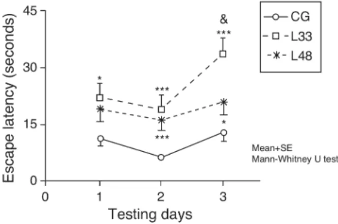
&P < .05; &&P < .01; &&&P < .001; differences between es-cape latencies in both model groups. CG: control group; L18 and L33: animals that began testing at 18 days and 33 days post-lesion. . . 24 3.7 Short-term memory impairments as a result of chronic cerebral
hypoperfusion. Points along the curve correspond to mean values ±standard error of the mean. ∗P < .05;∗ ∗ ∗P < .001: indicates that escape latencies in the model group were longer than in the control group. &P < .05: shows differ- ences between different model groups. CG: control group; L33 and L48: animals that began testing at 33 days and 48 days post-lesion. . . 25
5.1 Experimental configuration: The electrode recognize the bacil-lus of the sample. The biosensor signal is detected with an arduino which is connected to Raspberry Pi. The results is shown with a "Data Collector" program. . . 37 5.2 Electrodes signal Voltage vs time for B.thuringiensis additions. 39 5.3 Electrodes signal with Kalman filter and the running mean to
filter the signal . . . 39
LIST OF FIGURES
6.1 Scheme DOXO cell release . . . 43 6.2 Scheme for Apo preparation . . . 45 6.3 SEM image: left for Apo and right for Apo-Doxo. . . 46 6.4 Fluorescence spectra: left differents Apo-Doxo concentration
and right calibration curve for Doxo. . . 46 6.5 MTT assay for Doxo at 24 hrs. . . 48 6.6 MTT assay at 24 hrs. . . 48
A.1 Is shown the subunit A and B of Shiga toxin. (Taken from RCSB Protein Data Bank) . . . 59 A.2 In the fig 2A is shown the Stx1 and in the fig 2B Stx2. (Taken
List of Tables
4.1 Clinical characteristics of Norovirus and Rotavirus infection, in children under 5 years old. . . 32
1
Abbreviations
• POCCA: permanent occlusion of commun carotid arteries.
• SOD: superóxide dismutase.
• CAT: catalase.
• GFAP: glial fibrillary acidic protein.
• MWM: Morris water maze.
• ROS: reactive oxygen species.
• NoV: norovirus.
• RoV: rotavirus.
• AGE: acute gastroenteritis.
• DOXO: doxorubicin.
• BrC: breast cancer.
• Apo: apoferritin.
• ELISA: Enzyme-Linked Immunosorbent Assay.
• HIV: human immunodeficiency virus.
2
Introduction
Biochemistry, the “chemistry of life”, is synonymous of two older terms: phys-iological chemistry and bphys-iological chemistry. Biochemistry surge as scientific discipline around the early 19th century. Carl Neuberg, an early pioneer, was the fist to propose the term “biochemistry” in 1903.
According to Biochemical Society, “Biochemistry explores the chemical processes within and related to living organisms. The Chemistry of Life con-centrates on handling what happens at a molecular level that, what’s happen-ing inside the livhappen-ing cells and organelles, studyhappen-ing components like proteins, enzymes, lipids, carbohydrates, DNA and RNA” [1]. Biochemistry describes, in molecular terms, all chemical processes in the living organisms. Biochem-istry contains itself a very broad range of scientific disciplines, including molecular biology, genetics, physiology, microbiology-virology, forensics, an-imal and plant sciences and medicine [2]. Although its ultimate concern is with the wonder of life itself, some of the most exciting questions emerged in the Biochemistry has developed greatly medicine and biology [3].
I’m Biochemistry graduated from the Biology faculty of the University of Havana, Cuba. I started my degree in 2005. My career consist, of five years of academic formation culminating with a thesis. During the first three years I had the opportunity to learn about mathematics, physics, chemistry, biol-ogy and all related with biochemistry. At this time I was gaining an interest for molecular biology. During my bachelor thesis, I had the opportunity to work from the molecular level up to the organism level. More specifically, I worked rat models of cerebral hypoperfusion at International Center for Neurological Restoration, Havana, Cuba. This work finished with a publica-tion [4], presented in chapter 3, and it was a big motivapublica-tion for me as young research.
2- Introduction
permanent occlusion model in rats. I was responsible for characterizing both molecular and behavioral animal models, to test the lesion and control rats group in the behavioral trial Morris water maze (MWM), and make dissec-tions of brain areas for molecular assays. I was responsible too for the ox-idative tests, catalase (CAT) and superoxide dismutase (SOD), in this work. This publication was a part of my thesis degree my responsibility, write a big part.
After finishing my undergrad studies, I had the opportunity to work in the Diagnostic Lab of the Institute of Tropical Medicine “Pedro Kourí” in La Habana, Cuba. It is a reference national laboratory in Cuba for confirma-tion a positive results for dengue, rabies, rubella, measles, mumps, rotavirus (RoV), norovirus (NoV) and human immunodeficiency virus (HIV). In this period I gain knowledge, about viruses and detection methods, including Ul-tramicro ELISA assay and molecular techniques as RT-PCR and SDS-PAGE. At this time, my main responsibility as young research, was molecular stan-dardization method (RT-PCR) for NoV and RoV detection. I learned how important, is to quickly identify NoV and RoV diseases in children. At this time, we made a study about infection in children caused by NoV and RoV in a cuban hospital, which ended in a published article [5], presented in chapter 4.
Later in 2012, I was accepted in the "Carrera de Especialización en Aplica-ciones Tecnológicas de la Energía Nuclear" (CEATEN) course at Atomic Cen-ter of Bariloche, Argentina, which aims to bring together multidisciplinary professionals into a single place specializing them in nuclear technologies. Here I learned how nuclear technology is used nowadays in medicine produc-tion and therapy of cancer cells.
In 2014, I started to work at INESC-TEC Porto. The first work in this project was a review about Shiga toxin. This toxin create, outbreak, with serious public health, so developing a sensor that could detect it in real time could have a large impact in the outbreak control. This review is presented in appendix A. The goal of this work, to know the state of the art in methods for bacterial detection using biosensors. With this review, we understood that biosensors promise to be rapid, reliable and sensitive detection platforms for bacterial detection. More specifically, during this time, I was involved in the development of a biosensor with the aim to detect bacteria in meals through the detection of bacillus thuringiensis and bacillus cereus. At this time I learned about the R programming language, through which the results were processed. I also had the opportunity to participate in two international events with a poster presentation. First, at the "21 Chemistry Conference"
with a poster: Bacterial threat remote detection using electronic tongues: modeling using Bacillus thuringiensis in Santiago de Cuba, Cuba. Second,
2- Introduction
was thethird SIPS Training School and"Third European conference on smart inorganic polymers" events hold in Porto, Portugal with a posterUsing dye-doped polymer arrays for food contaminant detection.
More recently in January 2018, I started to work at BioMark, Sensor Research, ISEP, Porto, a project supported by the grant, BIL/IBEROS-BioMark/03. The goal of the job the development of nanomaterials for car-rying an antitumor agent, targeting breast cancer. It couldn’t be more excit-ing to work in such cuttexcit-ing-edge research. Up to the current year, we have obtained a very promising preliminary results which points us the next imme-diate steps to do and also suggest more ambitious goals which we pretend to develop in future project. My role in this research is to develop a nanomate-rial carrying on its surface the Trastuzumab and in its interior an antitumor agent (doxorubicin). I was responsible for prepare vehicle which support the drug and encapsulate the DOXO inside of apoferritin. Optimization assays for testing the Dose/response of doxorubicin/vehicle.
This work is organized as follows.
First chapter contains my work in the ischemia model in rats to evaluate the molecular mechanisms that conduce to behavioral disorders, developed from June 2007 until April 2010.
Second chapter is devoted to my work viruses detection, specifically Noroviruses and Rotaviruses, developed from September 2010 until April 2012.
The third chapter contains the progress we made in the project about, development of an electronic tongue for bacterial detection, developed from February 2014 until November 2016.
3
Metabolism disorders in a rats model
This chapter is based on [4].
3- Metabolism disorders in a rats model
3.1
Introduction
It is a well-known fact that while stroke incidence increases with age, strokes are not necessarily fatal [6]. Ischaemic events occur due to occlusion of a major artery that causes a decrease in blood flow [7] The brain possesses specific traits which make its tissue highly vulnerable to the effects of ox-idative stress [8, 9]. An ischaemic event will result in a marked increase in reactive oxygen species (ROS). However, enzymatic antioxidant defence sys-tems found in the brain neutralise such highly reactive species. This defence system relies on cooperative action between intracellular enzymes, which in-clude superoxide dismutase (SOD) and catalase (CAT) [10]. Ischaemia has historically been considered untreatable, and there are no effective treatment options even today [11]. One facet of the search for new treatment thera-pies focusing on neuroprotection or neurorestoration of the damaged tissue is the development of experimental animal models [12]. The model for perma-nent occlusion of the common carotid arteries (POCCA) in rats reproduces both the ischaemic event in its early stages and the subsequent oligaemia once cerebral hypoperfusion has become chronic [13]. The hippocampus is part of the brain that has received the most attention from studies on POCCA-induced neuropathological alterations. Cerebral hypoperfusion has been associated with impaired memory and learning; both of these processes involve the hippocampus [12, 13]. It has been suggested that, in addition to the hippocampus, the striatum and the cortex are also highly susceptible to ischaemic events [10]. The purpose of this study is to describe alterations in oxidative metabolism in the cortex, striatum, and hippocampus, and the effect of these changes on memory and learning disorders in a rat model of cerebral hypoperfusion.
3.2
Materials and methods
3.2.1
Experimental subjects
Subjects were male Sprague-Dawley rats from the National Center for the Production of Laboratory Animals (Havana, Cuba). Rats were kept in stan-dard laboratory conditions at a mean temperature of 22o
C ± 2o
C with a 12/12 hour light-dark cycle and free access to food and water. The estab-lished ethical principles for animal research were followed throughout all ex-periments [14]. Rats had a mean weight of 311g ± 4.37 g at the time the lesion was induced.
3- Metabolism disorders in a rats model
3.2.2
Surgical procedure
The 42 rats in the model group were anaesthetised with an intraperitoneal injection of 350 mg chloral hydrate per kilogram body weight. The rats’ necks were incised and common carotid arteries were permanently ligated using suture silk 3-0. Rats in the control group (n = 30) were subjected to the same surgical procedure but did not undergo POCCA.
3.2.3
Study of oxidative metabolism
Obtaining biological samples
Following the surgical procedure, at 24 hours (10 experimental, 9 control) and at 22 days (10 experimental, 5 control), the animals were deeply sedated with an intraperitoneal injection of chloral hydrate (700 mg/kg of body weight) and decapitated. Brains were removed so as to dissect areas of the brain (cortex, hippocampus, and striatum) from both hemispheres. Tissues were preserved at -70o
C until they were used.
Tissue homogenisation
Areas of the brain were homogenised using a solution of Tris 1 mol/L sucrose 0.25 mol/L with a pH of 7.4 in a homogeniser at 1000 rpm in an ice bath. The product was placed in the centrifuge during 15 minutes at 4o
C and 14 000 rpm; the supernatant was preserved frozen at -70o
C until it was processed.
Determining superoxide dismutase activity
We used the Marklund method [15] based on the ability of SOD to inhibit the pyrogallol reaction. Delipidation of the samples was performed using an aliquot of 100 µ L of homogenate and adding 30 µ L chloroform and 50 µ L methanol. The mixture was shaken for 1 minute in a laboratory shaker and centrifuged at 4o
3- Metabolism disorders in a rats model
Determining the enzymatic activity of catalase
CAT activity was determined by using Aebi’s spectrophotometric method [16] to observe the decomposition of H2O2. Our trial used a phosphate buffer
(KH2P O4 0.12 mol/L-K2HP O4 0.12 mol/L) with a pH of 7.4 and a 13
mmol/L H2O2 substrate solution in a phosphate buffer. During the trial,
375 µL of phosphate buffer, 204 µL of substrate solution, and 21 µL of homogenate were mixed and shaken. OD values at 240 nm were measured every 2 seconds during 1 minute in a variable temperature cell set to 37o
C. OD values were obtained from duplicate measurements. One unit of enzyme activity was defined as the amount of enzyme necessary in order to transform 1µmol of H2O2 in 1 minute at 37
o C.
Measuring total proteins
One hundred microlitres each of water (blank sample), a reference sample, and our sample were then mixed with 2.0 mL Bradford reagent (0.1 mg/mL Coomassie blue in 8.5% H3P O4 and 4.75% ethanol). OD was measured
at 595 nm. Calibration was performed using bovine serum albumin at its extinction coefficient of 280 nm (k = 0.68mL/mg ).
3.2.4
Evaluation of cell damage
For purposes of the histological study, animals with POCCA (n = 3) and controls with mock lesions (n = 3) were anaesthetised with chloral hydrate 7 days after cerebral hypoperfusion surgery. They received a perfusion of 250 mL 0.9% sodium chloride solution and 250 mL of a 10% formalin flush. Rat brains were placed in an automatic tissue processor (Histokinette). Brains were dehydrated in a series of graded alcohol baths, the alcohol was removed with xylene, and the tissue was subjected to paraffin infiltration. Coronal slices 6 µm thick were stained with haematoxylin and eosin stain and alter-nately subjected to the procedure for immunohistochemical detection of glial fibrillary acidic protein (GFAP). Slices were treated with 20% fetal bovine serum/Triton X-100 0.25%in a saline phosphate buffer during 30 minutes and incubated overnight at 4o
C with polyclonal anti-GFAP antiserum (1 : 500), followed by incubation with biotin anti-mouse IgG1 antibody (1 : 500) for 1 hour at room temperature. Slices were submerged in a streptavidin-biotin-peroxidase conjugate (1 : 100) for 1 hour and the reaction was observed with 0.02%H2O2 and 0.05%diaminobenzidine as a chromogen. The reaction was
terminated by adding regular water. Slices were dehydrated with a series of graded alcohol baths, rinsed in xylene, and observed under an optical
3- Metabolism disorders in a rats model
microscope.
3.2.5
Evaluation of behaviour disorders
The Morris water maze (MWM) is a procedure in which researchers measure the time it takes animals to locate a platform measuring 11 cm in diameter, whether it is visible or hidden, in order to escape from a pool of water (escape latency). Each rat was given a maximum of 60 seconds to search for a way out of a circular tank 1.5 m in diameter and filled with water to a depth of 40 cm. A camera trained over the centre of the water tank enabled data acquisition, and data were processed using SMART software version 2.0, copyright Panlab, 2001.
Behavioural tests were conducted in 2 different phases: in 1 group of animals at 18 days post-lesion (model group n = 9, mock lesion group n = 8) and in the other group at 33 days post-lesion (model group n = 10, mock lesion group n = 5).
Evaluating sensorimotor and motivational deficits
The platform was placed so that it was visible, and each rat completed 8 trials at 22 and 37 days post-lesion. Each trial began by placing the rat at a randomly selected position along the perimeter of the tank.
Evaluating long-term or spatial reference memory
In this test, which was conducted between days 18-21 and days 33-36 post-lesion, the rats had to find the platform, now concealed, at its previous location. Each rat had 29 trials (8 trials in the first 3 days and 5 trials on the last day).
Evaluating short-term or working memory
Spatial reference memory was tested at 33-35 and at 48-50 days after injury. Animals were tested during 3 days, and the position of the hidden platform was changed every day. The 4 trials were conducted in 3 days; the time elapsed between a trial and the following one was 20 seconds at first, followed by 20 minutes, and finally followed by 2 hours.
3.2.6
Statistical method
3- Metabolism disorders in a rats model
the mean ±standard error of the mean. We determined whether or not the data were normally distributed using the Kolmogorov-Smirnov test. Since the Levene test did not reveal homogeneity of variance, we performed a non-parametric data analysis.
Comparisons of antioxidant enzymes between animals in the mock lesion group and those in the model group were performed using the Mann-Whitney U-test. The same test was used to compare results from behavioural tests in the control group and in the mock lesion group, and between animals in the experimental group at different points in time. The Wilcoxon rank-sum test was used to compare behavioural task performance by the same group of animals at different times.
3.3
Results
3.3.1
Oxidative metabolism
There were no significant differences in antioxidant enzyme activity between animals in different control groups, so all control groups were considered to be the same.
At 24 hours after occlusion, there were no variations in SOD activity in any of the brain areas that were studied (Fig. 3.1). There was a significant increase in SOD activity in the cortex and hippocampus at 22 days post-lesion (P <0.001). On the other hand, animals with cerebral hypoperfusion at 22 days post-lesion also showed a significant increase in enzymatic activity in the 3 areas of the brain that were studied (P <0.05) compared to animals that were euthanised at 24 hours post-lesion.
The POCCA procedure led to increased CAT activity at 24 hours, but this increase was only significant in the cor- tex (P < 0.01) (Fig. 3.2). At 22 days after carotid occlusion, this increase was no longer statistically significant.
3.3.2
Evaluation of cell damage
We observed pronounced vacuolisation in the cortex and striatum. However, decreases in neuronal density were only recorded in the striatum and in the CA1 and CA3 populations of the hippocampus (Fig. 3.3).
Animals with cerebral hypoperfusion displayed an increase in glial mark-ers in the cortex and the striatum,with decreased glial response in CA1 and CA3 cells compared to control animals (Fig. 3.4).
3- Metabolism disorders in a rats model
Figure 3.1: Activity of superoxide dismutase in the cortex, hippocampus, and striatum of rats subjected to permanent occlusion of common carotid arteries. The graph shows enzyme activity among model group animals at 22 days post-lesion compared to the control group (∗ ∗ ∗P < 0.001) and the model group at 24 hours post-lesion (&P < 0.05; &&P < 0.01; &&&P < 0.001). Bars represent mean values±standard error of the mean. CTX: cortex; STR: striatum; CG: control group; HPC: hippocampus; L-1d: group at 24 hours post-lesion; L-22d: group at 22 days post-lesion; SOD: superoxide dismutase.
3- Metabolism disorders in a rats model
Figure 3.3: H&Estains of coronal slices of the cortex (A and B), the striatum (C and D), CA1 (E and F), and CA3 (G and H) at 40X magnification. Columns on the left show slices taken from control-group animals; columns on the right show sections from animals with cerebral hypoperfusion at 7 days post-lesion.
Figure 3.4: Microphotographs showing coronal slices from rat brains with glial fibrillary acid protein immunostaining in the cortex (A and B) the stria-tum (C and D), CA1 (E and F), and CA3 (G and H) at 40X magnification. Columns on the left show slices taken from control-group animals; columns on the right show sections from animals with cerebral hypoperfusion at 7 days post-lesion.
3- Metabolism disorders in a rats model
Figure 3.5: Influence of chronic cerebral hypoperfusion on sensorimotor and motivational deficits. For this task, the escape platform was visible. ∗ ∗ ∗P <
0.001: escape latency in the model group at 22 days post-lesion was longer than in the control group. &P < 0.05: escape latency in the model group at 37 days lesion was shorter than in the model group at 22 days post-lesion. Bars represent mean values ± standard error of the mean. CG: control group; L-22d: group at 22 days post-lesion; L-37d: group at 37 days post-lesion.
3.3.3
Behavioural trials
Control groups matched to model groups for different times post-lesion dis-played no significant differences on any of the behavioural tests. All of these control groups were therefore considered to be a single control group.
Sensorimotor and motivational deficits
3- Metabolism disorders in a rats model
Figure 3.6: Evaluating long-term or spatial reference memory. In this trial, the platform remained at the same location, but was hidden. Values show mean escape latencies ± standard error of the mean. ∗P < 0.05 : ∗ ∗ ∗P <
0.001: escape latencies from both model groups compared to control group.
&P < .05; &&P < .01; &&&P < .001; differences between escape latencies in both model groups. CG: control group; L18 and L33: animals that began testing at 18 days and 33 days post-lesion.
Long-term or spatial reference memory
A long-term memory assessment revealed that animals in the study decreased their escape latency period as the days passed (P < 0.05). Performance by rats with cerebral hypoperfusion at 18 days post-lesion differed significantly from that of control-group animals (P < 0.001). Escape latency in animals at 33 days post-lesion was longer than in control-group animals and the differences were statistically significant for the first, third, and last testing days (P < 0.05). Therefore, animals with cerebral hypoperfusion in earlier post-lesion stages showed escape latencies that were significantly longer than animals at 33 days post-lesion on the second, fourth, and last testing days (P <0.05) (Fig. 3.6).
Short-term or working memory
Escape latencies in rats at 33 days post-lesion was significantly longer than latencies in the mock-lesion group for all 3 testing days (Fig. 3.7) (P < 0.05). Animals evaluated at 47 days post-lesion showed significant differences in the final 2 testing days (P < 0.05). Comparison of escape latencies among the model groups revealed that latencies were shorter in the group at the most advanced post-lesion stage. However, this difference was only statistically
3- Metabolism disorders in a rats model
Figure 3.7: Short-term memory impairments as a result of chronic cerebral hypoperfusion. Points along the curve correspond to mean values±standard error of the mean. ∗P < .05;∗ ∗ ∗P < .001: indicates that escape latencies in the model group were longer than in the control group. &P < .05: shows differ- ences between different model groups. CG: control group; L33 and L48: animals that began testing at 33 days and 48 days post-lesion.
significant for testing day 3 (P <0.05).
3.4
Discussion
In ischaemic and oligaemic processes, the generation of reactive oxygen species takes place as a result of decreased blood flow to the brain [17,18]. Our studies show that SOD activity did not change in response to acute cerebral hypop-erfusion; however, the enzyme was extremely sensitive to chronic cerebral hypoperfusion at 22 days post-lesion. At 24 hours post-lesion, the 3 areas of the brain studied here experienced a marked decrease in blood flow, [19] which was accompanied by an abrupt decrease in oxygen supply. This fact, added to reperfusion in this model being gradual and very slow, results in the production of only small amounts of superoxide radicals. It is therefore likely that xisting SOD is sufficient to eliminate those superoxide radicals. However, at 22 days after POCCA was induced, most of the cerebral blood flow had been restored. As a result, the brain had a better oxygen supply and increased formation of superoxide radicals, which was sufficient to induce SOD activity.
The protective effect of SOD can only be achieved with the consecutive participation of another 2 enzymes that degradeH2O2 : CAT and GPX [20].
3- Metabolism disorders in a rats model
24 hours after the lesion. This increase in CAT activity took place despite the lack of a parallel increase in SOD activity, which would normally occur with reperfusion [10]. This behaviour can be explained if we consider the possible presence of an accumulation of H2O2 which would be accompanied by an
increase in CAT activity. IfH2O2 is able to accumulate, it may demonstrate
that this molecule is stable and able to cross membranes. Furthermore, since reperfusion does not take place suddenly, circulatingH2O2 is not eliminated
and continues accumulating.
Despite the marked increase in SOD activity in the 3 areas of the brain we studied, we did not identify any significant increases in CAT activity at 22 days post-lesion. Although some in vitro studies show that CAT is essential for intraneuronal H2O2 detoxification, the activity of this enzyme
in the nervous system has been proven to be very low [21,22]. It is therefore likely that GPX metabolises most of the H2O2 that forms as a product of
SOD activity.
The haematoxylin and eosin stain is the most commonly used staining method in histology. The extensive vacuolisation observed in the cortical tissue and striatum of animals that were euthanised 1 week after POCCA resembles that described in other studies published in the scientific litera-ture [23,24]. Furthermore, we observed a decrease in neuronal density in the striatum. Neuronal loss in the CA1 and CA3 cell populations of the hip-pocampus in our study, induced by cerebral hypoperfusion, is coherent with results from other studies [25–27].
The reactive astroglioisis found in the cortex of rats with cerebral hy-poperfusion supports findings from the study by Schmidt-Kastner et al. [28]. These authors believe that reduced blood flow in the cortex results in a defi-cient supply of substrates to the neurons, and that neurons may then induce an astrocyte response. On the other hand, reactive gliosis may occur in an-swer to a neuronal loss that favours the stimulation of restorative processes in brain tissue, such as the synthesis and release of neurotrophic factors [29]. In keeping with this line of reasoning, we discovered a considerable increase in both the size and the number of glial cells in the striatum of the injured rats.
The decrease in GFAP expression associated with the CA1 and CA3 cell populations in the hippocampus corresponds to findings published previously in the literature [30]. Astrocytes present different responses to ischaemic events depending on damage magnitude and duration [31]. Some have the-orised that astrocytes proliferate during early stages of ischaemia, but when the period of hypoperfusion is prolonged, they suffer progressive degenera-tion [32].
The rat model of cerebral hypoperfusion has mainly been associated with
3- Metabolism disorders in a rats model
neurodegeneration of the hippocampus. The escape latencies for the lesion group in the trial with the visible platform at 22 days post-lesion suggest an impaired ability to locate visual stimuli. This hypothesis is supported by a number of studies showing that cerebral hypoperfusion results in degenera-tion of the optic tract, retinopathy, and loss of pupillary reflex [12, 27, 33].
Animals with chronic cerebral hypoperfusion at 37 days post-lesion had no difficulty in locating visual stimuli, as demonstrated by the lack of signif-icant differences between the latency periods of animals in the experimental and control groups. This indicates that visual impairment improves as time elapses following the lesion. This finding may be associated with the activa-tion of adaptive mechanisms that restore blood flow levels. Scientists have demonstrated that cerebral hypoperfusion leads to an increase in diameter in the arteries linked to the circle of Willis, which include the basilar artery, the posterior cerebral artery, and the posterior communicating artery [28, 34].
Analysis of results from the MWM tasks evaluating spa- tial reference memory reveals that animals in both groups were able to learn the location of the platform. This is corroborated by the significant decrease in escape latency over the days during the testing period. Our results indicate that chronic reduction of cerebral blood flow compromises spatial reference mem-ory, as other authors have also concluded [30,35,36]. We must not lose sight of the fact that the animals’ performance on MWM tasks depends on their being able to locate visual clues outside of the maze itself. This is why the visual system impairment occurring in this model elicits poorer performance of this task. However, in this study we did observe an improvement in spa-tial reference memory among animals in the model group as post-lesion time elapsed. Improvements were parallel to those observed for the visible plat-form task. This finding suggests that, in tests perplat-formed after a longer period of oligaemia, behavioural results may reflect changes in memory and learning processes.
3- Metabolism disorders in a rats model
3.5
Conclusions
One hypothesis is that the formation of free radicals is a mechanism con-tributing to neural impairment [37]. Our study demonstrates the existence of an oxidative imbalance in response to chronic cerebral hypoperfusion in the hippocampus, striatum, and cortex. These regions are especially vulner-able to the generation of ROS [38]. This finding suggests that the formation of free radicals may be partially responsible for the ongoing cellular dam-age which characterises the areas of the brain studied in our experimental model [30].
On the other hand, we also know that cognitive functions are housed in specific neural circuits which in some cases may span wide areas of the brain. A number of cortical zones, together with the amygdala and the striatum, participate in gathering the information. Some of this information is transferred to the hippocampus. The CA1 and CA3 areas participate in creating short-term memories and transfer this information to the neocortex, which stores it for longer time periods [29]. On this basis, we might say that neuropathological changes associated with cerebral hypoperfusion in the POCCA model underlie the deterioration of memory and spatial learning processes.
Animal models for cerebral ischaemia began to be used in the 1970s. The purpose of such models was to enable study of damage caused by cerebral ischaemia under physiologically controlled, repeatable conditions [39].
The pathophysiological processes in cerebral ischaemia result from a series of cellular and molecular phenomena taking place over both the short-term and the long-term. They converge in 2 modalities of cell death: necrosis and apoptosis (programmed cell death). Our understanding of these mechanisms is growing, and this is fundamental to the implementation of neuroprotec-tive strategies in clinical practice. Basic knowledge of the pathophysiology of ischaemia and of microglial and macroglial responses is necessary in or-der to plan neuroprotective strategies. Such strategies must be designed to prevent both acute cell death and later-onset cell death modalities, and also strengthen surviving tissue. Promising neuroprotective drugs are becoming available, and they are being studied in both experimental animal models and in clinical trials in humans [7].
Different phases have been suggested for neuroprotective strategies, de-pending on which molecular event is the target of the intervention. Ex-perimental studies enabling molecular characterisation and establishing time windows for such interventions are available and may be consulted for further information. Neuroprotective strategies are based on detailed knowledge of each of these phases, and scientists search for key events that may be
3- Metabolism disorders in a rats model
geted by pharmacological or physical interventions aimed at limiting neu-ronal damage and facilitating recovery [7]. With this in mind, the rat model of induced cerebral hypoperfusion may be useful for evaluating the efficacy of neurorestorative, neuroprotective, and antioxidant therapeutic strategies that promote recovery of cognitive functions.
4
Norovirus and Rotavirus detection
This chapter is based on [5].
4.1
Introduction
Norovirus (NoV) and Rotavirus (RoV) are the major cause of outbreaks and sporadic gastroenteritis worldwide. NoV has become the leading cause of acute gastroenteritis (AGE) in all age groups [40]. The aim of the study was to identify NoV infection, to know the clinical characteristics and its asso-ciation with RoV infection in children aged less than five years hospitalized at the Havana Centre Paediatric Hospital, Havana, Cuba, due to AGE, from March 2010 to February 2011.
4.2
Materials and methods
Eighty-eight stool samples collected from children aged less than five years were studied. RoV and NoV antigens were detected by ELISA (IDEIA TM Rotavirus, OXOID Ltd., United Kingdom) and RT-PCR respectively [41]. The SPSS programme (SPSS Inc., Chicago, IL) was used for the statistical analysis.
4.3
Results
Fifty-four samples (61.4%) turned out positive for RoV and 31.8% (28/88) were positive for NoV (p > 0.05). Infections of RoV were seen in 51.9%
of male children (28/54), whereas NoV infections were observed in 53.6%
4- Norovirus and Rotavirus detection
Table 4.1: Clinical characteristics of Norovirus and Rotavirus infection, in children under 5 years old.
Among positive patients, NoV genogroup GII was the most prevalent, detected in78.6%(22/28). Mixed infections were identified in20.5%patients (18/88).
The peak incidence of infection (RoV /81.5%;N oV /82.1%) was in children under 12 months. No death was reported.
The clinical features of patients with RoV and NoV infections are dis-played in Table: 4.1. RoV positive cases had more episodes of diarrhoea in 24 h (81.5%) than cases of NoV infection (78.6%).
4.4
Discussion
Clinical sings and symptoms were not different between RoV and NoV case-patients. However, fever (41/75.9%) and vomiting (37/68.5%) were more common in RoV compared to NoV infection (fever- 20/71.4%;
vomiting-14/50%) (p > 0.05). The classic triad of symptoms (diarrhoea, vomiting, and fever) occurred in62.9% of RoV and 46.4% of NoV infected children.
RoV and NoV were detected year round; however, the infection peaked in the months from December to February: 62.9% (34/54) and 60.7% (17/28)
4- Norovirus and Rotavirus detection
of RoV and NoV respectively.
In conclusion, it was found that RoV continues to be a common etiological agent of diarrhoea in hospitalized children aged less than five years, as has been reported previously [42, 43]. However, the laboratory limitations to detect sporadic cases of NoV in paediatric hospitals, underestimate diagnosis of this infection.
Of the NoV genogroups, GII was predominant, which was consistent with studies conducted in other settings [44].
A seasonal pattern of RoV and NoV was not noted, because samples were not collected every month. Clear seasonality in RoV infection in the country has not been demonstrated, although a high frequency has been found during winter months [40, 42].
4.5
Conclusions
In the study, RoV and NoV clinical characteristics were mostly indistinguish-able, although some authors consider vomiting associated to NoV infection more frequently [40, 44].
Mixed infection among RoV and NoV was observed in children aged less than one year. Different factors can cause infection in these children, as early breastfeed replacement and the hygienic measures taken by adults who work with children [44].
5
Bacterial threat remote detection using
electronic tongues
Bacteria, viruses and other microorganisms are responsible of many infec-tious disease which are widely distributed in all world. The incidence of human disease caused by foodborne pathogens increase annually. The tradi-tional methods to bacterial detection are not effective, are laborious, relative time-consuming, expensive equipment and trained personal. Alternatively to conventional platforms for pathogens detection have been recently consid-ered the biosensors, due to high grade of sensitivity and detection specificity, miniaturization and portability to real-time monitoring. Specifically, the concept of electronic tongues is gaining attention through the possibility of, multiples sensors to minimize the errors regarding the use of sensor. The
Bacillus thuringiensis is an insect pathogen with application in biological control and are not known to cause disease in human. Therefore, has been used as non pathogenic organism in the development of biohazard models. In our workB. thuringiensis strains, as model ofB. cereusto construction of electrodes multiples with similarities among them in chemical composition. The electrodes response detection was with an arduino connected to micro PC (Raspberry Pi), which have a program capable to show the measure from each electrode. The data access is remotely through a normal internet con-nexion from a control center. Thus, is possible, create an epidemiological monitorization network without the use of periodic sampling at distant sites.
5.1
Introduction
5- Bacterial threat remote detection using electronic tongues
The microbial disease is a major cause of death in many developing countries. The incidence of human disease caused by foodborne pathogens increase an-nually. The effective detection of bacteria requires methods of analysis where the time and sensitivity is the fundamental limitation for these testing [45].
It is considered that an appropriate response to bacterial outbreaks con-sist, in its early detection and the use of adequate antibiotics to control them. Typically, the traditional methods involve a series of steps: pre-enrichment, selective pre-enrichment, biochemical screening and serological con-firmation. This process is laborious, relative time-consuming, expensive equipment and highly qualified technical staff are required [46]. Pathogen diagnostics requires of rapid detection, quantification and report of a mul-titude of different analytes simultaneously. The development of new tech-niques is important to provide an efficient and quick diagnostics. In this context, biosensors can be particularly useful giving its versatility and it is important to build biosensors capable to obtain multiple and larges amounts of datas and with a low signal-to-noise ratio. Biosensors require traduc-tion elements capable to process multiple analytes separately in fact these biosensors demand sophisticated fabrication technology. Nevertheless, many studies suggest only to detect single compounds [47].
Biosensors have been recently considered as attractive alternative to con-ventional platforms for pathogens detection because they have superior char-acteristics. High grade of sensitivity and detection specificity, minimum ef-fort in sample preparation, profitability, miniaturization and portability to real-time monitoring, while reducing the total time requested for detection. Therefore, a great effort has been allocated, the development of rapid biosen-sors of diverse nature, as they are considered promising devices for pathogenic bacteria detection [46, 48].
Biofabrication technologies for biosensors applications have been advanc-ing and especially involvadvanc-ing novel materials or combinations of materials. The advances most applicable to biosensors are print, and deposition technolo-gies. For the building of biosensors of high-throughput, typically is deposited a molecule of interest a strate, and is measured the bind, of another molecule or cell. Three elements biosensors: the biological signal (analyte), the trans-ducer(s), and the detector/readout. First occurs the biological signal, and the engineering parts (transducer and detector) transforms a biological changes in a measured signal for analysis [47].
Specifically, the concept of electronic tongues is gaining attention through the possibility of multiples sensors to minimize the use errors regarding the use of unique sensor. The electronic tongue is a new concept in the world of chemical biosensors. According to Del Valle, 2011 electronic tongues are defined a multisensor system, which consists of a number of low-selective
5- Bacterial threat remote detection using electronic tongues
Figure 5.1: Experimental configuration: The electrode recognize the bacillus of the sample. The biosensor signal is detected with an arduino which is connected to Raspberry Pi. The results is shown with a "Data Collector" program.
sors and uses advanced mathematical procedures for signal processing based on Pattern Recognition and/or Multivariate data analysis—Artificial Neural Networks (ANNs), Principal Component Analysis (PCA), and so forth [49].
Bacillus anthracis, Bacillus cereus and Bacillus thuringiensis have ge-nomic content in correspondence with a single species but possess diverse virulence properties [50]. The Bacillus thuringiensis is an insect pathogen with application in biological control and are not known to cause disease in human. Therefore, has been used as non pathogenic organism in the devel-opment of biohazard models, more specifically have been used as an outdoor simulant of anthrax. This is not a novel idea but has been adopted because it facilitate the experimental studies with biohazard organism [51]. We used B. thuringiensis strains as model of B. cereus to construction of electrodes multiples with similarities among them in chemical composition.
The aim of our work is to obtain a preliminary configuration to detect
5- Bacterial threat remote detection using electronic tongues
5.2
Materials and methods
5.2.1
Bacillus Thuringiensis Preparation
Bacillus thuringiensis (Bt): 4 mg of Bt is resuspended in 5 ml of distilled water. Working solution of 0.2mg/ml equivalent to 3x10⁷
spores/ml.
For bacteria/spore recognition the imprinted is made at the surface. Briefly, on a acetate dish, of the same diameter of the electrode is poured a drop of gelatin. After gelatin solidification the solution of the target is poured on top of the obtained gelatin structure. The prepared surface is poistioned on a electrode containing the polymeric mixture.
5.2.2
Electrode Preparation
The working electrodes were prepared onto of copper disc of 100 µm thick. The electrodes were fabricated as follows: the activated carbon and super-glue were mixed and with this carbon paste was coated the copper disc and was formed the electrode body. The electrode body made with the carbon paste it was left to dry at 50o
C in an oven for overnight. Over carbon was deposited a vinyl solution prepared with: 0.15 ml of Vinyl Acetate(C4H6O2),
0.15 ml of Ethyl Vinyl Ether (C4H8O), 0.4 ml of Styrene (C8H8), 0.15 ml
of 4-Vinylpyridine (C7H7N), and as reactor activator the 1 mg of Benzoyl
peroxide(C14H10O4), all the reactive was obtained from Aldrich.
Immediately after to deposited the vinyl solution theBacillus thuringien-sis preparation was immobilizated on the electrode surfaces. The electrodes was drying and after was washing with distilled water.
5.2.3
Data Acquisition
Data acquisition was made with an low cost acquisition card Arduino Leonardo (www.arduino.cc). This card was programmed with an sketch with convert it into a serial listener client for a Raspberry Pi (www.raspberrypi.org) small format personal computer. The sketch written fo the Arduino card answer the query sent by the PC, which in this case is just to measure the sensor(s) voltage. The Raspberry runs a server written in node.js wich sends a com-mand to the Arduino card, receiving its data and then the data are saved to a sqlite3 database and at the same time broadcasted through a TCP port to the network.
5- Bacterial threat remote detection using electronic tongues
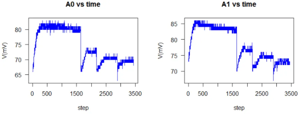
Figure 5.2: Electrodes signal Voltage vs time for B.thuringiensis additions.
Figure 5.3: Electrodes signal with Kalman filter and the running mean to filter the signal
5.3
Results
The fig. 5.2 shown two electrodes signals (A0 left, A1 right). The graph of voltage of electrodes vs time shows a jump at the addition time.
The arduino board has an unique A/D conversor. For electronical reasons, reading 8 port with only one conversor introduces some noise. To supress that noise, a Kalman filter and the running mean were use to filter (smooth) the signal.
5- Bacterial threat remote detection using electronic tongues
5.4
Discussion
We have preliminary results in the electrodes response for B.thuringiensis
detection. This particular disposal was able to find the spore additions.The staircase graph is the plotting of the concentration against time and shown each jump was the signal was in response of spores additions. The sensor A0 and A1 correlates well each other, allowing to evaluate the reproducibility of the method.
The data was observed remotely, its was able to be observed from several computers at the same time. These results were visualized with an http interface which can be shown through any browser as Mozilla Firefox or Google Chrome in Windows or Linux without problems.
This interface was build using the popular JavaScript library Ext JS and currently allows to see real time graph of voltage of electrodes vs time, and to save, rename and delete the measured data.
This setup offers the possibility to acquire data from several arduino cards at the same time, being able to construct a network of hundred of sensors inexpensively. In fact, some tests were also conducted with an STM32F4 dis-covery kit, as substitutes of the Arduino card, using the same node.js server obtaining promissing results. The amount of sensors connected simultane-ously to the arduino card were from four to six for each experiment.
5.5
Conclusions
These preliminary results, shown that a simple and cheap configuration can be used for the pathogen reproducibility of the electrodes and how a simple processing tool: the running mean, is able to find the spore additions.
The Data Collector program is able to maximize the accesibility of the data through mutiple devices and operative systems, as the client only need a web browser installed to function.
6
Smart nanomaterials for drug-delivery
Breast cancer is leading the statistics of cancer diseases in women. Although chemical treatments are improving within time, their well-known secondary effects sustain the need for (novel) targeted therapies. Cancer cell targeting research is therefore a field under continuous expansion. The targeting ele-ment is obviously critical in this context. Humanized monoclonal antibodies primary choice in this context, currently used in clinical context. As proof-of-concept, this approach is applied to track HER2+ breast cancer receptors, employing a carrier of apoferritin (Apo) containing doxorubicin (DOXO).
6.1
Introduction
Breast cancer (BrC) is the most common cancer diagnosed among women [54], being the second cause of death around the world [55]. Despite the exist-ing techniques for its early detection, the global mortality remains high. BrC can be classified by the expression of cell-specific biomarkers into differents groups: estrogen receptor (ER), progesterone receptor (PR) and human epi-dermal growth factor receptor-2 (HER2) [56]. Although HER2-positive can-cer is not the most abundant form of BrC, it is aggressive and fast-growing, requiring quick and effective responses. This form of cancer is character-ized by an overexpression of the HER2 receptor [57], which is related to its severity and drug response [58]
6- Smart nanomaterials for drug-delivery
cell, leaving normal healthy cells unharmed. This approach became more effective and less toxic for cancer patients than conventional therapy [56].
DOXO is a anthracycline antibiotic used frequently alone or with other drugs as treatments in several differents types of cancer, and have showed, be effective [60]. Its usage as conventional medication limited due to the car-diotoxicity adverse effect. A novel idea for decreased DOXO-related toxicity is the use of carrier systems. Thus, development of drug-delivery systems rep-resents a successful strategy to limited the exposure in sites frequently asso-ciated with conventional anthracycline toxicity, such as the myocardium [61]. Many authors report, that the determinants in the DOXO cardiomyopathy is the cumulative dose. The use of smaller and divided doses decrease the likelihood of developing heart failure [62].
Many therapeutic solutions have been focused in the development of nanoparticles for drug delivery targeting [59]. As targets for drugs delivery are used cell-specific biomarkers, like monoclonal antibodies, non-antibody proteins and other molecules [56].
The vehicle supporting the drug and the targeting element should offer biodegradable and biocompatible features and undergo a rapid and complete metabolism and elimination after their in vivo activity. The most common drug delivery, are recurrently based on inorganic materials, organic matri-ces or hybrid (inorganic/organic, core/shell) structures. Alternatively, the protein-cage molecules based on ferritins is emerging as an effective way to carry drugs. Ferritins have exceptional characteristics, namely biodegrad-ability, solubility, functionalization versatility, and remarkable capacity to bind different types of drugs [63, 64].
Apoferritin (Apo) has the ability to self-assemble: disassembles into sub-units at pH < 3.0 and reassembles as an intact sphere at pH > 5 [63]. Thus, using the iron empty cavities of Apo nanocage [65], DOXO can be loaded inside the nanocarriers by a reassemble route involving pH variations [63].
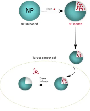
In the present work we intend to develop a nanomaterial carrying in its interior DOXO as model antitumor agent. This is herein tested in HER2+ breast cancer (fig. 6.1).
6.2
Materials and methods
6.2.1
Materials and instruments
The biological material used was MCF7 cell line from human breast (adeno-carcinoma). The chemical, used were ferritin equine spleen Type I (Sigma Aldrich), Sodium acetate (Riedel-de-Haen), Acetic acid glacial (Analar
6- Smart nanomaterials for drug-delivery
Figure 6.1: Scheme DOXO cell release
pur), mercaptosuccinic acid (Sigma Aldrich), dialysis sacks MWCO 1200 Da (Sigma Aldrich), Sodium Chloride (NaCl, Panreac), Sodium hydroxide (NaOH, EKA), Hydrochloric acid (HCl, Panreac), Phosphate Buffered Saline (PBS, Amresco), DOXO hydrochloride (DOXO, Sigma Aldrich), Dimethyl Sulfoxide (DMSO), Modified Eagle’s Medium (DMEM), ethanol, fetal bovine serum (FBS), thiazolyl blue tetrazolium bromide (MTT). Measurements were realized in UV-vis spectrophotometer Evolution 220 from Thermo Scientific., F-4500 fluorescence spectrophotometer (Hitachi, Japan).
6.2.2
Preparation of Apo
Apo was prepared using, previuosly procedure, described in reference [65], with minor modifications. The ferritin equine spleen Type I (224 µl, 0.34 mmol) was diluted until 5 mL 0.1 M N aOAc buffer (pH 5.5). First, was dialyzed against 152 ml of N aOAc buffer (0.1 M) at pH 5.5 for 20 min. Then, was added to the ferritin solution mercaptosuccinic acid (371 µl, 0.1 M) and dialyzed for 2 h against N aOAcbuffer (0.1 M). Later, mercaptosuc-cinic acid (185 µl, 0.1 M) was added and dialysis continued for 1 h against
6- Smart nanomaterials for drug-delivery
maintained for 20 min. This process was repeated until reaching a complete decoloration of the ferritin solution.
6.2.3
DOXO encapsulation
Apo-Doxo were constructed by disassembly/reassembly method reported in reference [63]. Apo solution at 0.5 mg/ml prepared with NaCl (16µl in 5 ml of NaCl) was adjusted to pH 2.0 by addition of HCl (0.1 M) and incubated for 10 min. DOXO (0.01 mg/ml) was added to the solution and the pH was maintained for 5 min. Then, the pH was increased to 8.0 using NaOH and was stirred at room temperature for 30 min. The resulting solution was dialyzed against PBS to remove inbound DOXO for 2 h. After dialysis, solutions were centrifuged at 3500 rpm for 40 min at 4o
C and supernatant was collected.
6.2.4
Intrinsic emission fluorescence
The fluorescence spectra of Apo-Doxo and Doxo standard were analyzed using fluorescence spectrophotometer. Spectra were recorded on a 1 cm path-length cuvette with an excitation slit width of 20 nm and an emission slit width of 20 nm. The excitation wavelength was set at 480 nm and the emission spectra were recorded at 500 nm to 700 nm.
6.2.5
Cell culture
Human breast carcinoma cells (MCF-7) were culture in Dulbecco’s Modified Eagle Medium (DMEM) (Sigma, PT), supplemented with 10%Fetal Bovine Serum (FBS, Invitrogen Life technologies, UK), 1%penicillin/streptomycin (Invitrogen Life technologies, UK) and 3,7 g/L sodium bicarbonate and main-tained at 37o
C in a humidified atmosphere containing 5%CO2. The cells were
used between passages 17 to 29.
6.2.6
MTT viability Test
MCF7 were seeded in a 96-well plate at a density of 1x10⁵ cells/well and incubated in complete medium at 37o
C for 24h. Treatment was administered after 24h. DOXO was formulated into concentrations of 0, 0.05, 0.1, 0.2, 0.4, 0.8, 1, 1.6 and 3µg/ml, and 3 replicate wells were used for each concentration, with a final volume of 200µl. NP loaded with DOXO was add in triplicated and Apo was used as control of NP not loaded. Following 24h of treatment incubation washed with PBS and starting the MTT measurements, added
6- Smart nanomaterials for drug-delivery
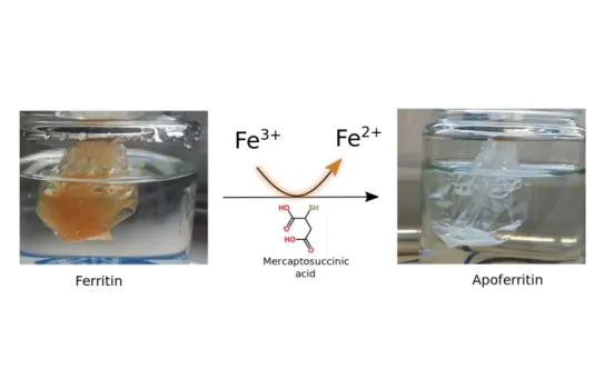
Figure 6.2: Scheme for Apo preparation
20 µL of 20 mM MTT (dissolved in PBS). After incubate the cells for 3 hours at 37o
C, the MTT solution was carefully removed and 200µL DMSO was added in order to solubilize the violet formazan crystals. The plates were then shaken on a horizontal shaker for 15 min to allow for complete dissolution. The optical density (OD) was read using a spectrophotometric microtiter plate reader set at a dual wavelength of 550/650 nm.
6.3
Results
Figure 6.2 show that the iron core is extracted. The transparency of the fer-ritin protein in solution evidence the absence of the iron core. In this manner we obtained the apoferritin nanoparticle prepared for loaded a DOXO.
The empty iron cavities of Apo nanocages are used for DOXO encap-sulation by a reassemble route involving pH variations. The Transmission electronic microscopy (TEM) images indicated the similar morphology and size of Apo-Doxo and Apo. The corresponding TEM image confirmed the formation of the expected protein nanocages (see fig: 6.3).
6- Smart nanomaterials for drug-delivery
Figure 6.3: SEM image: left for Apo and right for Apo-Doxo.
Figure 6.4: Fluorescence spectra: left differents Apo-Doxo concentration and right calibration curve for Doxo.
6- Smart nanomaterials for drug-delivery
According to the FDA, IC50 represents the concentration of a drug that is required for 50%inhibition in vitro, see Table:6.1.
DOXO µg/ml OD value IR (%)
0 0.703+
− 0.04 0
0.05 0.688+
− 0.05 2.13
0.1 0.655+
− 0.05 5.45
0.2 0.636+
− 0.03 9.53
0.4 0.539+
− 0.03 23.32 0.8 0.363+
− 0.08 48.36 1.6 0.104+
− 0.07 85.16
3 0.128+
− 0.003 81.69 Table 6.1: Table for Inhibitory rate for DOXO
The inhibition of cell growth (IR) was calculated using the following equa-tion:
IR(%) = (1−ODg
ODc
)∗100 (6.1)
where:
• R: Inhibitory rate (%)
• ODg = mean OD value in the experimental group.
• ODc = mean OD value in the control group.
Cell viability assay was through the MTT. MTT reagent is added to the medium and to incubate between 1 to 4 hours. When we performed this protocol in 24 and 12 hours treatment, the results were incongruous, we obtained more metabolic activity in higher concentrations that were against all previous published results.
We hypothesize, that the results were due to DOXO coloration and a possible interaction with the pH indicator of the medium, in this case, phenol red. Taking this in account, we decided to performed several washes before and do the incubation with MTT reagent with PBS. It was finally obtained the expected curve where is possible to obtained the IC50 value, fig: 6.5.
6- Smart nanomaterials for drug-delivery
Figure 6.5: MTT assay for Doxo at 24 hrs.
Figure 6.6: MTT assay at 24 hrs.
6- Smart nanomaterials for drug-delivery
6.4
Discussion
Iron storage protein ferritin, as a template for Apo-DOXO preparation. DOXO fluorescence is dependent of several variables, mainly if DOXO is con-jugated with other biological compounds, with proteins and membrane sys-tems exhibit variable effects on DOXO fluorescence. One way to understand the potential effects of the interaction with model cellular compounds was to compare the fluorescence spectra of the final compound with the charac-teristic spectra obtained from DOXO fluorescence. The fluorescence spectra standard and Apo similar indicated, the structural integrity was not affected by the encapsulation procedure. Using DOXO as a single form of treatment, we could conclude that the most efficient concentration 0,8 µg/mL, IC50 value (half maximal inhibitory concentration).
MCF-7 cell used in many application as model to investigate the applica-tion in antitumour drugs. In this study, it was shown that the encapsulated DOXO has an antiproliferative effect in MCF-7. This confirmed the reported, in the literature [66,67], that loading ferritin with DOXO is successful strate-gies, and particularly their release into the cell.
6.5
Conclusions
7
Conclusions
In this report, I have reviewed my professional path as biochemist. I fin-ished my Biochemistry degree in 2010. During this period I learned about ischemia process and I gained skilled in many laboratory techniques, such as spectrophotometer, animal surgery and behavioral trials. Work in something that could lead to a better understanding about how ischemia happens, and how it can be treated. The purpose of this study was to describe alterations in oxidative metabolism in the cortex, striatum, and hippocampus, and the effect of these changes on memory, and learning disorders in a rat model of cerebral hypoperfusion. One of the things I liked more at this time was the fact that this may potentially have an impact in real lives.
After obtaining my degree, I started as junior researcher in September 2010 at IPK institute, Havana, Cuba. Here I was involved in a study to identify NoV by RT-PCR, to know the clinical characteristics and associ-ation with RoV infection in children aged less than five years. I learned HIV microelisa, detection of HBsAg, ELISA, SDS-PAGE, DNA and RNA extraction, RT-PCR, Real-Time PCR, immunofluorescence, agarose gel elec-trophoresis and cell culture.
Later in Portugal I continued my research career at INESC-TEC devel-oping a biosensor for bacterial detection. We used B. thuringiensis strains as model of B. cereus in the construction of electrodes. The goal was to built a sensor for bacteria/spore recognition, using the molecular imprinted technique. In this opportunity, I learned how the development of biosensors can have an impact in the early detection of outbreaks, and about how can they prevent its propagation. I gained skill in electrode construction.
7- Conclusions
be used not only as a drug but also as the smart delivery targeting system. We continue working in this direction for obtained an effective system for smart drug delivery. Until now, I learned more about cell culture, electrochemical measurements, building of immunosensor, breast cancer and nanomaterials.
7- Conclusions
A
Shiga Toxin Detection : A Short Review
Shiga toxin is secreted by certain types of bacteria, like Shigella dysenteriae and certain strains of Escherichia coli bacterias. Shiga toxin and the closely related Shiga-like toxins represent a group of very similar of cytotoxins that may play an important role in diarrheal diseases and in the hemolytic-uremic syndrome. The outbreaks caused by this toxin create serious public health crisis with significant economical cost. In the past years there has been research relative to finding specific protective measures or therapy against the infections provoked by STEC. The supportive therapy, which includes antibiotics or antidiarrheals, may potentially lead to more severe manifesta-tions of the disease, as some recent clinical result have shown, thus looking for alternatives approaches is required. On the other hand, the knowledge of the molecular structure of the toxin will provide useful information that may be used in therapies and vaccine candidates for certain types of can-cer and infectious diseases. At the present are necessary techniques of rapid identification, simple and sensitive which can be employed in the country-side with minimally-sophisticated instrumentation. Biosensors have shown tremendous promise to overcome these limitations and are being aggressively studied to provide rapid, reliable and sensitive detection platforms for such applications.