Abstract Chemical therapy for the treatment of leishmaniasis is still inadequate, and a number of drugs and therapeutic programs are being tested. Besides treatment, the ultimate goal is an effective cure, and histopathological analyses of the lesion cicatrices constitute an important measure of treatment success, or otherwise, in this respect. In this paper, we describe histopathological patterns in cases of American cutaneous leishmaniasis in 32 patients from the municipality of Caratinga, Minas Gerais, Brazil, before and after treatment with the following therapeutic methodos: l) leishvacin + glucantime; 2) leishvacin + BCG associated with glucantime; 3) glucantime; 4) leishvacin + BCG. Lesion fragments were collected from all patients by biopsy prior to, and approximately 30 days after, each treatment which resulted in a clinical diagnosis of cure. Following the analysis of slides, the preparations were described from a histopathological point of view and grouped taking into account the prevalence or significance of the characteristic elements. This process resulted in the following classification: 1. exsudative reaction (ER); 2. exsudative giant cell reaction (EGCR); 3. exsudative productive reaction (EPR); 4. exsudative productive giant cell reaction (EPGCR); 5. exsudative productive necrotic reaction (EPNR); 6. necrotic exsudative reaction (NER); 7. productive exsudative reaction (PER), 8. productive giant cell reaction (PGCR); 9. productive exsudative giant cell reaction (PEGCR); 10. productive exsudative giant cell granulomatous reaction (PEGCGR); 11. productive reaction (PR) and 12. productive cicatricial (cure) reaction (PCR). After this analysis, it was noted that clinical cure did not always coincide with histopathological cure.
Key-words:Cutaneous leishmaniasis. Histopathology. Treatment. Criteria for cure.
Histopathology of human American cutaneous
leishmaniasis before and after treatment
Departamento de Parasitologia, Instituto de Ciências Biologicas, Universidade Federal de Minas Gerais, Belo Horizonte, MG, Brasil.
Address to:Prof ª A.C.C. Botelho. Deptº de Parasitologia/ICB/UFMG. Av. Antonio Carlos 6627, Pampulha, 31270-901 Belo Horizonte, MG. Fax (031) 441-6909.
Recebido para publicação em 06/03/97.
Resumo A quimioterapia para a leishmaniose não é satisfatória e existem hoje, várias drogas e esquemas terapêuticos em teste. Além do tratamento ideal, busca-se um critério de cura efetivo, onde a análise da histopatologia da cicatriz poderá ser de grande valia. Este trabalho propõe caracterizar o padrão histopatológico de casos humanos de leishmaniose tegumentar americana, em 32 pacientes do município de Caratinga-MG, antes e após o tratamento com os seguintes métodos terapêuticos: 1) leishvacin + glucantime; 2) leishvacin + BCG associado ao glucantime; 3) glucantime; 4) leishvacin + BCG. Foram colhidos fragmentos das lesões de todos os pacientes, através de biópsias, antes e após o tratamento, com diagnóstico de cura. Após análise das lâminas, as preparações foram descritas, do ponto de vista histopatológico, e agrupadas levando em conta a prevalência e a significância do elemento característico. Tal processo resultou na classificação: 1. reação exsudativa; 2. reação exsudativa giganto-celular; 3. reação exsudativa produtiva; 4. reação exsudativa produtiva giganto-celular; 5. reação exsudativa produtiva necrótica; 6. reação necrótica exsudativa; 7. reação produtiva exsudativa; 8. reação produtiva giganto-celular; 9. reação produtiva exsudativa giganto-celular; 10. reação produtiva exsudativa giganto-celular granulomatosa; 11. reação produtiva e 12. reação produtiva cicatricial (cura histopatológica). Observamos após tal análise, que nem sempre a cura clínica coincide com a cura histopatológica. Palavras-chaves:Leishmaniose cutânea. Histopatologia. Tratamentos. Critérios de cura.
Histopatologia de leishmaniose tegumentar americana
humana, antes e após o tratamento
Leishmaniasis is a disease caused by a digenetic protozoan of the genus Leishmania Ross, 1903, which infects vertebrate hosts of a number of mammalian species, including humans, and invertebrate hosts, which in the New World are insects of the genus Lutzomyia. It is a disease which has a wide spectrum of clinical manifestations. In this paper we deal with the cutaneous form, referred to as american cutaneous leishmaniasis (ACL).
The introduction of the use of Tartar Emetic by the Brazilian physician Gaspar Viana in 1912 was an important advance in the treatment of leishmaniasis. However, due to its toxicity and the frequent relapse it has been substituted by the use of pentavalent antimony, used to the present day. Antimony, although less toxic, is still problematic and its use is not encouraged in cases such as cardiopathic and nephropathic patients nor for the aged or during pregnancy.
Besides the search for the most effective drugs and therapy for leishmaniasis, it is necessary to establish accurate criteria for evaluating its cure. Most authors have adopted clinical criteria based on the complete healing of the lesion. However, in 1915, D’ultra e Silva4 observed that the closing up of the lesion was not sufficient evidence for a complete cure. Perhaps agreeing with this finding, Schubach16 found leishmaniasis to be still present in a lesion which had healed over eight years previously. Likewise, Guerra5 not found the parasite in unaltered nasal mucous membranes in 147 patients with no history of leishmaniasis, but the i n t r a d e r m a l t e s t w a s p o s i t i ve i n 2 1 , 8 % showing a subclinical infection. Marsden10also recorded that some patients were still infected despite a clinical diagnosis declaring them cured. These findings underline the importance of histopathological studies of leishmaniasis, not only as an aid to diagnosis but also in the establishment of criteria to determine the success, or otherwise, of therapy.
Histopathological studies of leishmaniasis begun in Brazil when the disease was first identified in the country, and since then have worked towards establishing the characteristic morphological patterns of the disease. The first histopathological classification was suggested by Azulay1. Investigating cases of Old World diffuse leishmaniasis, Bryceson3 proposed a classification of six histopathological groupings. Nicolis13, also studying forms of Old World leishmaniasis, proposed a simplified scheme
with three histopathological phases. Ridley15, relating 60 biopsies with the prognosis of the lesions, proposed a classification of five groups, but later, added sub-groups which made the scheme difficult to use. Magalhães6studied 487 skin fragments of patients from Três Braços, in the State of Bahia, Brazil, and, while taking into account previous classifications, proposed his own of three groups. Magalhães7subsequently recommended a five group classification based on 162 skin fragments, and their correspondence with the clinical evolution of the disease which the author considered relevant in aiding doctors in their diagnosis and treatment. In another paper published the same year, Magalhães8 classified a further 378 biopsies of ACL carriers from the region of Três Braços into his five groupings of histopathological patterns. Finally Bittencourt and Barral2, taking into account the work of Ridley15and Magalhães7 8; proposed a simplified classification of just three patterns.
Based on this literature, we describe here our findings concerning the histopathology of skin fragments of patients from the municipality of Caratinga, in the State of Minas Gerais, Brazil, both before and after medical treatment, and characterize alterations in the basic histopathology and the evolution of the leishmaniasis lesion. We also analyze the processes involved in the cure of each of the therapeutic schemes under consideration.
We consider this study justifiable in that, as far we know, no systematic histopathological study of the leishmaniotic lesion has been carried out before and after treatment with four different therapeutic methods, with the additional aim of carrying out a comparative study of the development of the lesions and the kinetics of their cure. This enabled us to associate the clinical signs of cure with the histopathological aspects of the healed lesion, by comparing descriptions of the histopathological patterns with the respective treatments applied.
MATERIALS AND METHODS
were studied. Scars were biopsied on various dates, most usually 30 days after patients were considered clinically cured.
Diagnosis was made by a direct smear stained with Giemsa, and by the Montenegro test. The fragments were subjected to routine histological processing. The material was separated into four groups, according to the treatments given: group I- leishvacin + glucantime; group II-leishvacin + BCG associated with glucantime; group III- glucantime; group IV- leishvacin + BCG.
Leishvacin is a leishmaniasis vaccine developed in the Depar tment of Parasitology, UFMG (Mayrink11 12). It is a mixture of extracts of five dead, sonicated dermatotrophic strains of Leishmania: L. (L.) mexicana(MHOM/BR/60/BH6);L. L. amazonensis (IFLA/BR/67/PH8); L. L. sp (mexicana complex) (MHOM/BR/73/BH121); L.(L.) sp(L. major- like) (MHOM/BR/71/BH49);L. (Viannia) guyanensis(MHOM/BR/73/M1176). The p r o t e i n c o n c e n t r a t i o n o f l e i s h v a c i n i s 1500µg/ml.
Group I.Seven patients: two females and five males. The average age was 28 years, ranging from 12 to 42. Most patients had only one lesion of an average duration of 75 days (range 45-90 days). In all patients the parasitological examinations were positive before treatment. Treatment:The vaccine was given by intramuscular injection according to the scheme of Mayrink12: Day 1 - 100µl, Day 2 - 200µl, Day 3 - 300µl, Day 4 - 400µl, and Days 5-10 - 500µl. This scheme was repeated after an interval of 10 days.
Glucantime was administered by intramuscular injection in daily doses of 0.5ml/5kg weight, not exceeding 5ml/day, and given at the same time as the leishvacin injection, also with an interval o f 1 0 d ay s b e t we e n t r e a t m e n t s e s s i o n s. Treatment of this group of patients varied between 48 and 150 days.
Group II. Eight patients: three females and five males. The average age was 36 years, ranging from 14 to 66. Six patients presented just one lesion. The average duration of lesions was 90 days (range 30-180 days). In all patients, parasitological examinations were positive before treatment. Treatment: leishvacin + BCG in a single dose by intrader mal injection. Fifteen days later, glucantime was given, by intramuscular injection at a daily dose of 1ml/5kg weight, not exceeding 10ml/day, for 10 days, followed by a treatment interval of 10
days. The protein concentration of the vaccine was 600µg/0.2ml. The treatment period for this group varied between 43 and 108 days.
Group III. Eight patients: five females, three males. The average age was 39 years, ranging from 13 to 64. Seven of the patients had only one lesion. The average duration of lesions was 118.5 days (range 38-210). In three patients the parasitological examinations were proved negative before treatment.Treatment:glucantime was administered by intramuscular injection in daily doses of 1ml/5kg, not exceeding 10ml/day, for 10 days, with treatment intervals of 10 days. The treatment period for this group was 30-110 days.
Group IV. Nine patients: three females, six males. The average age was 20 years, ranging from 3 to 56. Six patients presented with only one lesion. The average duration of lesions before treatment was 79.4 days (range 40-180). In one patient the parasitological examination proved negative prior to treatment. Treatment: leishvacin + 100µg BCG, administered monthly until a clinical diagnosis of cure or up to a m a x i m u m o f f i v e m o n t h s . T h e p r o t e i n concentration of the leishvacin in this group was 600µg/0.2ml. In this group, treatment lasted 34 to 138 days.
The criteria used to access successful treatment (cure) were clinical: complete healing of lesions.
In all groups, each “series” (period of administration of treatment + intervals: see Table 1) were repeated until a patient was considered clinically cured.
RESULTS
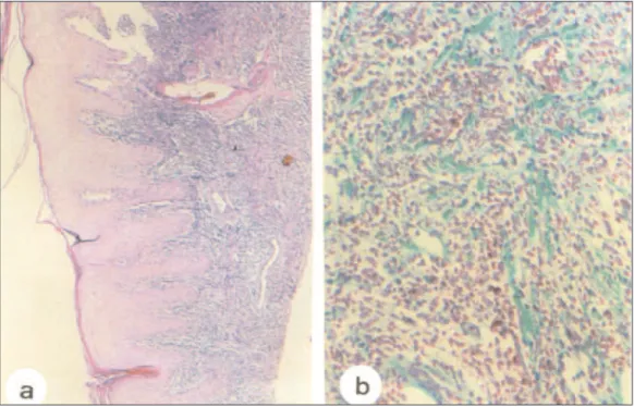
two cases were associated with parasites. Necrotic areas were rare. A few cases showed granulomatous reactions. Accentuated vascular neoformation. Collagen bundle neoformation, with broad and irregular fibers, sometimes disassociated. (Figure 1).
After treatment the principal alterations were 1) epidermis: persistence of hyperkeratosis, but less accentuated. Acanthosis, horn pearls and papilomatosis persisted in some cases. Persistence of the vacuolization of the cells of the Malpighian layer. 2) dermis: mononuclear exsudation, predominantly perivascular in m u l t i p l e fo c i o f va r i a bl e i n t e n s i t y. T h e predominance of lymphocytes, and the presence of giant cells of the Langhans and foreign body
types persisted. Capillary neoformation was of variable intensity (discrete and pronounced). Intense collagen neoformation, with fibers oriented in various directions or in one direction, and others uniformly oriented parallel to the basal epidermis, — or otherwise thick, poorly oriented, hyalinized, and located in the deepest part of the dermis. Typical granulomas were found extremely rare (Figure 2).
Following a complete and systematic analysis of all the slides to provide better understanding of lesion dynamics, we conventionally classified the inflammatory process by the prevalance and significance of the phenomena under consideration into 12 developmental phases (Table 2):
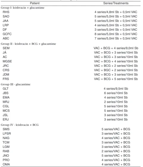
Table 1 - Treatment scheme for each therapeutic group.
Patient Series/Treatments
Group I- leishvacin + glucantime
RHS 4 series/4,8ml Sb + 0,5ml VAC
SAO 5 series/5,0ml Sb + 0,5ml VAC
JAA 5 series/5,0ml Sb + 0,5ml VAC
LRP 4 series/5,0ml Sb + 0,5ml VAC
DF 3 series/5,0ml Sb + 0,5ml VAC
GCFC 8 series/5,0ml Sb + 0,5ml VAC
ABC 7 series/5,0ml Sb + 0,5ml VAC
Group II - leishvacin + BCG + glucantime
SEM VAC + BCG + 4 series/9,0ml Sb
JÁ VAC + BCG + 3 series/10ml Sb
AC VAC + BCG + 3 series/10ml Sb
MGSE VAC + BCG + 4 series/10ml Sb
JRC VAC + BCG + 2 series/10ml Sb
CRS VAC + BGC + 2 series/10ml Sb
JDM VAC + BCG + 3 series/10ml Sb
FRS VAC + BCG + 5 series/10ml Sb
Group III - glucantime
GLT 4 series/9,5ml Sb
JBS 6 series/10ml Sb
EMA 4 series/10ml Sb
MRJ 2 series/10ml Sb
CGL 5 series/10ml Sb
MCS 5 series/10ml Sb
JSL 3 series/10ml Sb
ERJ 3 series/10ml Sb
Group IV - leishvacin + BCG
SMS 5 series/VAC + BCG
LPSR 3 series/VAC + BCG
NAG 4 series/VAC + BCG
TCM 3 series/VAC + BCG
LGM 3 series/VAC + BCG
RAB 2 series/VAC + BCG
JNO 5 series/VAC + BCG
PRO 4 series/VAC + BCG
Figure 1 - a,b: skin of the ankle of patient ABC, taken before treatment. Intense diffuse exsudation. Infiltrate of lymphocytes and plasma cells on the superficial dermis. Papillomatosis, hyperkeratosis and parakeratosis. (a) HE 40x, (b) Tricrômico de Gomori 200x.
1. exsudative reaction (ER); 2. exsudative giant cell reaction (EGCR); 3. exsudative productive reaction (EPR); 4. exsudative productive giant cell reaction (EPGCR); 5. exsudative productive necrotic reaction (EPNR); 6. necrotic exsudative reaction (NER); 7. productive exsudative reaction (PER); 8. productive giant cell reaction (PGCR); 9. productive exsudative giant cell reaction (PEGCR); 10. productive exsudative giant cell granulomatous reaction (PEGCGR); 11. productive reaction (PR); 12. productive cicatricial (cure) reaction (PCR).
The non-parametric Kruskall-Wallis test was used for the statistical analysis of the results i n w h i c h va l u e s w e r e a t t r i bu t e d t o t h e histopathological findings before and after treatment. The numbers applied after treatment were subtracted from those before treatment. The resulting value eliminated differences between the initial clinical situation of each patient (Table 2). The results showed that there was no significant statistical difference between the four treatments in the process towards cure (χ2 = 7.815, T = 1.537, T < χ2).
Table 2 - Main histopathological characteristics shown by the patients before (BT) and after treatment (AT) for each therapeutic group.
Patient Inflammatory process Difference Granuloma Giant cells
BT AT BT-AT BT AT BT AT
Group I - leishvacin + glucantime
RHS NER PEGCR 3 - TF - L
SAO ER PCR 11 - - -
-JAA EPGCR PCR 8 - - L
-LRP EPNR PR 6 - - -
-DF EPR EPR 0 - - -
-GCFC EPR PCR 9 - - -
-ABC NER PGCR 2 - - - FB
Group II- leishvacin + BCG + glucantime
SEM NER PCR 6 - - -
-JA EPR PR 8 - - -
-AC EPR PER 4 - - -
-MGSE ER PCR 9 - - -
-JRC EPGCR EPGCR 0 - - L L, FB
CRS EPR EPGCR 1 - TF - L
JDM EPGCR EPGCR 0 TF - L, FB L
FRS ER PER 6 - - -
-Group III - glucantime
GLT ER PCR 11 - - -
-JBS ER PER 6 - - -
-EMA EPR PR 8 - - -
-MRJ ER PEGCR 8 - TF - L
CGL EGCR PEGCG 8 - + L L, FB
MCS EPR PR 8 - - -
-JSL EPR EPR 0 - - -
-ERJ EGCR EPR 1 TF - L
-Group IV - leishvacin + BCG
SMS EPGCR PER 3 - - L
-LPSR EPGCR PEGCR 5 - + L L, FB
NAG EPR PER 4 - - -
-TCM EPR PEGCGR 7 - NTF - L
LGM EPGCR PEGCGR 6 - + L L
RAB ER EPR 2 - - -
-JNO ER EPGCR 3 - + - L, FB
PRO ER PER 6 - - -
-OMA ER PR 10 - - -
-Exsudative Reaction (ER); -Exsudative Giant Cell Reaction (EGCR); -Exsudative Productive Reaction (EPR); -Exsudative Productive Giant Cell Reaction (EPGCR); Exsudative Productive Necrotic Reaction (EPNR); Necrotic Exsudative Reaction (NER); Productive Exsudative Reaction (PER); Productive Giant Cell Reaction (PGCR); Productive Exsudative Giant Cell Reaction (PEGCR); Productive Exsudative Giant Cell Granulomatous Reaction (PEGCGR); Productive Reaction (PR); Productive Cicatricial (cure) Reaction (PCR)
Differences between treatments in the rate of cure were also tested (Kruskall-Wallis applied to the number of series/treatment) and likewise were not significant (χ2= 7.815, T = 5.538, T < χ2).
DISCUSSION
T h e a s s o c i a t i o n b e t w e e n t h e l e s i o n histopathology and the disappearance of leishmania parasites has been studied by a number of authors, identifying patterns in the evolution of the lesions or correlating them with the clinical situation and the immunological response. Observation of the histopathological picture before and after treatment, could allow us to say that a clinical cure of a patient was accompanied by an evolution of histopathological c u r e . H o w eve r, t h e t w o d i d n o t a l w ay s coincide. The number of series/treatment was not always directly proportional to the picture in terms of a histopathological cure. The ideal c u r e s h o u l d i n v o l v e b o t h a c l i n i c a l a n d histopathological cure, and studies of the latter cannot only aid diagnosis but can also be used as a criterion of cure.
In Group I, we observed coincident clinical and histopathological cures in three patients, and a histopathological picture with an active exsudative process even after the clinical cure was most pronounced in three patients.
In Group II, a clinical cure coincided with histopathological cure in two patients, and an active exsudative process, even after a clinical cure, was very marked in five patients.
In Group III, a clinical cure coincided with a histopathological cure in only one patient; there w a s r e c r u d e s c e n c e i n o n e , a n d n o coincidence in three patients.
In Group IV, none of the patients showed coincidence between clinical and histopathological assessment of cure. One patient was close to achieving this, but active exsudative processes after a clinical cure were notable in six patients.
There was no significant statistical difference between the four treatments in the evolution of the cure, or in the time taken to achieve it.
T h e p a t i e n t s s t u d i e d a r e s t i l l b e i n g a c c o m p a n i e d a n d o n l y o n e ( G r o u p I I I ) s h owe d r e l a p s e f i ve m o n t h s a f t e r b e i n g c o n s i d e r e d c l i n i c a l l y c u r e d . I n t h e f i r s t treatment, the patient received three series of glucantime, but remained with an exsudative
productive reation (EPR-3). Treatment after recrudescence involved three more glucantine series, and the final histopathological picture was of productive reaction (PR-11).
Schubach16recorded a case of a patient treated in 1979, who, when re-examined in 1987, showed a biopsy culture of the cicatrices which was positive for Leishmania sp. As a result, we carried out scar tissue cultures for three patients, 383, 273, and 242 days after the last dose of treatment, respectively. In all the three cases leishmania parasites were not detected. The aim of the classification adopted herein was to verify the process of lesion healing, from the beginning of treatment until the anatomical/clinical diagnosis of cure. For this reason we did not follow the schemes of Ridley14, Magalhães6 7and Bittencourt2.
Ridley14reported usually successful prognoses in cases with necrotic foci in the central area of the lesion. In the cases described herein four showed some foci of necrosis before treatment, but, in each of these cases, clinical cure was achieved.
Twenty-eight of the 32 cases showed positive results for parasitological tests with smear stained with Giemsa, but parasites were observed in the histological sections of only two patients.
Knowing that the prevalent species in Caratinga is L. b. braziliensis9strains from three patients were characterized by isoenzymes. All three were infected with L. braziliensis.
These results confirm some observations made on scar biopsies of 20 patients analyzed by D r H u g o S i l v i a n o B ra n d ã o ( p e r s o n a l communication to W.M. in 1982), in which it was found that all cases showed inflammatory processes even after the patient had been released following a clinical diagnosis of cure; in addition all of them showed negative results in direct searches of parasites in histological sections.
Proliferative phenomena of the epidermis simulating epidermal carcinoma which can sometimes complicate differential diagnoses were not found in any of the patients studied here.
were progressively destroyed up to the moment when the cure was accompanied by fibrosis. The distribution, structure, orientation, and regressive phenomena of collagen were very similar to that of secondary repair cicatrices, although more retractible and proliferative.
ACKNOWLEDGMENT
To Prof Ivan Sampaio, Departamento de Zootecnia, Escola de Veterinária, Universidade Federal de Minas Gerais, for his help in the statistical analysis of the data.
REFERENCES
1. Azulay RD. Histopatologia da Leishmaniose
Tegumentar. Dermatologia Ibero Latino-Americana
2:7-15,1960.Apud:Bittencour t AL, Barral A.
Evaluation of histopathological classifications of A m e r i c a n C u t a n e o u s a n d M u c o c u t a n e o u s Leishmaniasis. Memórias do Instituto Oswaldo Cruz 86: 51-56, 1991.
2. B i t t e n c o u r t A L , B a r r a l A . E v a l u a t i o n o f histopathological classifications of American Cutaneous and Mucocutaneous Leishmaniasis. Memórias do Instituto Oswaldo Cruz 86:51-56, 1991.
3. Bryceson ADM. Diffuse Cutaneous Leishmaniasis in Ethiopia. I. The clinical and histopathological features of the disease. Transactions of the Royal Society of Tropical Medicine and Hygiene 63:708-737, 1969.
4. D ’ u l t r a e S i l va O. S o b r e a L e i s h m a n i o s e Tegumentar e seu tratamento. Memórias do Instituto Oswaldo Cruz 7:213-248, 1915.
5. Guerra MOP, Furtado T, Barros GC, Sessa, PA, Carias VRD. Infecção subclínica na Leishmaniose
Tegumentar Americana. Anais Brasileiros de
Dermatologia 60: 365-369, 1985.
6. Magalhães AV, Llanos A, Cuba CC, Araujo FB, Parreiras BM, Medeiros JM, Barreto AC, Marsden PD, Raick NA. Nova classificação histopatológica da
Leishmaniose Tegumentar. In: Sociedade
Brasileira de Patologistas (ed) Anais do VI Congreso Regional Centro-Leste da Sociedade Brasleira de Patologistas, 3-6 Novembro, Imprensa Universitária, Uberlandia, p. 62,1982.
7. Magalhães AV, Moraes MAP, Raick AN, Llanos-Cuentas A, Costa JML, Cuba CC, Marsden PD. Histopatologia da Leishmaniose Tegumentar por
Leishmania braziliensis braziliensis. 4. Classificação histopatológica. Revista do Instituto de Medicina Tropical de São Paulo 28:421-430, 1986a.
8. Magalhães AV, Moraes MAP, Raick AN, Llanos-Cuentas A, Costa JML, Cuba CC, Marsden PD. Histopatologia da Leishmaniose Tegumentar por Leishmania braziliensis braziliensis. 1. Padrões histopatológicos e estudo evolutivo das lesões. Revista do Instituto de Medicina Tropical de São Paulo 28:253-262, 1986b.
9. Magalhães-Rocha NM, Melo MN, Babá EH, W i l l i a m s P, D i a s M , M i c h a l i ck M S, C o s t a C A , M ay r i n k W, Tavo r a P T C. I s o e n z y m a t i c
characterization of Leishmaniaisolated from human
hosts in the Rio Doce Valley - MG. Memórias do Instituto Oswaldo Cruz 82:96, 1987.
10. Marsden PD, Netto EM, Badaró R, Cuba CAC, Costa JLM, Barreto AC. Correspondence. The American Journal of Tropical Medicine and Hygiene 35:449, 1986.
11. Mayrink W, Costa CA, Magalhães PA, Melo MN, Dias M, Oliveira-Lima A, Michalick MSM, Williams P. A field trial of vaccine against American Dermal Leishmaniasis. Transactions of the Royal Society of Tropical Medicine and Hygiene 73:385-387, 1979. 12. Mayrink W, Michalick MSM, Melo MN, Nascimento E,
Magalhães PA, Costa CA, Oliveira-Lima A, Dias M. Tratamento da Leishmaniose Tegumentar utilizando vacina. Anais Brasileiros de Dermatologia 6:55-59, 1991.
13. Nicolis GD, Tosca AD, Stratigos JD, Capetanakis JA. A clinical and histological study of cutaneous leishmaniasis. Acta Der mato-venereologica 58:521-525, 1978.
14. R i d l ey D S. H i s t o p a t h o g e n i c m e c h a n i s m s i n leishmaniasis. Pathologica 75:471-479, 1983. 15. Ridley DS, Marsden PD, Cuba CC, Barreto AC.
A histological classification of mucocutaneous leishmaniasis in Brazil and its clinical evaluation. Transactions of the Royal Society of Tropical Medicine and Hygiene 74:508-514, 1980.