Printed version ISSN 0001-3765 / Online version ISSN 1678-2690 www.scielo.br/aabc
http://dx.doi.org/10.1590/0001-3765201520140288
Correspondence to: Elaine Soares Coimbra E-mail: elaine.coimbra@ufjf.edu.br
Antileishmanial activity of some Brazilian plants, with particular reference to Casearia sylvestris
LUCIANA M.R. ANTINARELLI1, NÍCOLAS C. PINTO2, ELITA SCIO2and ELAINE S. COIMBRA1 1
Departamento de Parasitologia, Microbiologia e Imunologia/ICB, Universidade Federal de Juiz de Fora, Campus Universitário,
Bairro São Pedro, 36036-900 Juiz de Fora, MG, Brasil
2Laboratório de Produtos Naturais Bioativos,Departamento de Bioquímica/ICB, Universidade Federal de Juiz de Fora, Campus Universitário,
Bairro São Pedro, 36036-900 Juiz de Fora, MG, Brasil
Manuscript received on June 11, 2014; accepted for publication on September 6, 2014
ABSTRACT
Leishmaniasis is a complex of diseases caused by Leishmania protozoa which treatment is restricted to a limited number of drugs that exhibit high toxicity, collateral effects and are often costly. There are a variety of tropical plants distributed in Brazil, and for many poor people the therapy for several diseases is based mainly on the use of traditional herbal remedies. In this work, the cytotoxic activity of 17 plant methanol extracts was evaluated on several Leishmania species and murine macrophages. Among them, the extract of Casearia sylvestris, Piptocarpha macropoda, Trembleyaparviflora, Samanea tubulosa and Plectranthus neochilus showed a promissing leishmanicidal activity, exhibiting IC50values below of 20 µg/mL against
at least one species of Leishmania. Casearia sylvestris showed the most expressive activity against all promastigote forms of Leishmania species (IC50 values of 5.4 µg/mL, 5.0 µg/mL, 8.5 µg/mL and 7.7 µg/
mL for L. amazonensis, L. braziliensis, L. chagasi and L. major, respectively), being more effective than the reference drug miltefosine. In spite of the cytotoxic effect on macrophages (CC50 value of 5.2 µg/mL), C.
sylvestris exhibited a strong inhibition against intracellular amastigotes of L. braziliensis (IC50 value of 1.3 µg/
mL). Further studies, including bio-guided fractionation will be conducted to identify the active compounds. Key words: Brazil,Casearia sylvestris, leishmanicidal activity, medicinal plants, natural products.
INTRODUCTION
Leishmaniasis is a vector borne disease caused by protozoa parasites of the genus Leishmania (WHO 2010). According to the World Health Organization, leishmaniasis is considered a major Neglected Tropical Disease with expressive economic, social,
and political impacts. Leishmaniasis is distributed in more than 90 countries, with an annual incidence of 1.5 to 2.0 million cases, and 350 million people under the risk of infection (WHO 2013).
Leishmaniasis comprises a complex of clinical manifestations including ulcerative skin lesions,
destructive mucosal inflammation, and disseminated
et al. 2005). These clinical manifestations occur due to a complex interaction between the parasite and the immune response of the mammalian host.
Current chemotherapeutic agents for the treat-ment of all clinical manifestations of leishmaniasis are pentavalent antimonials compounds (e.g., sodium stibogluconate and meglumine antimoniate) and amphotericin B, which unfortunately are considerably toxic (Croft and Olliaro 2011, Tempone et al. 2011, Singh and Sundar 2012). Furthermore, these drugs exhibit several limitations, including high cost and the need for daily parenteral administration (Singh and Sundar 2012). More recently the
anti-tumor drug miltefosine was introduced as the first
and still the only oral therapeutic option for the treatment of visceral leishmaniasis in India (Sundar et al. 2002, Dorlo et al. 2012).
There are a variety of tropical plants distributed in Brazil and for many poor people the therapy for several diseases is based mainly on the use of traditional herbal remedies (Lorenzi and Matos 2002). In most cases, these plants are used without
any scientific base. In the last years, several works
showed the leishmanicidal effect of some Brazilian plant extracts or essential oils (Santin et al. 2009, Albernaz et al. 2010, Alviano et al. 2012, Brito et al. 2013). In a previous work, we reported the antileishmanial activity of extracts of 20 plants
from the Brazilian flora (Braga et al. 2007). Due
to our continuous search for new alternatives for the treatment of leishmaniasis, this study aimed to investigated the leishmanicidal activity of 17 plant methanolic extracts against four Leishmania species and murine macrophages.
MATERIALS AND METHODS
PLANT MATERIAL
Specimens of 17 species were collected in Juiz de Fora, Minas Gerais, Brazil. A voucher specimen was deposited at the Herbarium Leopoldo Krieger (CESJ) of the Federal University of Juiz de Fora.
Table I shows the botanical name, local name, voucher specimen number and the popular uses of the plants tested.
PREPARATION OF PLANT EXTRACTS
The dried parts of the plants (50 g each) were powdered and macerated with methanol (3x
200 ml) for five days at room temperature. After
evaporation of the solvent under reduced pressure, the respective methanolic extracts were obtained. All extracts were kept dried in tightly stoppered bottles under refrigeration (4 °C) until used for the biological tests.
ANTILEISHMANIAL ACTIVITY
Parasites
Four Leishmania species were used for in vitro screening: L. amazonensis (IFLA/Br/67/PH8), L. major (MRHO/SU/59/P), L. braziliensis (MHOM/ Br/75/M2903) and L. chagasi (MHOM/Br/74/PP75). Promastigotes of L. amazonensis were cultured in Warren’s medium [brain heart infusion- BHI (Himedia, Mumbai, Indian), plus hemin and folic acid (Sigma Chemical Co, St. Louis, MO, USA)], promastigotes of L. major and L. braziliensis were maintained in Medium BHI, and promastigotes of L. chagasi were maintained in Medium 199 (Cultilab, Campinas, São Paulo, Brazil), both supplemented with 10% fetal bovine serum (FBS; Cultilab, Campinas, São Paulo, Brazil) at 25 °C.
Antipromastigote assay: The antileishmanial activity was determined by the colorimetric 3-(4,5-dimethylthiazol-2-yl)-2,5-diphenyl-tetrazolium bromide (MTT; Sigma-Aldrich, St. Louis, MO, USA) method based on tetrazolium salt reduction by mitochondrial dehydrogenases
(Mossman 1983). Briefly, promastigotes from
The screening was performed in 96-well microtiter plates maintained at 25°C. The analyses were made in duplicate. The extracts were dissolved in dimethyl sulfoxide (DMSO; Sigma-Aldrich, St. Louis, MO, USA) as a stock solution. Parasites were exposed to increasing concentration of the extracts solutions
(at minimum five serial dilutions) for 72h at 25 °C.
Controls containing 0.5% DMSO and medium alone were also included. The viability of promastigotes was obtained by measuring the absorbance at 570 nm (Multiskan MS microplate reader, LabSystems Oy, Helsink, Finland). Amphotericin B (Cristalia, São Paulo, Brazil) and miltefosine (Cayman Chemical Company, Michigan, USA) were used as reference drugs.
Antiamastigote assay: Inflammatory macro
phages were obtained from the peritoneal cavity of BALB/c mice previously inoculated with 3% thioglycollate medium (Sigma Chemical Co; St. Louis, MO, USA). After 72h, the peritoneal exudate was collected by washing with cold Hank´s Balanced Sal Solution (Sigma Chemical Co; St.
Louis, MO, USA) (Silva et al. 2012). Briefly,
peritoneal macrophages added at 2x10⁶ cells/mL to coverslips (13 mm diameter) previously arranged in a 24-well plate in RPMI 1640 medium (Cultilab, Campinas, São Paulo, Brazil) supplemented with 10% inactivated FBS, and allowed to adhere at 37 °C in 5% CO2. Adherent macrophages were infected with L. braziliensis (MRHO/BR/75/M2903) or L. amazonensis (IFLA/Br/67/PH8) promastigotes in the stationary growth phase using a 1:10 ratio at 33 °C for 4h. After this time, the no phagocytosed promastigotes were removed washing twice in sterile Phosphate Buffered Saline (PBS) and the test samples were added at nontoxic concentrations to the macrophages (5.0, 1.0 and 0.1 µg/mL) and maintained at 33 ºC 5% CO2 for 72h. Cells were
washed, fixed with absolute ethanol, and stained
with Giemsa. Cells were then dehydrated in acetone followed by a gradient acetone-xylol (9:1; 1:1; 1:9)
and finally xylol. The slides were mounted with
Canada balsam for parasite counting at a optical
microscopy (1000x magnification). At least 100
infected cells were counted and the results were expressed as Index infection, obtained multiplying the percentage of infected cells by mean number of amastigotes per cell. Miltefosine was used as reference drug. All procedures were performed in agreement with the Ethical Principles in Animal Research and according to protocols approved by the "Pró-Reitoria de Pesquisa/UFJF - Ethical Committee for Animal Research" (#016/2012-CEEA).
CYTOTOXICITY ON MAMMALIAN CELLS
Murine macrophages were obtained and cultured as
described before. Briefly, the inflammatory peritoneal
macrophages were used for cytotoxicity assay in a concentration of 2x10⁶ cells/mL in 96-well culture plates in RPMI 1640 medium supplemented with 10% inactivated FBS, at 37 °C and 5% CO2 atmosphere. After 24h, the adherent macrophages were incubated with the extracts in a serial dilution, in duplicate at each concentration for 72h at 37 °C and 5% CO2 atmosphere. The viability of the macrophages was determined with the MTT assay using a multiwall scanning spectrophotometer (Multiskan EX microplate reader),
as described above, and was confirmed by comparing
the morphology with the control (macrophages incubated in RPMI 1640 medium supplemented with 10% inactivated FBS), via light microscopy. Dose response curves were plotted (values expressed as percentage of control optical density) and the values were expressed as CC50 values (50% cytotoxicity concentration). All procedures were performed in agreement with the Ethical Principles in Animal Research and according to protocols approved by the "Pró-Reitoria de Pesquisa/UFJF - Ethical Committee for Animal Research" (#015/2012-CEEA).
STATISTICAL ANALYSIS
For Leishmania and murine macrophages assays, the IC50 or CC50 values, respectively, were carried
calculated by Litchtfiet and Wilcoxon method using
the Probit analysis, and the graphs were plotted by the program GraphPad Prism 4 (GraphPad Software, San Diego, CA, USA). One-way ANOVA followed by Dunnett post test were used. Differences were
regarded as significant when p < 0.0001 (***) and p < 0.001 (**).
RESULTS AND DISCUSSION
In the present work, 17 methanolic plant extracts were evaluated for antileishmanial activity against promastigote forms of Leishmania and citotoxicity against murine macrophages. In addition the
efficacy of the most promising plant extract against
intracellular amastigotes of Leishmania sp was also evaluated. Table I shows the botanical name, local name, voucher specimen number and popular uses of the tested plants.
Table II shows the extracts effect on promastigote forms of different Leishmania species: three from the New World (L. braziliensis, L. chagasi and L. amazonensis) and one species from the Old World (L. major) (Santos et al. 2008). Due to this, a different sensitivity of theses parasites to the tested extracts was expected. Previous in vitro studies have also shown differences in sensitivity of Leishmania species to different reference drugs, including pentavalent antimonials, amphotericin B (Minodier and Parola 2007), miltefosine (De Morais-Teixeira et
al. 2011) and crude or purified plant extracts (Braga
et al. 2007, Fabri et al. 2012a, b). Among the 17 methanolic extracts tested, 11 showed leishmanicidal activity against at least one promastigote forms of Leishmania species with IC50 values ranging from 5.0 to 88.3 µg/mL. Interestingly, in a general biological evaluation, the tested extracts were more active against L. braziliensis, responsible for cutaneous and mucocutaneous leishmaniasis; and against L. chagasi promastigote forms,the causative agent of fatal visceral leishmaniasis in the American continent (Cruz et al. 2009).Promastigotes of L. amazonensis were less sensitive to the extracts assayed.
Phytochemical screening of most of the extracts assayed in this study was previously reported and a variety of secondary metabolites was related to these extracts such as alkaloids, triterpens, sterols,
tannins, saponins and flavonoids (Scio et al. 2012).
In this work, among the extracts assayed, Casearia sylvestris, Piptocarpha macropoda, Trembleya
parviflora, Samanea tubulosa and Plectranthus
neochilus showed strong leishmanicidal activity, exhibiting IC50 values below of 20 µg/mL, for at least one species of Leishmania. Biological activity of C. sylvestris, P. macropoda and S. tubulosa could be due to the presence of secondary metabolites such as alkaloids, sterols, tannins and
Family Botanical name
[Voucher number] Common name
Parts
useda Ethnomedical uses References
Asteraceae
Achillea millefolium L. [CESJ 46087]
Novalgina, erva-de-carpinteiro, aquiléia,
milefólio
L Fever, headaches and general aches,colds, indigestion
Lorenzi and Matos 2002
Anthemis cotula L.
[CESJ 48584] Camomila-do-campo L
Fever, gastrointestinal disorders, dysenteria, gouty arthritis
Corrêa and Penna 1984
Bidens segetum Mart.
ex Colla [CESJ 47437] Picão-do-mato L No use reported
Piptocarpha macropoda (DC.) Baker [CESJ 49448]
L No use reported
Vernonanthura divaricata (Spreng.) H.
Rob. [CESJ 49450]
Cambará-açu L No use reported
Euphorbiaceae
Alchornea triplinervia
(Spreng.) Müll. Arg. [CESJ 49442]
Tapiá-vermelho,
tapiágua-çu-branco, pau-óleo L Gastric disturbances
Lima et al. 2011
Fabaceae
Chamaecrista desvauxii (Collad.) Killip [CESJ 23372]
Sene, acácia, carquejado-tabuleiro, flordelilás,
capim reis
L Wounds in the uterus, worms, bowel, arthritis
Moreira and Guarim-Neto
2009
Samanea tubulosa
(Benth.) Barneby & J.W. Grimes [CESJ 49743]
Amendoim-de-veado, árvore-da-chuva e
pau-de-cangalha
L Eye disorders
Hajdu and Hohmann
2012
Flacourtiaceae Casearia sylvestris Sw. [CESJ 49218]
Guaçatonga, bugre-branco, bravo,
café-de-frade
L
Burns, cutaneous injuries, herpes, tonic, depurative, rheumatism,
inflammation, analgesic, hemostatic, gastritis Moreira and Guarim-Neto 2009 Lamiaceae
Leonurus sibiricus L.
[CESJ 46176] Macaé L
Intestinal problems, cough, bronchitis, gastric defect, rheumatism, fever diseases of eyes
Lorenzi and Matos 2002
Ocimum basilicum L.
[CESJ 46161] Manjericão, alfavaca L
Gastrointestinal disorders, fever, digestive, bacterial infections,
parasitosis
Lorenzi and Matos 2002
Plectranthus neochilus
Schltr. [CESJ 46580] Boldo L
Treatment of respiratory infections or related symptoms,
gastrointestinal disorders, skin infections, hepatic insufficiency
Caixeta et al. 2011
Melastomataceae
Trembleya parviflora
(D. Don) Cogn. [CESJ 49219]
Manacá L No use reported
Myrtaceae
Syzygium malaccense
(L.) Merr. and L.M.Perry [CESJ
46600]
Jambo L Inflammation Dustan et al.
1997 Poaceae Cymbopogon citratus (DC) Stapf. [CESJ 46582] Capim-cheiroso, erva-cidreira, Capim-erva-cidreira, capim-limão AP Calmative, gastrointestinal disorders, infectious diseases,
colic treatment, anxiety
Albuquerque 1989
Tropaeolaceae Tropaeolum majus L. [CESJ 46586]
Capuchinha, chaguinha, alcaparra-de- pobre, chagas, mastruço-do-peru
L
Scurvy, sepses, expectorant, urinary, gastrointestinal and dermatological disinfectant Lorenzi and Matos 2002 Verbenaceae Lippia rubella (Moldenke) T.R.S. Silva & Salimena
[CESJ 46178]
AP No use reported
TABLE I
Ethnomedical data on medicinal plants.
a
Plants
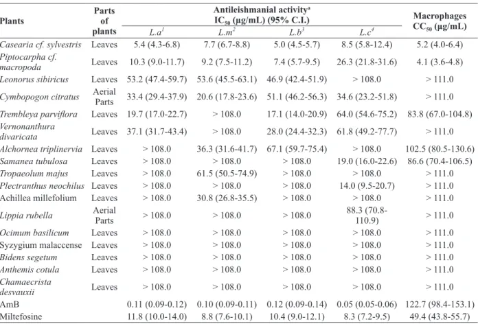
Parts of plants
Antileishmanial activitya
IC50 (µg/mL) (95% C.I.) Macrophages
CC50 (μg/mL)
L.a1 L.m2 L.b3 L.c4
Casearia cf. sylvestris Leaves 5.4 (4.3-6.8) 7.7 (6.7-8.8) 5.0 (4.5-5.7) 8.5 (5.8-12.4) 5.2 (4.0-6.4)
Piptocarpha cf.
macropoda Leaves 10.3 (9.0-11.7) 9.2 (7.5-11.2) 7.4 (5.7-9.5) 26.3 (21.8-31.6) 4.1 (3.6-4.8) Leonorus sibiricus Leaves 53.2 (47.4-59.7) 53.6 (45.5-63.1) 46.9 (42.4-51.9) > 108.0 > 111.0
Cymbopogon citratus Aerial
Parts 33.4 (29.4-37.9) 20.6 (17.8-23.6) 51.1 (46.2-56.3) 34.6 (23.2-51.8) > 111.0
Trembleya parviflora Leaves 19.7 (17.0-22.7) > 108.0 17.1 (14.0-20.9) 64.0 (54.6-75.2) 83.8 (67.0-104.8)
Vernonanthura
divaricata Leaves 37.1 (31.7-43.4) > 108.0 28.0 (24.4-32.3) 61.8 (49.2-77.7) > 111.0 Alchornea triplinervia Leaves > 108.0 36.3 (31.6-41.7) 67.1 (59.7-75.4) > 108.0 102.5 (80.5-130.6)
Samanea tubulosa Leaves > 108.0 > 108.0 > 108.0 19.0 (16.0-22.6) 86.6 (70.4-106.5)
Tropaeolum majus Leaves > 108.0 61.5 (50.5-74.9) > 108.0 > 108.0 > 111.0
Plectranthus neochilus Leaves > 108.0 > 108.0 > 108.0 14.0 (9.5-20.7) > 111.0 Achillea millefolium Leaves > 108.0 30.8 (26.8-35.5) > 108.0 > 108.0 > 111.0
Lippia rubella Aerial
Parts > 108.0 > 108.0 > 108.0
88.3
(70.8-110.9) > 111.0
Ocimum basilicum Leaves > 108.0 > 108.0 > 108.0 > 108.0 > 111.0 Syzygium malaccense Leaves > 108.0 > 108.0 > 108.0 > 108.0 > 111.0
Bidens segetum Leaves > 108.0 > 108.0 > 108.0 > 108.0 > 111.0
Anthemis cotula Leaves > 108.0 > 108.0 > 108.0 > 108.0 > 111.0
Chamaecrista
desvauxii Leaves > 108.0 > 108.0 > 108.0 > 108.0 > 111.0
AmB 0.11 (0.09-0.12) 0.10 (0.09-0.11) 0.12 (0.09-0.14) 0.05 (0.05-0.06) 122.7 (98.4-153.1) Miltefosine 11.8 (10.0-14.0) 8.8 (7.6-10.1) 10.4 (9.0-12.1) 8.3 (7.2-9.5) 49.4 (43.8-55.7)
TABLE II
Effect of plant extracts in promastigotes of
Leishmania species and murine macrophages.
a
Data are IC50 values in μg/mL and 95% confidence intervals are in brackets. 1
L.a.= Leishmania amazonensis; 2L.m.= L. major; 3L. b.=
L. braziliensis and 4L.c.= L. chagasi. These data represent the average of three independent experiments. AmB (amphotericin B) and miltefosine were used as reference drug. The highest concentration used of DMSO was 0.1% (v/v), which is not toxic to the parasites.
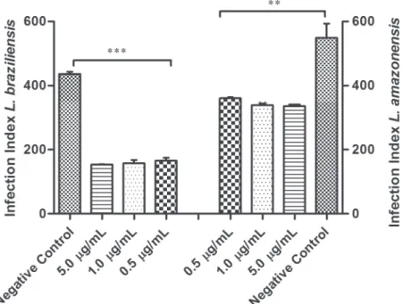
Promastigotes IC50 (µg/mL)
AmastigotesIC50
(µg/mL)
Macrophages CC50 (µg/mL)
aSI PRO
bSI AMA
cSP PRO/AMA
Casearia sylvestris 5.0 1.3 5.2 1.0 4.0 3.8
Miltefosine 10.4 2.4 49.4 4.7 20.6 4.3
IC50 (Inhibitory concentrationat 50% inhibition). CC50 (Cytotoxic concentrationat 50% inhibition). a
SI: selectivity index (CC50
of macrophages /IC50 of promastigotes). b
SI: selectivity index (CC50 of macrophages /IC50 of amastigotes).
cSP: specificity
index (ratio between promastigotes IC50 and intracellular amastigotes IC50). Miltefosine was used as reference drug.
TABLE III
Effect of Casearia cf. sylvestris extract on Leishmania
braziliensis, specificity and selectivity index.
parasite (Table III). In addition, Table III furnishes
information about the selectivity and specificity of
C. sylvestris extract. Regarding selectivity, when this value is greater than 1, the extract is more
was assayed against amastigotes, it was much more destructive to parasite than to the host cells, being four-fold more selective against the parasite. De Muylder et al. (2011) established a cut-off
regarding the specificity of compounds between
these two stages of the parasite Leishmania sp.
Specificity value > 2 was the cutoff point chosen to define a compound as being more active against the intracellular amastigote stage; while a specificity
value < 0.4 indicated a more active compound
against promastigotes; compounds with specificity
values between 0.4 and 2 were considered as being active against both stages. It is interesting
to point out that the extract was more specific for
intracellular form of L. braziliensis (specificity index= 3.8) and this result reinforces the potential leishmanicidal activity of C. sylvestris.
Previously, Mesquita et al. (2005) demonstrated the antileishmanial potential of C. sylvestris against promastigotes forms of L. donovani, of which
extracts displayed a significant activity with IC50 values ranging from 0.1 to 4.9 µg/mL depending on the plant parts used.
Regarding the cytotoxicity effects on macrophages, C. sylvestris and P. macropoda were the most cytotoxic to mammalian cells (Table II). Cytotoxicity tests with natural products are important because they represent a potential source for the isolation of compounds for the development of new antiprotozoal agents (Brenzan et al. 2007).
molluscicide (Alves et al. 2000) activities. Regarding antiprotozoal properties of C. sylvestris, some authors have reported activity against Trypanosoma cruzi (Mesquita et al. 2005). Phytochemical studies have attributed the high antitumor activity of C. sylvestris to the numerous secondary metabolites, especially diterpenes, which are likely based on its larger hydrophobicity and facility to penetrate across cell membrane and interact with intracellular targets (Ferreira et al. 2010). Secundary metabolites of C. sylvestris, such as alkaloids, sterols, saponins,
tannins and flavonoids may be associated with significant activity against Leishmania sp. However more investigations are necessary to determine the activity of each extract component separately and in combination to ensure whether they act alone or synergistically.
CONCLUSION
In conclusion, the methanolic extracts of Casearia sylvestris and Piptocarpha macropoda showed
significant leishmanicidal activity, specially C. sylvestris extract, which may be a potential source of active compounds for the development of novel therapeutic agents to treat leishmaniasis.
ACKNOWLEDGMENTS
The authors are grateful to Fundação de Amparo à Pesquisa do Estado de Minas Gerais (FAPEMIG),
Conselho Nacional de Desenvolvimento Científico e Tecnológico (CNPq) and UFJF for financial
support, and to Dr. Fatima Regina Salimena and
Dr.Tatiana Konno for the botanical identification
of the species.
RESUMO
As leishmanioses são um complexo de doenças causadas por protozoários Leishmania, cujo tratamento é restrito a um número limitado de fármacos que apresentam toxicidade elevada, efeitos colaterais e geralmente custos elevados. Existe uma enorme variedade de plantas tropicais distribuídas no Brasil e para muitas
pessoas pobres a terapia para várias doenças baseia-se principalmente no uso de remédios tradicionais obtidos de plantas. Neste trabalho, a atividade citotóxica de 17 extratos metanólicos de plantas foi avaliada em várias espécies de Leishmania e em macrófagos murinos. Dentre eles, os extratos de Casearia sylvestris,
Piptocarpha macropoda, Trembleya parviflora,
Samanea Tubulosa e Plectranthus neochilus mostraram atividade leishmanicida promissora, exibindo valores de CI50 abaixo de 20 µg/mL em pelo menos uma
das espécies de Leishmania. Casearia sylvestris
apresentou a atividade mais expressiva em todas as formas promastigotas de espécies de Leishmania
(valores de CI50 de 5,4 µg/mL, 5,0 µg/mL, 8,5 µg/mL
and 7,7 µg/mL em L. amazonensis, L. braziliensis,
L. chagasi e L. major, respectivamente), sendo mais
eficaz que o fármaco de referência miltefosina. Apesar
do efeito citotóxico em macrófagos (valor de CC50de
5,2 µg/mL), C. sylvestris exibiu uma forte inibição em formas amastigotas de L. braziliensis (valor de CI50 de 1,3 µg/mL). Mais estudos, incluindo
fracionamento bio-guiado, serão realizados para identificar os compostos ativos.
Palavras-chave: Brasil, Casearia sylvestris, atividade leishmanicida, plantas medicinais, produtos naturais.
REFERENCES
ALBERNAZ LC, PAULA JE, ROMERO ARS, SILVA MRR, GRELLIER P, MAMBU L AND ESPINDOLA LS. 2010. Investigation of plant extracts in traditional medicine of the Brazilian Cerrado against protozoans and yeasts. J Ethnopharmacol131: 116-121.
ALBUQUERQUE JM. 1989. Plantas medicinais de uso popular, Ministério da Educação. Brasília: ABEAS, 93 p.
ALVES TMA, SILVA AF, BRANDÃO M, GRANDI TSM, SMÃNIA EFA, SMÃNIA JUNIOR A AND ZANI CL. 2000. Biological screening of Brazilian medicinal plants. Mem Inst Oswaldo Cruz 95: 363-373.
ALVIANO DS, BARRETO ALS, DIAS FA, RODRIGUES IA, ROSA MSS, ALVIANO CS AND SOARES RMA. 2012. Conventional therapy and promising plant-derived compounds against trypanosomatid parasites. FMICB 3: 1-10.
BRENZAN MA, NAKAMURA CV, PRADO DIAS FILHO B, UEDA-NAKAMURA T, YOUNG MC AND APARÍCIO GARCIA CORTEZ D. 2007. Antileishmanial activity of crude extract and coumarin from Calophyllum brasiliense leaves against Leishmania amazonensis. Parasitol Res 101: 715-722.
BRITO AMG, DOS SANTOS D, RODRIGUES SA, BRITO RG AND XAVIER-FILHO L. 2013. Plants with anti-Leishmania
activity: Integrative review from 2000 to 2011. Pharmacog Rev 7: 34-41.
CAIXETA SC ET AL. 2011. Chemical composition and in vitro schistosomicidal activity of the essential oil of
Plectranthus neochilus grown in southeast Brazil. Chem Biodivers 8: 2149-2157.
CHIAPPETA ADA, DE MELLO JF AND MACIEL GM. 1983. Higher plants with biological activity– Plants of Pernambuco. Rev Inst Antibiot 21: 43-50.
CORRÊA MP AND PENNA LA. 1984. Dicionário de Plantas Úteis do Brasil e das Exóticas Cultivadas. Rio de Janeiro: Ministério da Agricultura, 747 p.
CROFT SL AND OLLIARO P. 2011. Leishmaniasis chemotherapy-challenges and opportunities. Clin Microbiol Infec 17: 1478-1483.
CRUZ AK, DE TOLEDO JS, FALADE M, TERRÃO M, KAMCHONWONGPAISAN S AND UTHAIPIBULL C. 2009. Current treatment and drug discovery against Leishmania spp. and Plasmodium spp.: a review. Curr Drug Targets 10: 178-192.
DA SILVA SL, CALGAROTTO AK, CHAAR JS AND MARANGONI S. 2008. Isolation and characterization of ellagic acid derivatives isolated from Casearia sylvestris Sw. aqueous extract with anti-PLA2 activity. Toxicon 52: 655-666.
DE MORAIS-TEIXEIRA E, DAMASCENO QS, GALUPPO MK, ROMANHA AJ AND RABELLO A. 2011. The in vitro
leishmanicidal activity of hexadecylphosphocholine (miltefosine) against four medically relevant Leish mania species of Brazil. Mem Inst Oswaldo Cruz 106: 475-478.
DE MUYLDER G, ANG KK, CHEN S, ARKIN MR, ENGEL JC AND MCKERROW JH. 2011. A screen against Leishmania
intracellular amastigotes: comparison to a promastigote screen and identification of a host cellspecific hit. PLos Negl Trop Dis 5: e-1253.
DORLO TPC, BALASEGARAM M, BEIJNEN J AND DE VRIES PJ. 2012. Miltefosine: a review of its pharmacology and therapeutic efficacy in the treatment of leishmaniasis. J Antimicrob Chemother 67: 2576-2597.
DUSTAN CA, NOREEN Y, SERRANO G, COX PA, PERERA P AND BOHLIN L. 1997. Evaluation of some Samoan and Peruvian medicinal plants by prostaglandin biosynthesis and rat ear oedema assays. J Ethno-pharmacol 57: 35-56.
FABRI RL, COIMBRA ES, ALMEIDA AC, SIQUEIRA EP, ALVES TM, ZANI CL AND SCIO E. 2012a. Essential oil of
Mitracarpus frigidus as a potent source of bioactive compounds. An Acad Bras Cienc 84: 1073-1080.
FABRI RL, GRAZUL RM, CARVALHO LO, COIMBRA ES, CARDOSO GM, SOUZA-FAGUNDES EM, SILVA AD AND SCIO E. 2012b. Antitumor, antibiotic and antileishmanial properties of the Pyranonaphthoquinone Psychorubrin from Mitracarpus frigidus. An Acad Bras Cienc 84: 1081-1090.
FERREIRA PMP, COSTA-LOTUFO LV, MORAES MO, BARROS FWA, MARTINS AMA, CAVALHEIRO AJ, BOLZANI VS, SANTOS AG AND PESSOA C. 2011. Folk uses and pharmacological properties of Casearia sylvestris: a medicinal review. An Acad Bras Cienc 83: 1373-1384. FERREIRA PMP, SANTOS AG, TININIS AG, COSTA PM,
CAVALHEIRO AJ, BOLZANIVS MORAES MO, COSTA-LOTUFO LV, MONTENEGRO RC AND PESSOA C. 2010. Casearin X exhibits cytotoxic effects in leukemia cells triggered by apoptosis. Chem Biol Interact 188: 497-504. HAJDU Z AND HOHMANN J. 2012. An ethnopharmacological
survey of the traditional medicine utilized in the community of Porvenir, Bajo Paraguá Indian Reservation, Bolivia. J. Ethnopharmacol 139: 838-857.
LIMA ZP, BONAMIN F, CALVO TR, VILEGAS W, SANTOS LC, ROZZA AL, PELLIZZON CH, ROCHA LRM AND HIRUMA-LIMA CA. 2011. Effects of the ethyl acetate fraction of
Alchornea Triplinervia on healing gastric ulcer in rats. Pharmaceut4: 1423-1433.
LORENZI H AND MATOS FJA. 2002. Plantas Medicinais no Brasil: Nativas e Exóticas Cultivadas. São Paulo: Plantarum, 544 p. MENEZES PR, SCHWARZ EA AND SANTOS CAM. 2004. In
vitro antioxidant activity of species collected in Paraná. Fitoterapia 75: 398-400.
MESQUITA ML, DESRIVOT J, BORIES C, FOURNET A, PAULA JE, GRELLIER P, AND ESPÍNDOLA LS. 2005. Antileishmanial and trypanocidal activity of Brazilian Cerrado plants. Mem Inst Oswaldo Cruz 100: 783-787.
MINODIER P AND PAROLA P. 2007. Cutaneous leishmaniasis treatment. Travel Med Infect Dis 5: 150-158.
MOREIRA DL AND GUARIM-NETO G. 2009. Usos Múltiplos de Plantas do Cerrado: Um Estudo Etnobotânico na Comunidade Sítio Pindura, Rosário Oeste, Mato Grosso, Brasil. Polibotânica 27: 159-190.
MOSSMAN T. 1983. Rapid colorimetric assay for cellular growth and survival: application to proliferation and cytotoxicity assays. J Immunol Methods 65: 55-58. MURRAY HW, BERMAN JD, DAVIES CR AND SARAVIA NG.
2005. Advances in leishmaniasis. Lancet366: 1561-1577. SANTIN MR, DOS SANTOS AO, NAKAMURA CV, DIAS FILHO BP, FERREIRA ICP AND NAKAMURA TU. 2009. In vitro
activity of the essential oil of Cymbopogon citratus and its major component (citral) on Leishmania amazonensis. Parasitol Res 105: 1489-1496.
SANTOS DO ET AL. 2008. Leishmaniasis treatment – a challenge that remains: a review. Parasitol Res 103: 1-10.
SILVA ALN, ADADE CM, SHOYAMA FM, NETO CPS, PADRÓN TS, ALMEIDA MV, REZENDE CAM, SILVA CV AND SOUZA MA. 2012. In vitro leishmanicidal activity of N-dodecyl-1,2-ethanediamine. Biomed Pharmacother 66: 180-186. SINGH B AND SUNDAR S. 2012. Leishmaniasis: vaccine
candidates and perspectives.Vaccine 30: 3834-3842. SUNDAR S, JHA TK, THAKUR CP, BHATTACHARVA SK AND RAI
M. 2002. Oral miltefosine for Indian visceral leishmaniasis. N Engl J Med 347: 1739-1746.
TEMPONE AG, DE OLIVEIRA CM AND BERLINCK RS. 2011. Current Aproaches to Discover Marine Antileishmanial Natural Products. Planta Med 77: 572-585.
WHO. 2010. Control of the leishmaniasis: report of a meeting of the WHO Expert Committee on the Control of leishmaniases. In: WHO Technical Report Series. Geneva, p. 22-26.