Major Article
Corresponding author: Dra. Milena de Paiva Cavalcanti.
e-mail: mp@cpqam.iocruz.br; milena.cavalcanti@bol.com.br Received 31 January 2017
Accepted 2 June 2017
Standardization and evaluation of a duplex real-time
quantitative PCR for the detection of
Leishmania infantum
DNA: a sample quality control approach
Lays Adrianne Mendonça Trajano-Silva
[1],[2], Rômulo Pessoa-e-Silva
[1],[3],
Suênia da Cunha Gonçalves-de-Albuquerque
[1],[3], Rayana Carla Silva de Morais
[1],
Cíntia Nascimento da Costa-Oliveira
[1], Tayná Correia de Goes
[1]and Milena de Paiva-Cavalcanti
[1][1] Departamento de Microbiologia, Centro de Pesquisas Aggeu Magalhães, Recife, PE, Brasil. [2] Centro de Ciências Biológicas, Universidade Federal de Pernambuco, Recife, PE, Brasil. [3] Setor de Sorologia, Laboratório Central de Saúde PúblicaDr. Milton Bezerra Sobral, Recife, PE, Brasil.
Abstract
Introduction: Molecular techniques have been shown to be alternative methods for the accurate detection of infectious and parasitic diseases, such as the leishmaniases. The present study describes the optimization and evaluation of a duplex real-time quantitative PCR (qPCR) protocol developed for the simultaneous detection of Leishmania infantum DNA and sample quality control. Methods: After preliminary tests with the newly designed TaqMan® probes for the two targets (L. infantum and
glyceraldehyde 3-phosphate dehydrogenase (G3PD) gene), the duplex qPCR protocol was optimized. For the evaluation of the standardized protocol, human blood samples were tested (n=68) and the results were compared to those obtained by reference diagnostic techniques. Statistical analyses included percentage agreement and the Kappa (k) coeficient. Results: The detection limit of L. infantum DNA reached 2x10² fg (corresponding to ~1 parasite) per µL of blood (ε: 93.9%). The percentage agreement
obtained between the duplex VL qPCR and the reference techniques was individually obtained as follows: molecular: 88.3% (k=0.666; 95% CI 0.437–0.894, good), and serological: 81.7% (k=0.411; 95% CI 0.125–0.697, moderate). Between the reference techniques, the percentage agreement was 86.7% (k=0.586; 95% CI 0.332–0.840, moderate). Conclusions: The new duplex VL qPCR protocol indicated good potential for the accurate, fast, and reliable detection of L. infantum DNA, when applied as a complement to the classical diagnostic tools already available, especially in health or research reference centers.
Keywords: Visceral leishmaniasis. Diagnosis. HIV/VL co-infection. Sample quality control. Duplex qPCR.
INTRODUCTION
The leishmaniases are parasitic diseases caused by protozoans from the genus Leishmania (Kinetoplastida, Trypanosomatidae)1. In the Latin America, the development of visceral leishmaniasis is often associated with the species Leishmania infantum (L. infantum)2.
To control the advancement of disease in infected individuals, early detection and fast implementation of treatment are crucial for successful outcomes. However, classic diagnostic methods have several limitations, such as low sensitivity and high invasiveness (parasitological tests), the possibility of cross-reactivity (as with Leptomonas seymouri, a monoxenous
trypanosomatid)3, and the lack of accuracy in diagnosing immunosuppressed patients (serology), such as those co-infected with human immunodeiciency virus and visceral leishmaniais (HIV/VL)4,5. Thus, these diagnostic methods can
yield false positive and negative results, thus impairing the appropriate therapeutic intervention.
Given the limitations of classic diagnostic methods, molecular methods, especially polymerase chain reaction (PCR), have become popular alternatives for the diagnosis and control of Visceral Leishmaniais (VL)4,6-10. Speciically,
To enhance the eficiency and reliability of diagnostic techniques, sample quality controls are often included that are based on the ampliication of a host’s constitutive genes, such as the β-actin, β-globin, albumin, and glyceraldehyde 3-phosphate dehydrogenase (G3PD) genes in mammals. Quality control measures have been routinely used in laboratories, but these additional steps generate more costs and prolong the time to result interpretation and reporting8,9,11. However, during qPCR,
it is possible to simultaneously amplify both the sample quality control and the target DNA in the same tube by applying multiplex protocols that use probes directed at a speciic target and marked with different luorochromes12,14.
The aim of this study was to standardize and evaluate the inclusion of a sample quality control (internal control) into a qPCR protocol for the detection of L. infantum DNA, thus enabling the simultaneous tracking of possible false negative results.
METHODS
Ethical considerations
Prior to sample collection, written informed consent was obtained from all subjects and/or their legal guardians. This work was approved by the Research Ethics Committee (CEP/ CPqAM/FIOCRUZ-PE, 42/2010) in consonance with the National Research Ethics Committee (CONEP-BR; CAAE: 0041.0.095.000-10). All procedures were in accordance with the ethical standards of the institutional and/or national research committee and with the 1964 Declaration of Helsinki and its later amendments or comparable ethical standards.
Study design
This study evaluated diagnostic methods based on the steps proposed by Sackett and Haynes15, which consists of the
following three phases: I) analytical sensitivity analysis; II) reproducibility analysis; and III) concordance analysis between results of the new test and results obtained by reference tests, using samples from patients.
Sample collection, processing, and group deinitions
Samples were obtained by convenience (Non-Probabilistic Sample)16. Blood samples (2-4 mL) were collected from healthy
individuals living in non-endemic areas, who had not previously submitted for a blood transfusion and who were negative for immunological and molecular tests (negative control group). Blood was also collected from patients living in endemic areas and presenting with suggestive VL symptomatology. The patients were treated in the following reference hospitals in the Pernambuco state of Brazil: The Professor Fernando Figueira Integral Medicine Institute (IMIP); Correia Picanço Hospital (HCP); Oswaldo Cruz University Hospital (HUOC); Clinics Hospital (HC); and Barão de Lucena Hospital (HBL). All specimens were processed in laboratories of the Aggeu Magalhães Research Center (CPqAM-FIOCRUZ; Recife, PE, BR). Blood samples were extracted using the QIAamp® DNA Blood Mini Kit (QIAGEN Sample and Assay Technologies), according to the manufacturer’s instructions.
Immunological and molecular tests
All individuals included in the study were submitted to the following reference assays: an immunological test for anti-Leishmania antibody detection through recombinant kinesin 39 - immunochromatographic test (rK39-ICT) (InBios, Seattle, WA, USA) was performed following manufacturer’s instructions; while a qPCR molecular blood test for L. infantum kinetoplast DNA (kDNA) minicircle detection was performed as per the protocol previously described by Paiva-Cavalcanti et al17.
Parasitological test
Some patients suspected of having VL underwent a parasitological test that included a bone marrow biopsy. Six bone marrow smears were prepared from collected biological specimens and tested for amastigote forms (methodology preconized by the Ministry of Health, Brazil). The aspirates were obtained by trained physicians from the respective reference hospitals and only under prescription.
Sample positivity criteria
Positive results for at least two of the reference techniques – VL qPCR, rk39-ICT, and bone marrow aspiration – were deined as the set of diagnostic criteria (characterizing VL cases). As criteria of positivity of the Singleplex qPCR assay, the ampliication curve had to surpass the threshold before cycle 36, as recommended by Applied Biosystems18. The quality
assurance of each sample was achieved in separated reactions by mammalian G3PD constitutive gene ampliication, employing primers G1F (5’-ATC TTC CAG GAG CGA GAT CCC-3’) and G1R (5’-AGG GAT GAC CTT GCC CAC-3’)8.
Development of the duplex qPCR assay
The duplex qPCR system was developed through the combination of the L. infantum primers (LINF 1B)17 and the
G3PD1 set8 for the simultaneous detection of the L. infantum
kDNA and the G3PD gene from mammals (internal control), respectively. All experiments were performed using the ABI Prism 7500 (Applied Biosystems®, CA, USA) equipment. The software ABI Prism 7500 SDS was used for the analysis, interpretation, and registration of results.
TaqMan® probes design:using the software PrimerQuest
(http://www.idtdna.com/scitooes), speciic probes for the sets G3PD1 (probe A) and LINF 1B (probe B) were designed. To compose the duplex qPCR assay, the probes were chosen following the manufacturer’s instructions for the TaqMan probe (Applied Biosystems®) technology. The probes’ speciicity was preliminarily analyzed by multiple alignments of sequences, using the nucleotide Basic Local Alignment Search Tool (BLASTn) (http://blast.ncbi.nlm.nih.gov).
Individual optimization of the sets LINF 1B and G3PD1:
using the respective probe (A or B) at 12.5pmol/reaction. A standard amount of 1x10⁶fg of L. infantum (syn. L. chagasi)
DNA (MHOM/BR/1974/PP75) or DNA extracted from whole blood (negative control group) was added to the respective reactions. The inal volume per reaction was as follows: 50µL, consisting of 25µL TaqMan® Universal Master Mix (Applied Biosystems®, CA, USA) and 5µL of template. All samples were produced in duplicates. The cycling conditions used were the standardized cycles used by Paiva-Cavalcanti et al.17: 95ºC/15
s and 60ºC/1 min, at 40 cycles. The lowest amounts of forward and reverse primers that yielded a minimum Ct (threshold cycle) and a maximum ΔRn (normalized reporter) were chosen as optimal. Second, between 2.5 and 12.5pmol of the probes A and B per reaction were tested by using the optimal amount of G3PD1 and LINF 1B primers found in the previous experiments. The same cycling and reaction conditions of the previous step, as well as the amount of the standard DNA, were utilized. The lowest amounts of the probes that yielded a minimum Ct were chosen as optimal.
Optimization of duplex qPCR system: the LINF 1B set was
combined with G3PD1 set (primers + probe). The system formed (duplex VL qPCR) was evaluated in preliminary experiments by using the amounts of primers and probes optimized in the previous step and in the same cycling conditions that were standardized by Paiva-Cavalcanti et al.17. The detection limit
was determined by using dilution curves prepared from the blood of healthy individuals (negative control group): concentrations between 2x10⁻¹ and 2x10⁵fg (from 0.001 to 1,000 parasites,
according to Grimaldi et al.19, with a serial dilution factor of 10)
per µL of whole blood genomic DNA from L. infantum (MHOM/ BR/1974/PP75) were used. When necessary, changes were performed in the cycling conditions (annealing and extension temperatures), as well as in the concentration of reagents according to the Applied Biosystems® protocol20.
Reproducibility analysis: for reproducibility evaluation of
the new test, intra- and inter-assay analyses were performed. After optimization, DNA from two aliquots of three different concentrations (2x10², 2x10³ and 2x10⁴fg of L.infantum DNA per µL of blood) from the dilution curve was extracted. The duplex VL qPCR was performed with the duplicates (the two aliquots) of each selected concentration. The experiment was then repeated twice. The points in which the ampliication curve surpassed the threshold (Ct values) were used to calculate the coeficients of variation (CV) between the replicates.
Comparative analyses
The samples were subjected to the duplex protocol and the results were compared with those obtained from the set of criteria (see item 2.6 Sample positivity criteria), which was deined in this study as the set of diagnostic criteria. Comparative analysis between the techniques and concordance analysis was performed using descriptive statistics in absolute and percentage distribution values. Concordance was also evaluated by applying the Kappa (k) coeficient to the Conidence Interval (CI) set at 95%, and the agreement between the tests was judged using the Cohen19 framework as follows: k=0.0, no agreement;
0.0≤k≤0.20, poor; 0.21≤k≤0.40, fair; 0.41≤k≤0.60, moderate; 0.61≤k≤0.80, good; and 0.81≤k≤1.00, very good. All analyses were performed with the BioEstat software (version 5.0; Mamirauá/CNPq, Belém, PA, Brazil). No template controls (NTC) and quantitative standards were included in all reactions.
RESULTS
Patient and group deinitions
Blood samples from 68 patients who presented with symptoms indicative of VL (fever, hepatomegaly and/or splenomegaly, anemia, fatigue, and weight loss) were included in the analyses. Among them, 50 patients had been previously diagnosed with HIV/AIDS. Samples from 61 patients were subjected to the, rK39-ICT test. From these sample indings, only five patients underwent bone marrow aspiration for parasitological analysis (positive: 4; negative: 1). To maintain the reliability of the results, samples negative for the G3PD gene in the singleplex qPCR reaction (n=1) were excluded from the analysis of the duplex VL qPCR (total included, 60). According to the established gold standard, 8 (13.3%) patients were considered positive for VL cases and 52 (86.7%) patients were considered non-cases or negative patients.
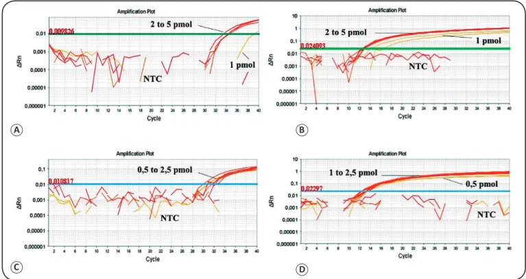
Optimization of the duplex real-time PCR system In regards to the requirements for the use of the probes, the sequences to each set of primers were designed. G3PD1 (5’-ATC ACT GCC ACC CAG AAG ACT GTG-3’) was designed with the following characteristics: size, 24bp; GC, 54%; Tm, 68°C; and reporter fluorochrome, VIC®.LINF 1B (5’-AAA TGG GTG CAG AAA TCC CGT TCA AA-3’) was designed with the following characteristics: size, 26bp; GC, 42.3%; Tm, 59.4°C; and reporter luorochrome, FAM™. These sequences were analyzed in silico, demonstrating the impossibility of self-annealing and annealing with non-speciic targets. Through singleplex qPCR reactions, the amounts (per reaction) of primers and probes were selected as follows: for G3PD1, 15pmol of each primer and 2.5pmol of probe A (Ct=30.88), and for LINF 1B, 10pmol of each primer and 7.5pmol of probe B [(Ct=13.04); Figure 1].
The duplex protocol was formed and evaluated in preliminary experiments that demonstrated the good performance of the two sets of primers, LINF 1B and G3PD1, for the simultaneous ampliication of both targets. The added solution volumes of the primers and probes were adjusted to maintain the optimum amounts of reagents per reaction, for the multiplex format. An optimization procedure, referred to as the Limiting Primer Matrix, was executed according to the ABI Prism® 7700 Sequence Detection System (User Bulletin #5)20, in an effort to
improve the analytical sensitivity by minimizing the competition between the sets. The detection limit was then reassessed. After the modiications, the detection limit of the duplex VL qPCR was established: 2x10²fg of L. infantum DNA (~1 parasite,
according to Grimaldi et al.19) per µL of blood; eficiency (ε),
93.9% (Figure 2). The duplex VL protocol was maintained in the same cycling conditions, as standardized by Paiva-Cavalcanti
FIGURE 1 - Individual deinition of the optimal amounts (per reaction) of primers and probe for each set. (A): G3PD1 primers: 15pmol. (B): probe A: 2.5pmol. (C): LINF 1B; primers: 10pmol, and (D): probe B, 7.5pmol. NTC: negative control. Optimal amounts of primers and probes were deined considering those with earlier threshold cycles (Ct). ΔRn: Reporter – normalized luorescence; NTC: no template control (negative control); G3PD1: glyceraldehyde 3-phosphate dehydrogenase 1 primer set; LINF: Leishmania infatum primers set.
were developed as follows: G3PD1: 15pmol; LINF1B: 10pmol; probe G3PD1: 2.5pmol; and probe LINF1B: 7.5pmol. The volume of the TaqMan® Universal Master Mix 2X (Applied Biosystems) was 25µL. A volume of 5µL of DNA template was added. In total, the volume was 50µL.
Reproducibility analysis
The reproducibility analysis was conducted to determine the detection limit (2x10²fg/µL) was maintained in both the
intra- and inter-assays. The inter-assay CV, as calculated from the average Ct values of the duplicates of three different curve concentrations (2x10², 2x10³, and 2x10⁴fg per µL of blood) from three independent experiments, were as follows: 3.8%, 2.2%, and 2.8%, respectively. The intra-assay CV, as calculated from the Ct of the duplicates (from one experiment) of the same three curve concentrations, were as follows: 0.3%, 0.2%, and 4.4%, respectively.
Comparative analysis
The concordance analysis between the new protocol (duplex VL qPCR) and the set of diagnostic criteria (rK39-ICT + VL qPCR) showed an 81.7% agreement. Nevertheless, the k coeficient was considered fair: 0.373 (95% CI 0.081-0.665). The concordance analysis performed with the data submitted to the set of the diagnostic criteria (rK39-ICT + VL qPCR) and the bone marrow aspiration showed a 60% agreement. Since the number of patients who underwent bone marrow aspiration was low (n=5), the k analysis was not allowed.
To conduct a broader and more discriminative evaluation, comparative analyses were also individually performed with the reference techniques of the criteria set (Table 1). The duplex VL PCR and the rK39-ICT presented an 81.7% agreement with
a k coeficient of 0.411 (95% CI 0.125-0.697). The percentage
agreement between the duplex VL qPCR and VL qPCR was 88.33%, with a k coeficient of 0.666 (95% CI 0.437-0.894). The percentage agreement between VL qPCR and rK39-ICT (reference techniques was 86.7%, with a k coeficient of 0.586 (95% CI 0.332-0.840).
DISCUSSION
In recent years, molecular biology has been used in the development of alternative methods for the study and diagnosis of various infectious and parasitic diseases. The PCR technique and its variations have aided in the advancement of diagnosis accuracy in both clinical forms of leishmaniasis because the method enables a more sensitive and speciic detection of the etiological agent’s DNA in various samples, such as blood and urine5,17,21-23.
As previously discussed, molecular techniques have numerous advantages, but they still contain some limitations, such as the occurrence of false negative results, as a result of using inadequate samples: presence of Taq polymerase enzyme inhibitors, such as proteinase K (used in DNA extraction process), high concentration of salts and ethylenediamine tetraacetic acid (EDTA), and poor storage. Through the habitual
A B
FIGURE 2 - Detection limit of the duplex VL qPCR. (A): Simultaneous ampliication of H. sapiens genomic DNA by a set of G3PD1 primers, and L. infantum genomic DNA (MHOM/BR/1974/PP75) in the concentration of 2x10²fg per µL of blood by the LINF 1B primer. NTC: negative control.
(B): Standard curve of L. infantum, resulting from the detection limit experiment of the duplex VL qPCR. Quantities between 1fg and 1x10⁶fg of DNA per
reaction (50µl) were used: Slope, -3,479; coeficient of determination (R2), 0,991; and eficiency (ε), 93.9%. CT: Cycle threshold; L.: Leishamania; G3PD1:
glyceraldehyde 3-phosphate dehydrogenase 1 primer set; NTC: no template control; VL: visceral leishmaniasis; qPCR: quantitative polymerase chain reaction; H.: Homo; DNA: deoxyribonucleic acid; LINF: Leishmania infatum primers set.
A
Tests rK39-ICT Total
Positive Negative
VL qPCR NegativePositive 86 442 1050
Total 14 46 60
κ: moderate
0.586 (CI 95% 0.332–0.840)
Tests rK39-ICT Total
Positive Negative
Duplex VL PCR Positive 6 4 10
Negative 7 43 50
Total 13 47 60
κ: moderate
0.411 (95% CI 0.125–0.697)
Tests VL qPCR Total
Positive Negative
Duplex VL PCR Positive 10 4 14
Negative 3 43 46
Total 13 47 60
κ:good
0.666 (95% CI 0.437–0.894)
TABLE 1
Concordance analysis between the new duplex VL qPCR protocol and each reference technique employed for the diagnosis of VL.
VL: visceral leishmaniasis; qPCR: quantitative polymerase chain reaction; rK39: recombinant kinesin 39; ICT: immunochromatographic test;
PCR: polymerase chain reaction.
use of sample quality controls, predominantly based on the ampliication of the host’s constitutive genes, the chances of erroneous results in molecular diagnosis become smaller8,24.
During real-time PCR reaction, the simultaneous ampliication of a sample quality control and the target DNA in the same tube is only possible through the application of multiplex protocols that use probes that are marked with different luorochromes and directed at the speciic target, thus helping to reduce processing time and costs8,12,14. Therefore, this study aimed to develop and
evaluate a duplex real-time PCR assay for VL diagnosis that could simultaneously detect L. infantum kDNA and the G3PD gene in blood samples to ensure the high quality of results through the association of accuracy and reliability.
From the individual optimization of the LINF 1B and G3PD1 sets, these probes excellently ampliied their respective targets. Between the resulting Cts and ΔRn of the amounts of primers and probe evaluated in each set, minimal differences were observed (Figure 1). The optimization process of the duplex qPCR protocol promoted a good detection limit without major changes in reaction or cycling conditions in the protocol standardized by
Paiva-Cavalcanti et al.19. Modiications in reagent concentrations and in
cycling temperatures were performed to optimize the duplex VL qPCR; however, the detection limit was maintained (2x10²fg of
parasite DNA per µL of whole blood) and the analytical eficiency (ɛ=93.85%) had no signiicant improvement. As evidenced in Figure 2, the ampliication of the parasite DNA is favored, and this is associated with the design of the probes, as well as with the chosen targets. The large amount of the host’s genetic material that is simultaneously puriied in the extraction step could impair the detection of the etiological agent DNA, mainly because of
competition between the primer sets for the PCR reagents9,25. In
larger parasite DNA concentrations, there is no ampliication of the G3PD gene (as from the concentration of 2x10³fg per µL of sample), but this does not affect the validation of the results, since the intention of the reaction is to favor the target DNA appearance.
In this study, we evidenced and reinforced the importance of including sample quality control measures because in the non-ampliication of this target, false-negative results are possible to track, thus avoiding the misinterpretation of results and increasing test reliability. Bezold et al.13 detected potential false negative results in 20% of the samples tested in the molecular diagnosis of herpes simplex virus and the varicella-zoster virus using swabs; thus emphasizing the importance of using internal controls, especially when analyzing DNA from different types of clinical specimens. Gonçalves
et al.8 used the same sample quality control (G3PD gene) in
multiplex reactions to detect VL through conventional PCR (cPCR) reactions, and demonstrated that it was possible to detect potential false negative results in 33% of the samples tested (no ampliication of the G3PD gene). Gonçalves-de-Albuquerque et al.25 standardized multiplex cPCR reactions
The classical diagnosis for VL is based on parasitological and immunological techniques, and despite these techniques’ widespread use, their existing limitations and potential for erroneous results demonstrate the need for a more thorough diagnostic scheme. According to Cota et al.26 and Srividya et al.27, the reliability of parasitological diagnostic techniques depends on numerous factors. The method is very speciic, but sensitivity depends on good sample collection, quality, and preparation, in addition to the analyst’s expertise. Singh and Sundar28 indicate the dificulty and invasiveness of the collection procedure. Further, the parasitological diagnostic technique is not included in primary health care centers (PHC), making it dificult to access a high number of well diagnosed and characterized samples for evaluation. In this study, to increase the number of samples for the tests, the positivity criteria had to be elaborated upon based on well-established serological and molecular methods. The new duplex VL qPCR protocol showed a reasonable percentage agreement (60%) with microscopic examination of bone marrow aspiration. However, one of the patients had taken 18 doses of the antileishmanial N-methylglucamine antimoniate (Glucantime®,among 10 and 20 mg/Sb+5/Kg/day) just after the positive result of the parasitological examination was found, thus causing both the singleplex VL qPCR and duplex VL qPCR results to be negative22,29. The three remaining patients who were positive for LV upon parasitological examination underwent treatment prior to sample collection; however, these patients had only taken one to two doses of the drug. In addition, the patient who was negative for LV upon bone marrow examination was positive for LV in both molecular techniques and rK39-ICT. By evaluating the parasitological test results within the predeined set of diagnostic criteria, the percentage agreement obtained remained 60%. In the comparison of the duplex VL real-time PCR results and the results of the set of diagnostic criteria (rK39-ICT + VL qPCR), a good percentage agreement was reached (81.7%), despite the fair agreement obtained by the k coeficient (0.373; 95% CI 0.081-0.665). When compared to the original qPCR protocol standardized by Paiva-Cavalcanti et al.17, the duplex VL real-time PCR technique had a great percentage agreement (88.33%), with a k coeficient indicating a good agreement (0.666; 95% CI 0.437-0.894) (Table 1). Only after the individual analysis was performed via the immunological technique (rK39-ICT) was it was possible to identify the likely reasons as to why there was a slight decrease in concordance between the duplex technique and the set of diagnostic criteria. Despite having low costs and quick rate of diagnosis, immunochromatographic rapid tests have some limitations that may promote erroneous diagnostic interpretation when the test is applied individually, such as low accuracy in immunosuppressed patients and cross-reactions with other trypanosomatids4,30,31. Thus, despite a moderate k agreement (0.411; 95% CI 0.125-0.697), only six out of the 13 samples were positive in both the duplex VL qPCR and the rK39-ICT (Table 1). All seven samples that presented negative in the immunological test, but positive in the duplex test, were from patients with symptomatic HIV. Further, the results of both the duplex and singleplex VL qPCRs were in agreement with one another in ive of these seven samples.
In addition, there were important divergences between the results of the reference techniques, with k indicating
moderate agreement [(0.586; 95% CI: 0.332-0.840); Table 1].
Naturally, methods with different principles (molecular and immunological) present discordant results when evaluated in the same population, thus highlighting the importance of adopting a reliable set of diagnostic criteria (associated with epidemiology plus clinical signs). In this context, the new duplex technique combined with classical diagnostic tools may help to develop accurate criteria for assessing positivity and minimize the occurrence of misdiagnosis.
Elmahallawy et al.30 described the importance of using qPCR
techniques because of the method’s sensitivity, speciicity, and quantitative ability, which enables the evaluation of the parasite load and treatment eficacy, especially in patients co-infected with HIV. Patients who are positive for HIV are particularly vulnerable to VL because the disease accelerates the replication and progression of HIV to AIDS and there is a higher risk of treatment failure and relapse32. In this study, we standardized and
evaluated a qPCR protocol with greater safety that displayed good potential for incorporation, as a complement, into the diagnostic scheme of VL within reference diagnostic centers. Through the monitoring of a greater number of patients (co-infected or not) before, during, and after treatment, the applicability of this technique for the monitoring of parasite load may be established. In conclusion, the evaluation of the new duplex VL qPCR technique indicated good potential for the accurate, fast, and reliable detection of L. infantum DNA, when applied as a complement to the classical diagnostic tools already available and as an alternative for clarifying possible inconclusive cases, especially in health or research reference centers.
Acknowledgements
The authors appreciate Dr. Zulma Maria Medeiros, Dr. Maria Almerice Lopes da Silva, and Elis Dionisio da Silva from Aggeu Magalhães Research Center (CPqAM/FIOCRUZ), for helping to supply samples from VL patients. We also thank Dr. Sinval Pinto Brandão Filho and Dr. Maria Edileuza Felinto de Brito, coordinators of the Reference Service in Leishmaniasis FIOCRUZ/ Pernambuco, for the operational support in the sample processing. Finally, we thank the Program of Technological Development in Health Supplies [Programa de Desenvolvimento Tecnológico em Insumos para Saúde (PDTIS/
FIOCRUZ)] for allowing us to use its facilities.
Conlict of interest
The authors declare that they have no conlict of interest.
Financial support
This work was supported by the Research Program for SUS [Programa Pesquisa para o SUS e Secretaria de Ciência, Tecnologia e Insumos Estratégicos do Ministério da Saúde (PPSUS/Decit/SCTIE/MS)] through the National
Council of Technological and Scientiic Development [Conselho Nacional de
Desenvolvimento Cientíico e Tecnológico (CNPq)], Foundation for Science and Technology Support of Pernambuco [Fundação de Amparo à Ciência e Tecnologia de Pernambuco (FACEPE)] and Pernambuco State Secretary of
REFERENCES
1. Chappuis F, Sundar S, Hailu A, Ghalib H, Rijal S, Peeling RW, et al. Visceral leishmaniasis: what are the needs for diagnosis, treatment and control? Nat Rev Microbiol. 2007;5(11):873-82.
2. World Health Organization (WHO). Control of the leishmaniasis. WHO Technical Report Series. Geneva: WHO; 2010. 202p. Available at: http://apps.who.int/iris/bitstream/10665/44412/1/ WHO_TRS_949_eng.pdf. Accessed 09 abr. 2013.
3. Ferreira LR, Kesper N, Teixeira MM, Laurenti MD, Barbieri CL, Lindoso JA, et al. New insights about cross-reactive epitopes of six trypanosomatid genera revealed that Crithidia and Leptomonas have
antigenic similarity to L. (L.) chagasi. Acta Trop. 2014;131(1):41-6.
4. Srivastava P, Mehrotra S, Tiwary P, Chakravarty J, Sundar S. Diagnosis of visceral leishmaniasis. Trans R Soc Trop Med Hyg. 2011;105(1):1-6.
5. Paiva-Cavalcanti M, Morais RCS, Pessoa-e-Silva R, Trajano-Silva LAM, Gonçalves-de-Albuquerque SC, Tavares DHC, et al. Leishmaniases diagnosis: an update on the use of immunological and molecular tools. Cell Biosci. 2015;5:31.
6. Singh S. Changing trends in the epidemiology, clinical presentation, and diagnosis of Leishmania-HIV co-infection in India. Int J
Infect Dis. 2014;29:103-12.
7. Alvar J, Aparicio P, Aseffa A, Den Boer M, Cañavate C, Dedet JP, et al. The relationship between leishmaniasis and AIDS: the second 10 years. Clin Microbiol Rev. 2008;21(2):334-59.
8. Gonçalves SC, Régis-da-Silva CG, Brito MEFC, Brandão-Filho SP, Paiva-Cavalcanti M. Application of the mammalian glyceraldehyde-3-phosphate dehydrogenase gene for sample quality control in multiplex PCR for diagnosis of leishmaniasis. J Venom Anim Toxins incl Trop Dis. 2012;18(2):188-97.
9. Gonçalves-de-Albuquerque SC, Pessoa-e-Silva R, de Morais RCS, Trajano-Silva LAM, Régis-da-Silva CG, Brandão-Filho SP, et al. Tracking false-negative results in molecular diagnosis: proposal of a triplex-PCR based method for leishmaniasis diagnosis. J Venom Anim Toxins incl Trop Dis. 2014;20:1-6.
10. de Ruiter CM, Van der Veer C, Leelang MMG, Deborggraeve S, Lucas C, Adams ER. Molecular tools for diagnosis of visceral leishmaniasis: systematic review and meta-analysis of diagnostic test accuracy. J Clin Microbiol. 2014;52(9)3147-55.
11. Castilho TM, Camargo LMA, McMahon-Pratt D, Shaw JJ, Floeter-Winteret LM. A rreal-time polymerase chain reaction assay for the identiication and quantiication of AmericanLeishmaniaspecies
on the basis of glucose-6-phosphate dehydrogenase.Am J Trop Med Hyg. 2008;78:122-32.
12. Arya M, Shergill IS, Williamson M, Gommersall L, Arya N, Patel HR, et al. Basic principles of real-time quantitative PCR. Expert Rev Mol Diagn. 2005;5(2):209-19.
13. Bezold G, Volkenandt M, Gottlöber P, Peter RU. Detection of herpes simplex virus and varicella-zoster virus in clinical swabs: frequent inhibition of PCR as determined by internal controls. Mol Diagn. 2000;5(4):279-84.
14. Lombardo G, Pennisia MG, Lupo T, Migliazzo A, Caprìa A, Solano-Gallego L. Detection of leishmaniainfantum DNA by
real-time PCR in canine oral and conjunctival swabs and comparison with other diagnostic techniques. Vet Parasitol. 2012;184(1):10-7. 15. Sackett DL, Haynes RB. The architecture of diagnostic research.
BMJ. 2002;324(7336):539-41.
16. Etikan I, Musa SA, Alkassim RS. Comparison of Convenience Sampling and Purposive Sampling. AJTAS. 2016;5(1):1-4.
17. de Paiva-Cavalcanti M, Felinto de Brito ME, de Souza WV, de Miranda Gomes Y, Abath FG. The development of a real-time PCR assay for the quantiication of Leishmania infantum in canine blood.
Vet J. 2009;182(2):356-8.
18. Applied Biosystems. Handbook. Applied Biosystems StepOne™ and StepOnePlus™ Real-Time PCR Systems. Getting Started Guide for Relative Standard Curve and Comparative CT Experiments. California: Part Number 4376785 Rev F 06/2010. 320p.
19. Grimaldi Jr G, Teva A, Porrozzi R, Pinto MA, Marchevsky RS, Rocha MGL, et al. Clinical and parasitological protection in a Leishmania infantum-macaque model vaccinated with adenovirus and the
recombinant A2 antigen. PLoS Negl Trop Dis. 2014;8(6):e2853. 20. Applied Biosystems. Handbook. Applied Biosystems ABI PRISM®
7700 Sequence Detector (Real Time Thermal Cycler). User Bulletin #5. System, Waltham. California: Conquer Scientiic Lab Equipment; 2001; 20p. Available at: http://www3.appliedbiosystems.com/sup/ URLRedirect/index.htm?xDoD=4306236. Accessed: 8 mar 2014. 21. Mohammadiha A, Mohebali M, Haghighi A, Mahdian R, Abadi AR,
Zarei Z, et al. Comparison of real-time PCR and conventional PCR with two DNA targets for detection of Leishmania (Leishmania) infantum infection in human and dog blood samples. Exp Parasitol.
2013;133(1):89-94.
22. Fisa R, Riera C, López-Chejade P, Molina I, Gállego M, Falcó V, et al. Leishmania infantum DNA detection in urine from patients
with visceral leishmaniasis and after treatment control. Am J Trop Med Hyg. 2008;78(5):741-4.
23. Galaï Y, Chabchoub N, Ben-Abid M, Ben-Abda I, Ben-Alaya-Bouaif N, Amri F, et al. Diagnosis of mediterranean visceral leishmaniasis by detection of Leishmania antibodies and Leishmania DNA in oral
luid samples collected using an Oracol device. J Clin Microbiol. 2011;49(9):3150-3.
24. Alaeddini R. Forensic implications of PCR inhibition--A review. Forensic Sci Int Genet. 2012;6(3):297-305.
25. Gonçalves-de-Albuquerque SC, Pessoa-e-Silva R, Trajano-Silva LAM, de Morais RCS, Brandão-Filho SP, Paiva-Cavalcanti M. Inclusion of quality controls on leishmaniases molecular tests to increase diagnostic accuracy in research and reference laboratories. Mol Biotechnol. 2015;57(4):318-24.
26. Cota GF, de Sousa MR, Demarqui FN, Rabello A. The diagnostic accuracy of serologic and molecular methods for detecting visceral leishmaniasis in HIV infected patients: meta-analysis. PLoS Negl Trop Dis. 2012;6(5):e1665.
27. Srividya G, Kulshrestha A, Singh R, Salotraet P. Diagnosis of visceral leishmaniasis: developments over the last decade. Parasitol Res. 2012;110(3):1065-78.
28. Singh OP, Sundar S. Developments in diagnosis of visceral leishmaniasis in the elimination era. J Parasitol Res. 2015;(2015): doi: 10.1155/2015/239469.
29. Pourabbas B, Moghadam AG, Pouladfar G, Rezaee Z, Alborzi A. Quantiication of Leishmania infantum kinetoplast DNA for
monitoring the response to meglumine antimoniate therapy in visceral leishmaniasis. Am J Trop Med Hyg. 2013;88(5):868-71. 30. Elmahallawy EK, Martínez AS, Rodriguez-Granger J,
Hoyos-Mallecot Y, Agil A, Mari JMN, et al. Article Review. Diagnosis of leishmaniasis. J Infect Dev Ctries. 2014;8(8):961-72.
31. Ferreira SA, Almeida GG, Silva SO, Vogas GP, Fujiwara RT, de Andrade ASR, et al. Nasal, oral and ear swabs for canine visceral leishmaniasis diagnosis: new practical approaches for detection of
Leishmania infantum DNA. PLoS Negl Trop Dis. 2013;7(4):e2150.